

Search Count: 20
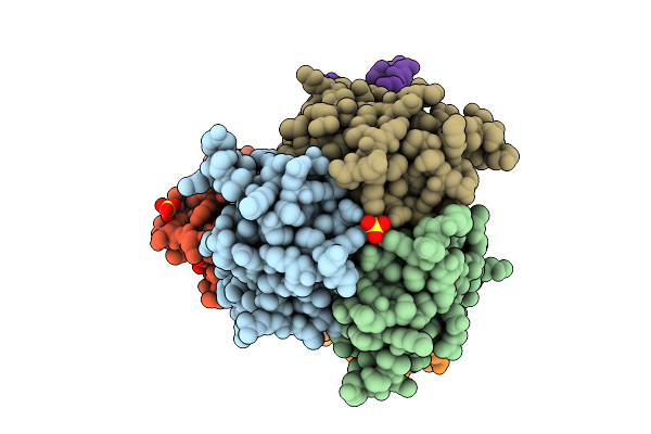 |
X-Ray Crystal Structure Of Tnfa-Vnar C4 Complex
Organism: Homo sapiens, Squalus acanthias
Method: X-RAY DIFFRACTION Resolution:1.92 Å Release Date: 2025-11-05 Classification: Cytokine/Immune System Ligands: EDO, SO4, NI |
Organism: Homo sapiens, Squalus acanthias
Method: X-RAY DIFFRACTION
Release Date: 2025-11-05
Ligands: EDO, SO4, NI
 |
X-Ray Crystal Structure Of Tnfa-Vnar D1 Complex
Organism: Squalus acanthias, Homo sapiens
Method: X-RAY DIFFRACTION Resolution:3.43 Å Release Date: 2025-09-10 Classification: TOXIN Ligands: GOL, SO4, CL |
Organism: Squalus acanthias, Homo sapiens
Method: X-RAY DIFFRACTION
Release Date: 2025-09-10
Ligands: GOL, SO4, CL
 |
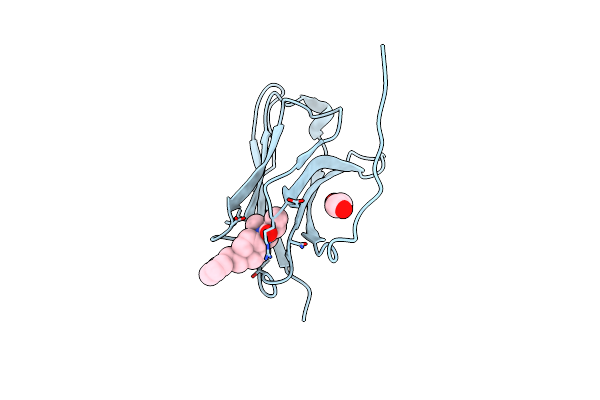
X-Ray Crystal Structure Of Jgfn4 Complexed With Fentanyl
Organism: Camelidae
Method: X-RAY DIFFRACTION Resolution:1.68 Å Release Date: 2024-08-07 Classification: ANTITOXIN Ligands: 7V7, EDO |
Organism: Camelidae
Method: X-RAY DIFFRACTION
Release Date: 2024-08-07
Ligands: 7V7, EDO
 |
X-Ray Crystal Structure Of Jgfn4 Complex With Fentanyl
Organism: Camelidae
Method: X-RAY DIFFRACTION Resolution:1.55 Å Release Date: 2024-08-07 Classification: ANTITOXIN Ligands: 7V7, EDO |
Organism: Camelidae
Method: X-RAY DIFFRACTION
Release Date: 2024-08-07
Ligands: 7V7, EDO
 |
X-Ray Crystal Structure Of Nanobody Jgfn4
Organism: Camelidae
Method: X-RAY DIFFRACTION Resolution:1.76 Å Release Date: 2024-08-07 Classification: ANTITOXIN |
Organism: Camelidae
Method: X-RAY DIFFRACTION
Release Date: 2024-08-07
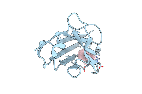 |
X-Ray Crystal Structure Of Jgfn4 N76D Complexed With Fentanyl In Monomer Form
Organism: Camelidae
Method: X-RAY DIFFRACTION Resolution:1.60 Å Release Date: 2024-08-07 Classification: ANTITOXIN Ligands: 7V7 |
Organism: Camelidae
Method: X-RAY DIFFRACTION
Release Date: 2024-08-07
Ligands: 7V7
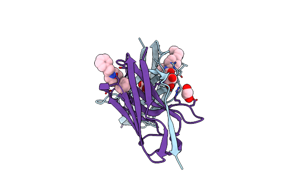 |
X-Ray Crystal Structure Of Jgfn4 N76D Complexed With Fentanyl In Dimer Form
Organism: Camelidae
Method: X-RAY DIFFRACTION Resolution:1.85 Å Release Date: 2024-08-07 Classification: ANTITOXIN Ligands: 7V7, PGE, EDO |
Organism: Camelidae
Method: X-RAY DIFFRACTION
Release Date: 2024-08-07
Ligands: 7V7, PGE, EDO
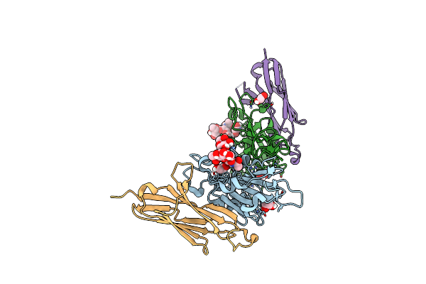 |
Crystal Structure Of The Sars-Cov-2 Receptor Binding Domain In Complex With Vnar 3B4
Organism: Severe acute respiratory syndrome coronavirus 2, Squalus acanthias
Method: X-RAY DIFFRACTION Resolution:1.92 Å Release Date: 2022-01-05 Classification: VIRAL PROTEIN/IMMUNE SYSTEM Ligands: EDO |
Organism: Severe acute respiratory syndrome coronavirus 2, Squalus acanthias
Method: X-RAY DIFFRACTION
Release Date: 2022-01-05
Ligands: EDO
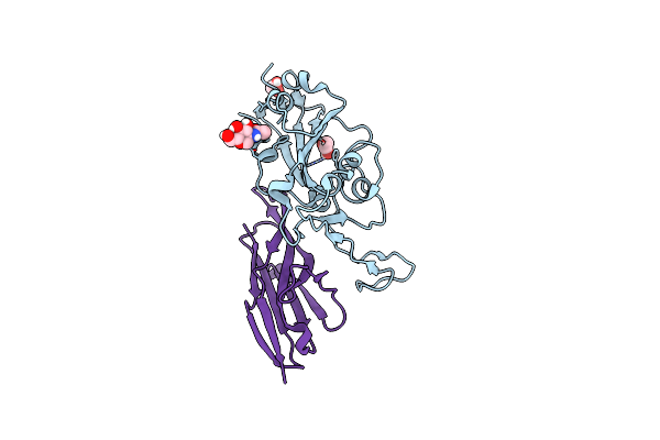 |
Crystal Structure Of The Sars-Cov-2 Receptor Binding Domain In Complex With Vnar 2C02
Organism: Severe acute respiratory syndrome coronavirus 2, Squalus acanthias
Method: X-RAY DIFFRACTION Resolution:1.96 Å Release Date: 2022-01-05 Classification: VIRAL PROTEIN/IMMUNE SYSTEM Ligands: EDO, NAG, CL |
Organism: Severe acute respiratory syndrome coronavirus 2, Squalus acanthias
Method: X-RAY DIFFRACTION
Release Date: 2022-01-05
Ligands: EDO, NAG, CL
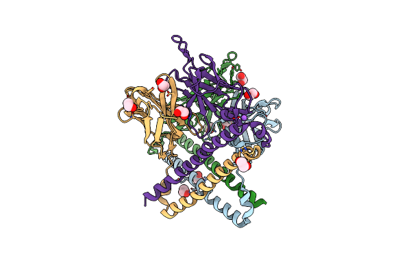 |
Apobec3B Antibody 5G7 Fv-Clasp
Organism: Homo sapiens
Method: X-RAY DIFFRACTION Resolution:1.67 Å Release Date: 2021-05-12 Classification: IMMUNE SYSTEM Ligands: EDO, CL, NA |
Organism: Homo sapiens
Method: X-RAY DIFFRACTION
Release Date: 2021-05-12
Ligands: EDO, CL, NA
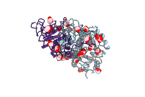 |
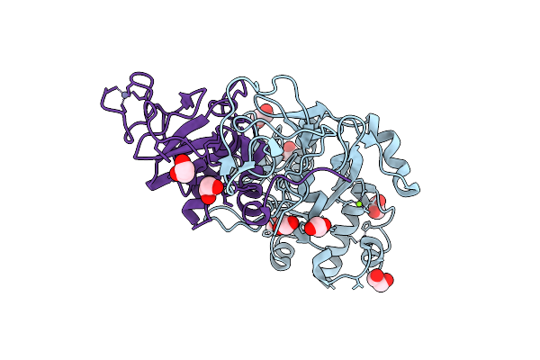
Crystal Structure Of The Sars-Cov-2 Exon-Nsp10 Complex
Organism: Severe acute respiratory syndrome coronavirus 2
Method: X-RAY DIFFRACTION Resolution:1.64 Å Release Date: 2021-05-05 Classification: VIRAL PROTEIN Ligands: ZN, EDO, TLA, CL |
Organism: Severe acute respiratory syndrome coronavirus 2
Method: X-RAY DIFFRACTION
Release Date: 2021-05-05
Ligands: ZN, EDO, TLA, CL
 |
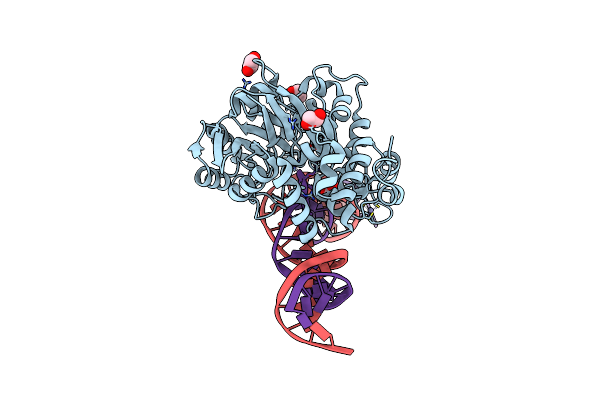
Crystal Structure Of The Sars-Cov-2 Exon-Nsp10 Complex Containing Mg2+ Ion
Organism: Severe acute respiratory syndrome coronavirus 2
Method: X-RAY DIFFRACTION Resolution:2.10 Å Release Date: 2021-05-05 Classification: VIRAL PROTEIN Ligands: ZN, EDO, CL, MG |
Organism: Severe acute respiratory syndrome coronavirus 2
Method: X-RAY DIFFRACTION
Release Date: 2021-05-05
Ligands: ZN, EDO, CL, MG
 |
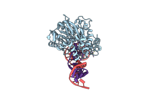
Crystal Structure Of Endonuclease Q Complex With 27-Mer Duplex Substrate With Du At The Active Site
Organism: Pyrococcus furiosus, Synthetic construct
Method: X-RAY DIFFRACTION Resolution:2.34 Å Release Date: 2021-03-17 Classification: HYDROLASE/DNA Ligands: ZN, MG, EDO |
Organism: Pyrococcus furiosus, Synthetic construct
Method: X-RAY DIFFRACTION
Release Date: 2021-03-17
Ligands: ZN, MG, EDO
 |
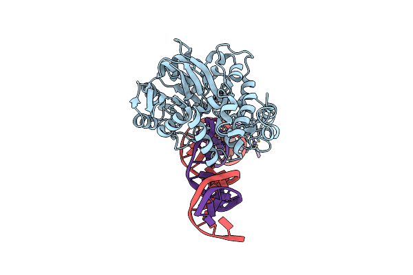
Crystal Structure Of Endonuclease Q Complex With 27-Mer Duplex Substrate With Di At The Active Site
Organism: Pyrococcus furiosus, Synthetic construct
Method: X-RAY DIFFRACTION Resolution:2.88 Å Release Date: 2021-03-17 Classification: HYDROLASE/DNA Ligands: EDO, ZN, MG, CL |
Organism: Pyrococcus furiosus, Synthetic construct
Method: X-RAY DIFFRACTION
Release Date: 2021-03-17
Ligands: EDO, ZN, MG, CL
 |
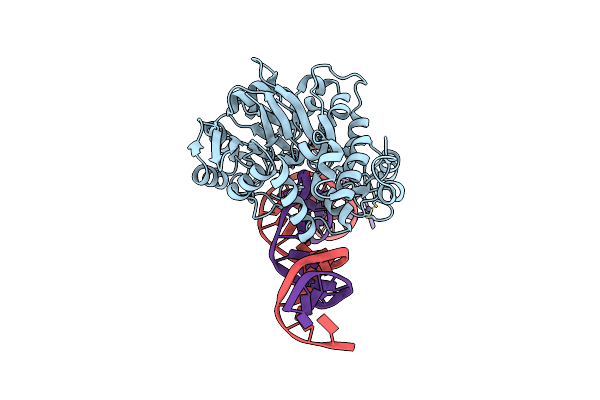
Crystal Structure Of Endonuclease Q Complex With 27-Mer Duplex Substrate With An Abasic Lesion At The Active Site
Organism: Pyrococcus furiosus, Synthetic construct
Method: X-RAY DIFFRACTION Resolution:3.11 Å Release Date: 2021-03-17 Classification: HYDROLASE/DNA Ligands: ZN, MG |
Organism: Pyrococcus furiosus, Synthetic construct
Method: X-RAY DIFFRACTION
Release Date: 2021-03-17
Ligands: ZN, MG
 |
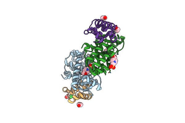
Crystal Structure Of Endonuclease Q Complex With 27-Mer Duplex Substrate With An Abasic Lesion At The Active Site
Organism: Pyrococcus furiosus, Synthetic construct
Method: X-RAY DIFFRACTION Resolution:3.11 Å Release Date: 2021-03-17 Classification: HYDROLASE/DNA Ligands: ZN, MG |
Organism: Pyrococcus furiosus, Synthetic construct
Method: X-RAY DIFFRACTION
Release Date: 2021-03-17
Ligands: ZN, MG
 |
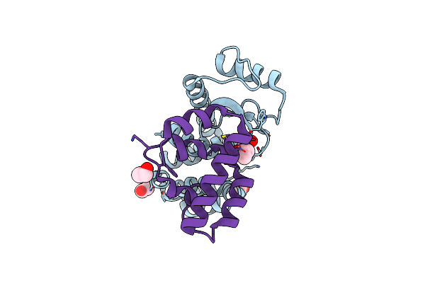
Crystal Structure Of Bacteriophage T4 Spackle And Lysozyme In Monoclinic Form
Organism: Escherichia virus t4
Method: X-RAY DIFFRACTION Resolution:1.78 Å Release Date: 2020-12-02 Classification: HYDROLASE Ligands: EDO, EPE, SIN, CL, FMT |
Organism: Escherichia virus t4
Method: X-RAY DIFFRACTION
Release Date: 2020-12-02
Ligands: EDO, EPE, SIN, CL, FMT
 |
Crystal Structure Of Bacteriophage T4 Spackle And Lysozyme In Orthorhombic Form
Organism: Escherichia virus t4
Method: X-RAY DIFFRACTION Resolution:1.92 Å Release Date: 2020-12-02 Classification: HYDROLASE Ligands: EDO, IPA |
Organism: Escherichia virus t4
Method: X-RAY DIFFRACTION
Release Date: 2020-12-02
Ligands: EDO, IPA
 |
Crystal Structure Of T4 Protein Spackle As Determined By Native Sad Phasing
Organism: Escherichia virus t4
Method: X-RAY DIFFRACTION Resolution:1.52 Å Release Date: 2020-09-16 Classification: VIRAL PROTEIN Ligands: CL |
Organism: Escherichia virus t4
Method: X-RAY DIFFRACTION
Release Date: 2020-09-16
Ligands: CL
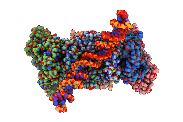 |
Cryo-Em Structure Of Htlv-1 Instasome
Organism: Saccharolobus solfataricus (strain atcc 35092 / dsm 1617 / jcm 11322 / p2), Human t-cell leukemia virus type i, Homo sapiens
Method: ELECTRON MICROSCOPY Release Date: 2020-07-01 Classification: DNA BINDING PROTEIN/DNA Ligands: ZN, MG |