

Search Count: 56
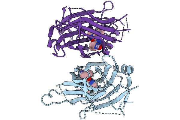 |
Tead2 Yap Binding Domain (Ybd)
Organism: Homo sapiens
Method: X-RAY DIFFRACTION Resolution:3.13 Å Release Date: 2026-03-04 Classification: TRANSCRIPTION Ligands: A1BXX |
Organism: Homo sapiens
Method: X-RAY DIFFRACTION
Release Date: 2026-03-04
Ligands: A1BXX
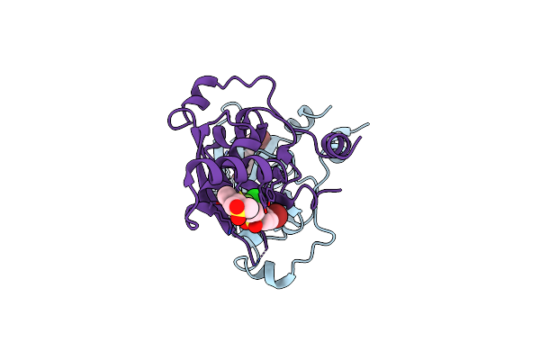 |
Covalent Inhibitor Vvd-442 Bound To The Ras Binding Domain (Rbd) Of Pi3Ka
Organism: Homo sapiens
Method: X-RAY DIFFRACTION Resolution:2.83 Å Release Date: 2025-01-01 Classification: PROTEIN BINDING Ligands: A1BF9 |
Organism: Homo sapiens
Method: X-RAY DIFFRACTION
Release Date: 2025-01-01
Ligands: A1BF9
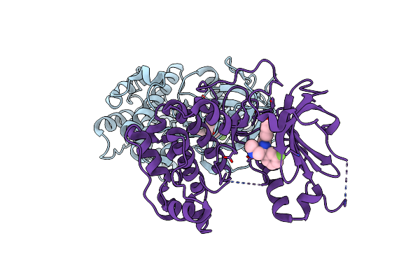 |
P70 S6K1 In Complex With Msc2317067A-1
Organism: Homo sapiens
Method: X-RAY DIFFRACTION Resolution:3.00 Å Release Date: 2022-07-06 Classification: TRANSFERASE/INHIBITOR Ligands: 1RJ |
Organism: Homo sapiens
Method: X-RAY DIFFRACTION
Release Date: 2022-07-06
Ligands: 1RJ
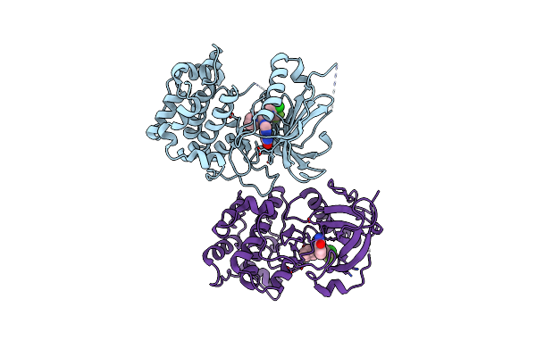 |
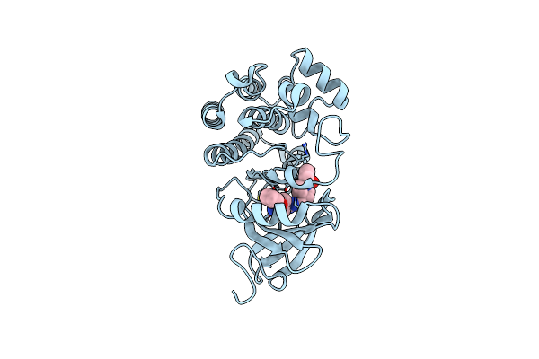
P70 S6K1 In Complex With Msc2363318A-1
Organism: Homo sapiens
Method: X-RAY DIFFRACTION Resolution:2.74 Å Release Date: 2022-07-06 Classification: TRANSFERASE/INHIBITOR Ligands: 1SK |
Organism: Homo sapiens
Method: X-RAY DIFFRACTION
Release Date: 2022-07-06
Ligands: 1SK
 |
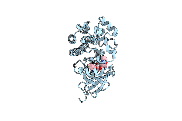
Structure Of Human Bruton'S Tyrosine Kinase In Complex With Evobrutinib
Organism: Homo sapiens
Method: X-RAY DIFFRACTION Resolution:1.41 Å Release Date: 2019-08-14 Classification: Transferase/Transferase Inhibitor Ligands: CL, MZJ |
Organism: Homo sapiens
Method: X-RAY DIFFRACTION
Release Date: 2019-08-14
Ligands: CL, MZJ
 |
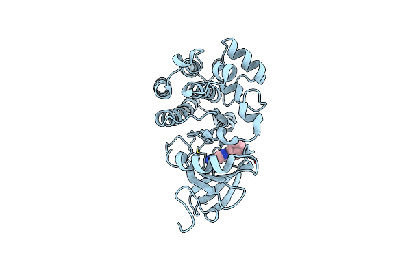
Discovery Of Affinity-Based Probes For Btk Occupancy Assay
Organism: Homo sapiens
Method: X-RAY DIFFRACTION Resolution:2.23 Å Release Date: 2019-02-06 Classification: IMMUNE SYSTEM Ligands: KHD |
Organism: Homo sapiens
Method: X-RAY DIFFRACTION
Release Date: 2019-02-06
Ligands: KHD
 |
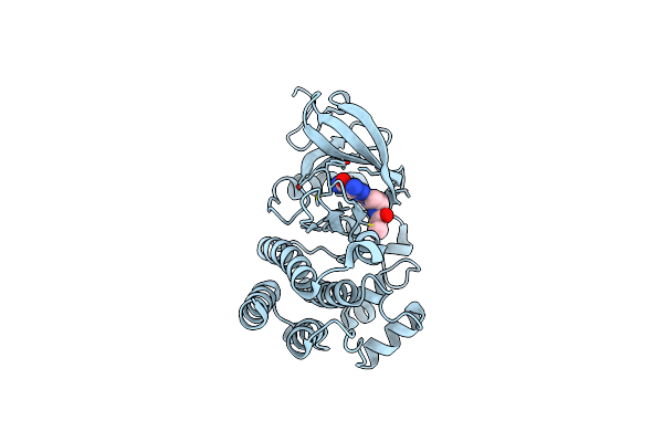
Crystal Structure Of Btk In Complex With Fragment Ligand
Organism: Homo sapiens
Method: X-RAY DIFFRACTION Resolution:1.30 Å Release Date: 2018-10-17 Classification: TRANSFERASE/TRANSFERASE Inhibitor Ligands: GJG |
Organism: Homo sapiens
Method: X-RAY DIFFRACTION
Release Date: 2018-10-17
Ligands: GJG
 |
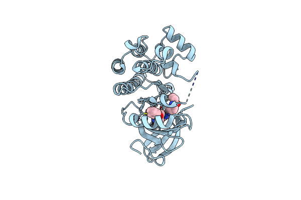
Crystal Structure Of Btk In Complex With Covalent Fragment Ligand
Organism: homo sapiens
Method: X-RAY DIFFRACTION Resolution:1.10 Å Release Date: 2018-10-17 Classification: TRANSFERASE/TRANSFERASE inhibitor Ligands: GJD |
Organism: homo sapiens
Method: X-RAY DIFFRACTION
Release Date: 2018-10-17
Ligands: GJD
 |
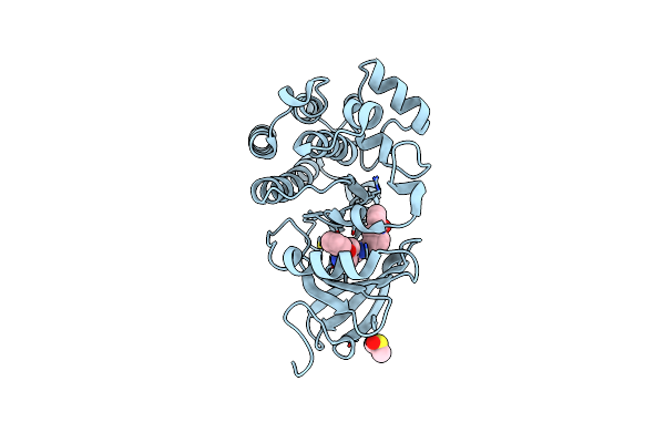
Crystal Structure Of Btk In Complex With Fragment Ligand
Organism: Homo sapiens
Method: X-RAY DIFFRACTION Resolution:2.00 Å Release Date: 2018-09-05 Classification: TRANSFERASE/TRANSFERASE Inhibitor Ligands: GJA |
Organism: Homo sapiens
Method: X-RAY DIFFRACTION
Release Date: 2018-09-05
Ligands: GJA
 |
Crystal Structure Of Btk In Complex With Covalent Inhibitor
Organism: Homo sapiens
Method: X-RAY DIFFRACTION Resolution:1.42 Å Release Date: 2018-09-05 Classification: TRANSFERASE/TRANSFERASE INHIBITOR Ligands: GJ7, DMS |
Organism: Homo sapiens
Method: X-RAY DIFFRACTION
Release Date: 2018-09-05
Ligands: GJ7, DMS
 |
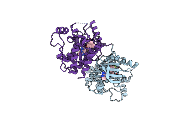
Crystal Structure Of Btk In Complex With Covalent Inhibitor
Organism: Homo sapiens
Method: X-RAY DIFFRACTION Resolution:1.25 Å Release Date: 2018-09-05 Classification: TRANSFERASE/TRANSFERASE Inhibitor Ligands: GJJ, DMS |
Organism: Homo sapiens
Method: X-RAY DIFFRACTION
Release Date: 2018-09-05
Ligands: GJJ, DMS
 |
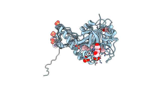
Crystal Structure Of Braf(V600E) In Complex With Ly3009120 Compnd
Organism: Homo sapiens
Method: X-RAY DIFFRACTION Resolution:2.70 Å Release Date: 2015-07-15 Classification: TRANSFERASE/TRANSFERASE INHIBITOR Ligands: 4Z5, CL |
Organism: Homo sapiens
Method: X-RAY DIFFRACTION
Release Date: 2015-07-15
Ligands: 4Z5, CL
 |
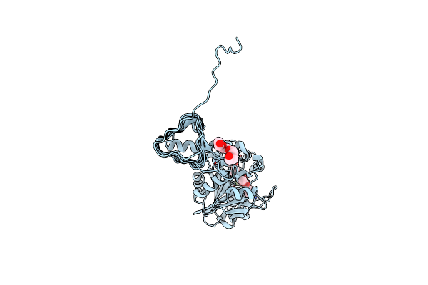
Crystal Structure Of Glmu From Haemophilus Influenzae In Complex With Quinazoline Inhibitor 1
Organism: Haemophilus influenzae
Method: X-RAY DIFFRACTION Resolution:1.99 Å Release Date: 2009-11-17 Classification: TRANSFERASE Ligands: LZR, PG4, PGE, SO4 |
Organism: Haemophilus influenzae
Method: X-RAY DIFFRACTION
Release Date: 2009-11-17
Ligands: LZR, PG4, PGE, SO4
 |
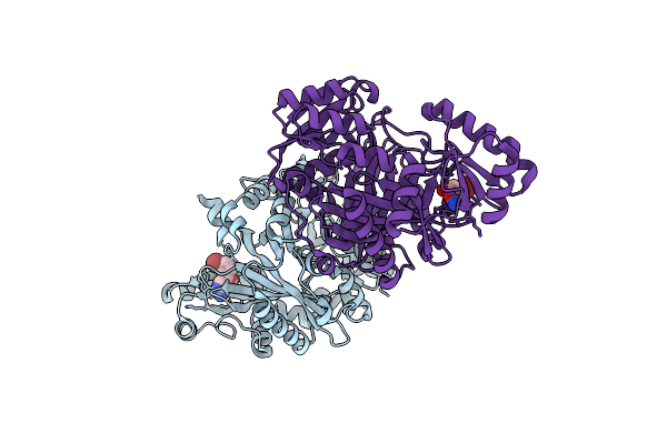
Crystal Structure Of Glmu From Haemophilus Influenzae In Complex With Quinazoline Inhibitor 2
Organism: Haemophilus influenzae
Method: X-RAY DIFFRACTION Resolution:2.59 Å Release Date: 2009-11-17 Classification: TRANSFERASE Ligands: LZS, PG4 |
Organism: Haemophilus influenzae
Method: X-RAY DIFFRACTION
Release Date: 2009-11-17
Ligands: LZS, PG4
 |
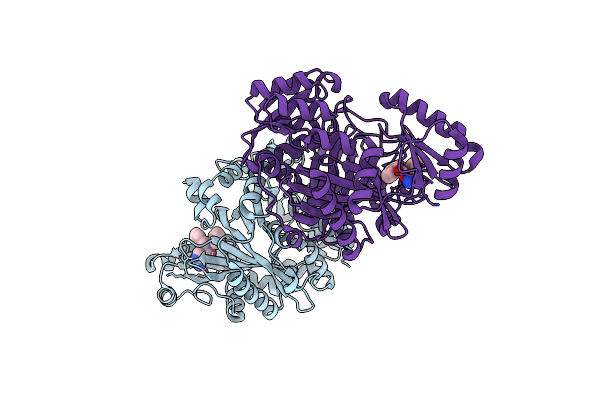
Crystal Structure Of Biotin Carboxylase From E. Coli In Complex With Amino-Oxazole Fragment Series
Organism: Escherichia coli
Method: X-RAY DIFFRACTION Resolution:2.00 Å Release Date: 2009-05-19 Classification: LIGASE Ligands: OA1, CL |
Organism: Escherichia coli
Method: X-RAY DIFFRACTION
Release Date: 2009-05-19
Ligands: OA1, CL
 |
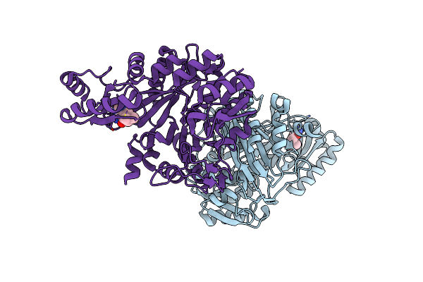
Crystal Structure Of Biotin Carboxylase From E. Coli In Complex With Amino-Oxazole Fragment Series
Organism: Escherichia coli
Method: X-RAY DIFFRACTION Resolution:1.87 Å Release Date: 2009-05-19 Classification: LIGASE Ligands: OA2, CL |
Organism: Escherichia coli
Method: X-RAY DIFFRACTION
Release Date: 2009-05-19
Ligands: OA2, CL
 |
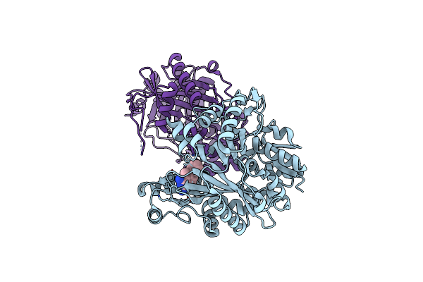
Crystal Structure Of Biotin Carboxylase From E. Coli In Complex With 4-Amino-7,7-Dimethyl-7,8-Dihydro-Quinazolinone Fragment
Organism: Escherichia coli
Method: X-RAY DIFFRACTION Resolution:2.50 Å Release Date: 2009-05-19 Classification: LIGASE Ligands: OA3, CL |
Organism: Escherichia coli
Method: X-RAY DIFFRACTION
Release Date: 2009-05-19
Ligands: OA3, CL
 |
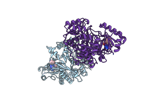
Crystal Structure Of Biotin Carboxylase From E. Coli In Complex With 5-Methyl-6-Phenyl-Quinazoline-2,4-Diamine
Organism: Escherichia coli
Method: X-RAY DIFFRACTION Resolution:1.85 Å Release Date: 2009-05-19 Classification: LIGASE Ligands: OA4 |
Organism: Escherichia coli
Method: X-RAY DIFFRACTION
Release Date: 2009-05-19
Ligands: OA4
 |
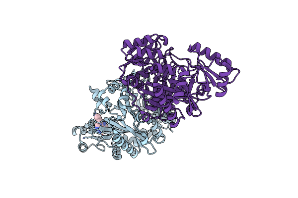
Crystal Structure Of Biotin Carboxylase From E. Coli In Complex With The Triazine-2,4-Diamine Fragment
Organism: Escherichia coli
Method: X-RAY DIFFRACTION Resolution:2.05 Å Release Date: 2009-05-19 Classification: LIGASE Ligands: OA5, CL |
Organism: Escherichia coli
Method: X-RAY DIFFRACTION
Release Date: 2009-05-19
Ligands: OA5, CL
 |
Crystal Structure Of Biotin Carboxylase From E. Coli In Complex With The 3-(3-Methyl-But-2-Enyl)-3H-Purin-6-Ylamine Fragment
Organism: Escherichia coli
Method: X-RAY DIFFRACTION Resolution:1.90 Å Release Date: 2009-05-19 Classification: LIGASE Ligands: L21, CL |
Organism: Escherichia coli
Method: X-RAY DIFFRACTION
Release Date: 2009-05-19
Ligands: L21, CL