

Search Count: 35
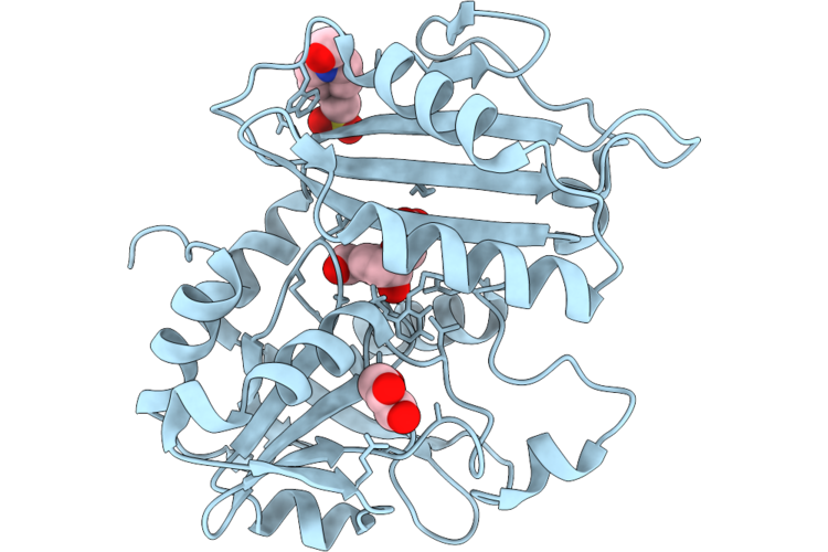 |
Crystal Structure Of 4-Amino-4-Deoxychorismate Lyase From Micromonospora Aurantiaca Atcc 27029 (Holo Form) In Complex With Chorismate
Organism: Micromonospora aurantiaca atcc 27029
Method: X-RAY DIFFRACTION Resolution:1.85 Å Release Date: 2026-05-27 Classification: LYASE Ligands: EPE, GOL, ISJ |
Organism: Micromonospora aurantiaca atcc 27029
Method: X-RAY DIFFRACTION
Release Date: 2026-05-27
Ligands: EPE, GOL, ISJ
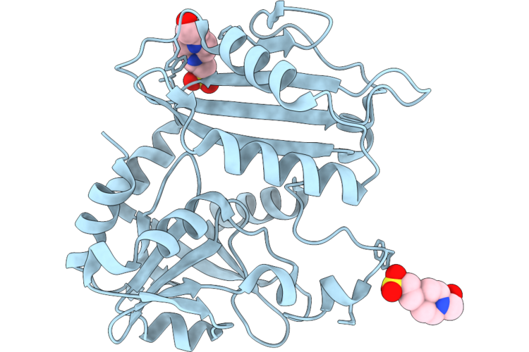 |
Crystal Structure Of 4-Amino-4-Deoxychorismate Lyase From Micromonospora Aurantiaca Atcc 27029 In Its Holo Form
Organism: Micromonospora aurantiaca atcc 27029
Method: X-RAY DIFFRACTION Resolution:2.00 Å Release Date: 2026-05-27 Classification: LYASE Ligands: EPE |
Organism: Micromonospora aurantiaca atcc 27029
Method: X-RAY DIFFRACTION
Release Date: 2026-05-27
Ligands: EPE
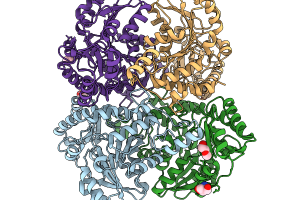 |
Cystathionine Beta-Lyase From Klebsiella Pneumoniae
Organism: Klebsiella pneumoniae
Method: X-RAY DIFFRACTION Resolution:1.90 Å Release Date: 2026-02-11 Classification: LYASE Ligands: PEG, TRS |
Organism: Klebsiella pneumoniae
Method: X-RAY DIFFRACTION
Release Date: 2026-02-11
Ligands: PEG, TRS
 |
Crystal Structure Of L-Asparaginase From Thermococcus Sibiricus
Organism: Thermococcus sibiricus
Method: X-RAY DIFFRACTION Resolution:1.90 Å Release Date: 2025-06-11 Classification: HYDROLASE Ligands: GLY |
Organism: Thermococcus sibiricus
Method: X-RAY DIFFRACTION
Release Date: 2025-06-11
Ligands: GLY
 |
Crystal Structure Of L-Asparaginase From Thermococcus Sibiricus Double Mutation D54G/T56Q
Organism: Thermococcus sibiricus
Method: X-RAY DIFFRACTION Resolution:1.90 Å Release Date: 2025-06-11 Classification: HYDROLASE Ligands: PEG, GOL, GLY |
Organism: Thermococcus sibiricus
Method: X-RAY DIFFRACTION
Release Date: 2025-06-11
Ligands: PEG, GOL, GLY
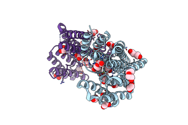 |
14-3-3 Zeta Chimera With The S202R Peptide Of Sars-Cov-2 N (Residues 200-213)
Organism: Homo sapiens, Severe acute respiratory syndrome coronavirus 2
Method: X-RAY DIFFRACTION Resolution:1.80 Å Release Date: 2025-05-21 Classification: SIGNALING PROTEIN Ligands: EDO, PEG |
Organism: Homo sapiens, Severe acute respiratory syndrome coronavirus 2
Method: X-RAY DIFFRACTION
Release Date: 2025-05-21
Ligands: EDO, PEG
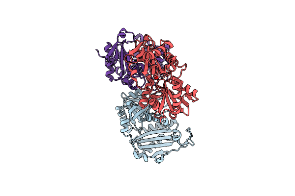 |
Crystal Structure Of Aminotransferase-Like Protein From Variovorax Paradoxus Mutant N174K
Organism: Variovorax paradoxus b4
Method: X-RAY DIFFRACTION Resolution:2.20 Å Release Date: 2025-01-29 Classification: TRANSFERASE Ligands: PEG |
Organism: Variovorax paradoxus b4
Method: X-RAY DIFFRACTION
Release Date: 2025-01-29
Ligands: PEG
 |
Crystal Structure Of Cysteine Synthase A From Limosilactobacillus Reuteri Lr1 In Its Apo Form
Organism: Limosilactobacillus reuteri
Method: X-RAY DIFFRACTION Resolution:2.20 Å Release Date: 2024-11-13 Classification: TRANSFERASE Ligands: PGE, EDO, PEG, SO4 |
Organism: Limosilactobacillus reuteri
Method: X-RAY DIFFRACTION
Release Date: 2024-11-13
Ligands: PGE, EDO, PEG, SO4
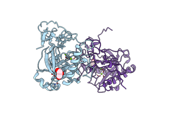 |
Crystal Structure Of Pyrophosphatase From Ogataea Parapolymorpha
Organism: Ogataea parapolymorpha
Method: X-RAY DIFFRACTION Resolution:1.80 Å Release Date: 2024-09-04 Classification: HYDROLASE Ligands: GOL, MG |
Organism: Ogataea parapolymorpha
Method: X-RAY DIFFRACTION
Release Date: 2024-09-04
Ligands: GOL, MG
 |
Crystal Structure Of The Biphotochromic Fluorescent Protein Moxsaasoti (F97M Variant) In Its Green On-State
Organism: Stylocoeniella armata
Method: X-RAY DIFFRACTION Resolution:2.00 Å Release Date: 2024-06-12 Classification: FLUORESCENT PROTEIN Ligands: NO3, EDO, PGE |
Organism: Stylocoeniella armata
Method: X-RAY DIFFRACTION
Release Date: 2024-06-12
Ligands: NO3, EDO, PGE
 |
The Structure Of Non-Activated Thiocyanate Dehydrogenase From Pelomicrobium Methylotrophicum (Pmtcdh)
Organism: Pelomicrobium methylotrophicum
Method: X-RAY DIFFRACTION Resolution:1.45 Å Release Date: 2024-05-08 Classification: OXIDOREDUCTASE Ligands: EDO, CL, CU, PEG |
Organism: Pelomicrobium methylotrophicum
Method: X-RAY DIFFRACTION
Release Date: 2024-05-08
Ligands: EDO, CL, CU, PEG
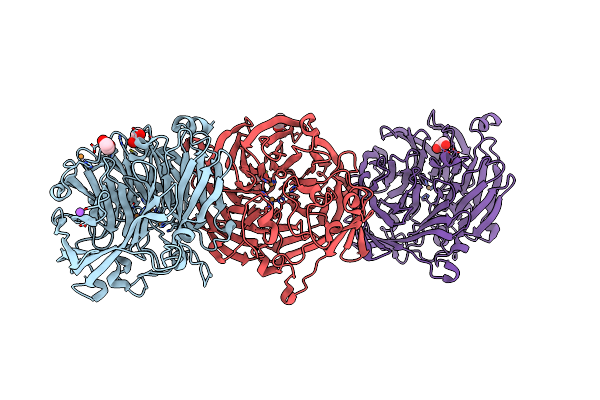 |
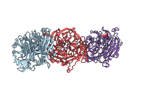
The Structure Of Thiocyanate Dehydrogenase From Pelomicrobium Methylotrophicum (Pmtcdh), Activated By Crystals Soaking With 1 Mm Cucl2 During 6 Months
Organism: Pelomicrobium methylotrophicum
Method: X-RAY DIFFRACTION Resolution:1.80 Å Release Date: 2024-05-08 Classification: OXIDOREDUCTASE Ligands: EDO, CU, NA |
Organism: Pelomicrobium methylotrophicum
Method: X-RAY DIFFRACTION
Release Date: 2024-05-08
Ligands: EDO, CU, NA
 |
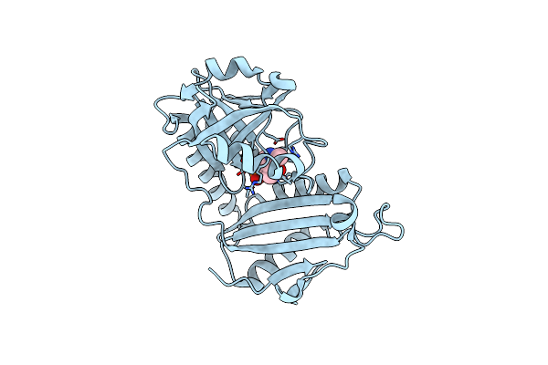
The Structure Of Thiocyanate Dehydrogenase From Pelomicrobium Methylotrophicum (Pmtcdh), Activated By Crystals Soaking With 1 Mm Cucl2 And Na Ascorbate During 12 Hours
Organism: Pelomicrobium methylotrophicum
Method: X-RAY DIFFRACTION Resolution:2.00 Å Release Date: 2024-05-08 Classification: OXIDOREDUCTASE Ligands: CU, EDO |
Organism: Pelomicrobium methylotrophicum
Method: X-RAY DIFFRACTION
Release Date: 2024-05-08
Ligands: CU, EDO
 |
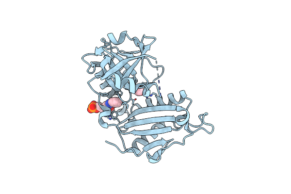
Crystal Structure Of D-Amino Acid Transaminase From Haliscomenobacter Hydrossis In The Holo Form Obtained At Ph 7.0
Organism: Haliscomenobacter hydrossis dsm 1100
Method: X-RAY DIFFRACTION Resolution:2.00 Å Release Date: 2024-04-17 Classification: TRANSFERASE Ligands: PLP |
Organism: Haliscomenobacter hydrossis dsm 1100
Method: X-RAY DIFFRACTION
Release Date: 2024-04-17
Ligands: PLP
 |
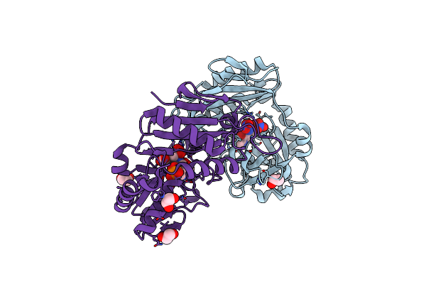
Crystal Structure Of D-Amino Acid Transaminase From Haliscomenobacter Hydrossis (Apo Form) After 15 Sec Of Soaking With Phenylhydrazine
Organism: Haliscomenobacter hydrossis dsm 1100
Method: X-RAY DIFFRACTION Resolution:2.00 Å Release Date: 2024-04-17 Classification: TRANSFERASE Ligands: ACT, ZXN |
Organism: Haliscomenobacter hydrossis dsm 1100
Method: X-RAY DIFFRACTION
Release Date: 2024-04-17
Ligands: ACT, ZXN
 |
Crystal Structure Of D-Amino Acid Aminotransferase From Aminobacterium Colombiense Point Mutant E113A Complexed With D-Glutamate
Organism: Aminobacterium colombiense
Method: X-RAY DIFFRACTION Resolution:1.85 Å Release Date: 2024-04-10 Classification: TRANSFERASE Ligands: EDO, NO3, PMP, PW0 |
Organism: Aminobacterium colombiense
Method: X-RAY DIFFRACTION
Release Date: 2024-04-10
Ligands: EDO, NO3, PMP, PW0
 |
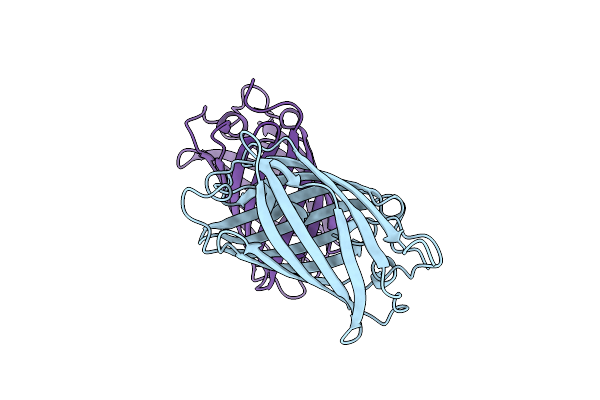
Structure Of Mbaojin2
Organism: Cytaeis uchidae
Method: X-RAY DIFFRACTION Resolution:1.90 Å Release Date: 2024-03-27 Classification: FLUORESCENT PROTEIN Ligands: SO4, GOL, CL |
Organism: Cytaeis uchidae
Method: X-RAY DIFFRACTION
Release Date: 2024-03-27
Ligands: SO4, GOL, CL
 |
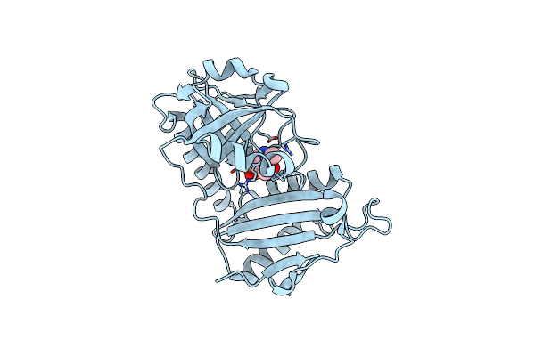
Crystal Structure Of The Biphotochromic Fluorescent Protein Saasoti (C21N/V127T Variant) In Its Green On-State
Organism: Stylocoeniella armata
Method: X-RAY DIFFRACTION Resolution:3.00 Å Release Date: 2024-01-17 Classification: FLUORESCENT PROTEIN |
Organism: Stylocoeniella armata
Method: X-RAY DIFFRACTION
Release Date: 2024-01-17
 |
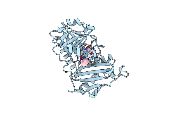
Crystal Structure Of D-Amino Acid Transaminase From Haliscomenobacter Hydrossis Point Mutant R90I (Holo Form)
Organism: Haliscomenobacter hydrossis dsm 1100
Method: X-RAY DIFFRACTION Resolution:2.00 Å Release Date: 2023-12-27 Classification: TRANSFERASE Ligands: PLP |
Organism: Haliscomenobacter hydrossis dsm 1100
Method: X-RAY DIFFRACTION
Release Date: 2023-12-27
Ligands: PLP
 |
Crystal Structure Of D-Amino Acid Transaminase From Haliscomenobacter Hydrossis Point Mutant R90I Complexed With Phenylhydrazine
Organism: Haliscomenobacter hydrossis dsm 1100
Method: X-RAY DIFFRACTION Resolution:2.00 Å Release Date: 2023-12-27 Classification: TRANSFERASE Ligands: ZXN, GOL |
Organism: Haliscomenobacter hydrossis dsm 1100
Method: X-RAY DIFFRACTION
Release Date: 2023-12-27
Ligands: ZXN, GOL