

Search Count: 90
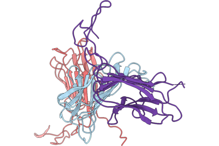 |
Crystal Structure Of Human Adamts-5 Cb And Spacer Domains
Organism: Homo sapiens
Method: X-RAY DIFFRACTION Resolution:2.60 Å Release Date: 2026-05-20 Classification: PROTEIN BINDING |
Organism: Homo sapiens
Method: X-RAY DIFFRACTION
Release Date: 2026-05-20
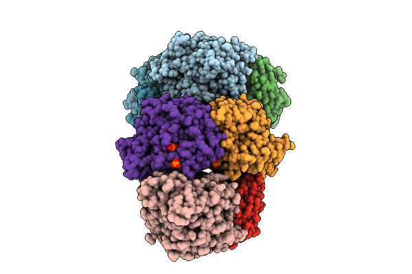 |
Crystal Structure Of Trans-O-Hydroxybenzylidenepyruvate Hydratase-Aldolase From Pseudomonas Fluorescens N3 Bound To Substrate Intermediate
Organism: Pseudomonas fluorescens
Method: X-RAY DIFFRACTION Resolution:1.76 Å Release Date: 2025-07-23 Classification: LYASE Ligands: KRN, PO4, GOL, NK |
Organism: Pseudomonas fluorescens
Method: X-RAY DIFFRACTION
Release Date: 2025-07-23
Ligands: KRN, PO4, GOL, NK
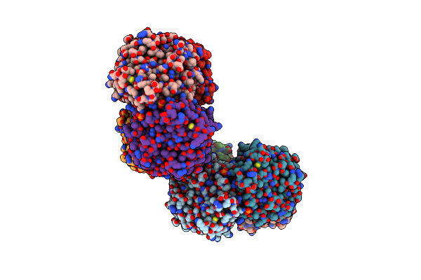 |
Crystal Structure Of Trans-O-Hydroxybenzylidenepyruvate Hydratase-Aldolase From Pseudomonas Fluorescens N3 Bound To Pyruvate
Organism: Pseudomonas fluorescens
Method: X-RAY DIFFRACTION Resolution:2.30 Å Release Date: 2025-07-02 Classification: LYASE Ligands: PYR, PO4, GOL |
Organism: Pseudomonas fluorescens
Method: X-RAY DIFFRACTION
Release Date: 2025-07-02
Ligands: PYR, PO4, GOL
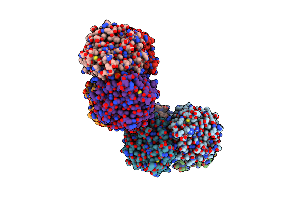 |
Crystal Structure Of Trans-O-Hydroxybenzylidenepyruvate Hydratase-Aldolase From Pseudomonas Fluorescens N3
Organism: Pseudomonas fluorescens
Method: X-RAY DIFFRACTION Resolution:1.96 Å Release Date: 2025-06-25 Classification: LYASE Ligands: GOL, PO4 |
Organism: Pseudomonas fluorescens
Method: X-RAY DIFFRACTION
Release Date: 2025-06-25
Ligands: GOL, PO4
 |
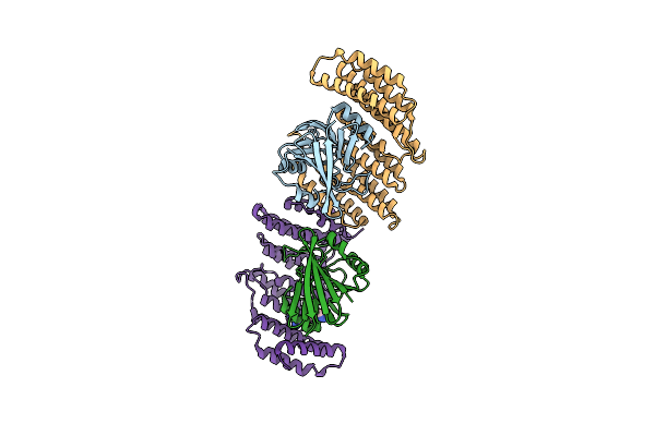
Crystal Structure Of Murine Aif Bound To N-Terminal Domain Of Chchd4
Organism: Mus musculus
Method: X-RAY DIFFRACTION Resolution:3.21 Å Release Date: 2024-05-08 Classification: PROTEIN BINDING Ligands: FAD, NAD |
Organism: Mus musculus
Method: X-RAY DIFFRACTION
Release Date: 2024-05-08
Ligands: FAD, NAD
 |
Crystal Structure Of The Retromer Complex Vps29/Vps35 With The Ligand Bis-1,3-Phenyl Guanylhydrazone, 2A
Organism: Homo sapiens
Method: X-RAY DIFFRACTION Resolution:2.50 Å Release Date: 2024-03-27 Classification: PROTEIN TRANSPORT Ligands: XFZ |
Organism: Homo sapiens
Method: X-RAY DIFFRACTION
Release Date: 2024-03-27
Ligands: XFZ
 |
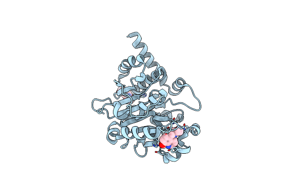
Crystal Structure Of The Retromer Complex Vps29/Vps35 With The Ligand Bis-1,3-Phenyl Guanylhydrazone, 2A
Organism: Homo sapiens
Method: X-RAY DIFFRACTION Resolution:2.40 Å Release Date: 2024-03-27 Classification: PROTEIN TRANSPORT Ligands: XFZ, TRS |
Organism: Homo sapiens
Method: X-RAY DIFFRACTION
Release Date: 2024-03-27
Ligands: XFZ, TRS
 |
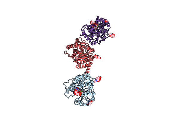
Catalytic Domain Of Udp-Glucose Glycoprotein Glucosyltransferase From Chaetomium Thermophilum In Complex With The 5-[(Morpholin-4-Yl)Methyl]Quinolin-8-Ol Inhibitor
Organism: Chaetomium thermophilum
Method: X-RAY DIFFRACTION Resolution:1.65 Å Release Date: 2023-04-26 Classification: TRANSFERASE Ligands: JM3, NAG, MAN |
Organism: Chaetomium thermophilum
Method: X-RAY DIFFRACTION
Release Date: 2023-04-26
Ligands: JM3, NAG, MAN
 |
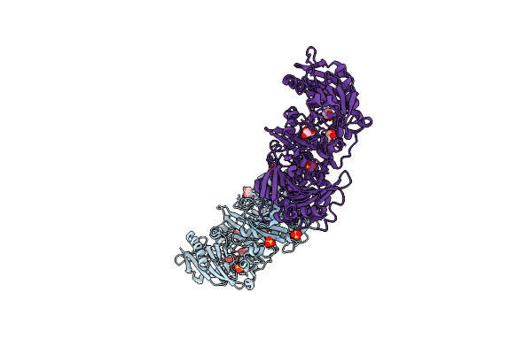
Catalytic Domain Of Udp-Glucose Glycoprotein Glucosyltransferase From Chaetomium Thermophilum In Complex With Udp-2-Deoxy-2-Fluoro-D-Glucose
Organism: Chaetomium thermophilum
Method: X-RAY DIFFRACTION Resolution:2.05 Å Release Date: 2022-06-22 Classification: TRANSFERASE Ligands: NAG, U2F, CA, PDO |
Organism: Chaetomium thermophilum
Method: X-RAY DIFFRACTION
Release Date: 2022-06-22
Ligands: NAG, U2F, CA, PDO
 |
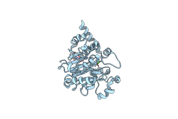
Crystal Structure Of Human Gelsolin Amyloid Mutant A551P
Organism: Homo sapiens
Method: X-RAY DIFFRACTION Resolution:3.00 Å Release Date: 2022-05-18 Classification: STRUCTURAL PROTEIN Ligands: SO4, GOL, CL |
Organism: Homo sapiens
Method: X-RAY DIFFRACTION
Release Date: 2022-05-18
Ligands: SO4, GOL, CL
 |
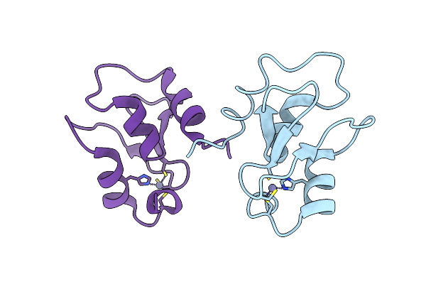
Catalytic Domain Of Udp-Glucose Glycoprotein Glucosyltransferase From Chaetomium Thermophilum (Apo Form)
Organism: Chaetomium thermophilum
Method: X-RAY DIFFRACTION Resolution:1.77 Å Release Date: 2022-04-20 Classification: TRANSFERASE Ligands: CA |
Organism: Chaetomium thermophilum
Method: X-RAY DIFFRACTION
Release Date: 2022-04-20
Ligands: CA
 |
Structure Of The Bir1 Domain Of Ciap2
Organism: Homo sapiens
Method: X-RAY DIFFRACTION Resolution:3.30 Å Release Date: 2022-01-12 Classification: APOPTOSIS Ligands: ZN |
Organism: Homo sapiens
Method: X-RAY DIFFRACTION
Release Date: 2022-01-12
Ligands: ZN
 |
Crystal Structure Of The Pathological N184K Variant Of Calcium-Free Human Gelsolin
Organism: Homo sapiens
Method: X-RAY DIFFRACTION Resolution:2.73 Å Release Date: 2019-11-27 Classification: STRUCTURAL PROTEIN Ligands: CL, GOL, SO4, TRS |
Organism: Homo sapiens
Method: X-RAY DIFFRACTION
Release Date: 2019-11-27
Ligands: CL, GOL, SO4, TRS
 |
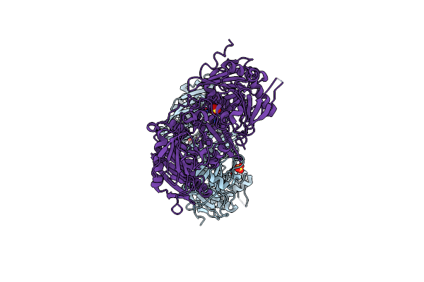
Crystal Structure Of The Pathological G167R Variant Of Calcium-Free Human Gelsolin,
Organism: Homo sapiens
Method: X-RAY DIFFRACTION Resolution:3.80 Å Release Date: 2019-11-27 Classification: STRUCTURAL PROTEIN Ligands: SO4, GOL |
Organism: Homo sapiens
Method: X-RAY DIFFRACTION
Release Date: 2019-11-27
Ligands: SO4, GOL
 |
Crystal Structure Of The Pathological D187N Variant Of Calcium-Free Human Gelsolin.
Organism: Homo sapiens
Method: X-RAY DIFFRACTION Resolution:3.50 Å Release Date: 2019-11-27 Classification: STRUCTURAL PROTEIN Ligands: SO4, GOL, NA |
Organism: Homo sapiens
Method: X-RAY DIFFRACTION
Release Date: 2019-11-27
Ligands: SO4, GOL, NA
 |
Calcium-Bound Gelsolin Domain 2
Organism: Homo sapiens
Method: X-RAY DIFFRACTION Resolution:1.30 Å Release Date: 2019-08-28 Classification: STRUCTURAL PROTEIN Ligands: CA |
Organism: Homo sapiens
Method: X-RAY DIFFRACTION
Release Date: 2019-08-28
Ligands: CA
 |
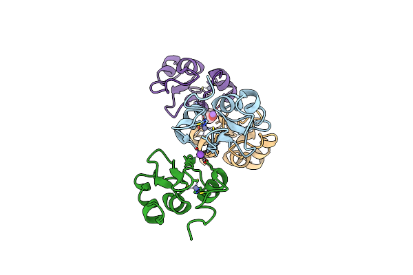
Structure Of Xiap-Bir1 Domain In Complex With An Nf023 Analog
Organism: Homo sapiens
Method: X-RAY DIFFRACTION Resolution:1.90 Å Release Date: 2019-05-29 Classification: APOPTOSIS Ligands: ZN, F2H |
Organism: Homo sapiens
Method: X-RAY DIFFRACTION
Release Date: 2019-05-29
Ligands: ZN, F2H
 |
Structure Of Xiap-Bir1 V86E Mutant
Organism: Homo sapiens
Method: X-RAY DIFFRACTION Resolution:2.30 Å Release Date: 2019-05-01 Classification: APOPTOSIS Ligands: ZN, NA, PEG |
Organism: Homo sapiens
Method: X-RAY DIFFRACTION
Release Date: 2019-05-01
Ligands: ZN, NA, PEG
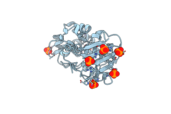 |
Crystal Structure Of The Periplasmic Domain Of Tgpa From Pseudomonas Aeruginosa
Organism: pseudomonas aeruginosa pao1
Method: X-RAY DIFFRACTION Resolution:1.60 Å Release Date: 2019-04-10 Classification: TRANSFERASE Ligands: PO4, CL |
Organism: pseudomonas aeruginosa pao1
Method: X-RAY DIFFRACTION
Release Date: 2019-04-10
Ligands: PO4, CL
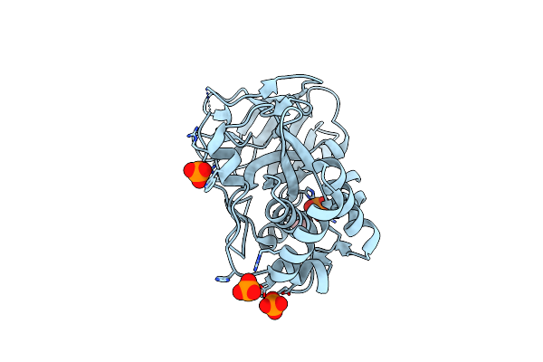 |
Crystal Structure Of The Periplasmic Domain Of Tgpa From Pseudomonas Aeruginosa Bound To Ethylmercury
Organism: Pseudomonas aeruginosa pao1
Method: X-RAY DIFFRACTION Resolution:1.80 Å Release Date: 2019-04-10 Classification: TRANSFERASE Ligands: PO4, EMC |
Organism: Pseudomonas aeruginosa pao1
Method: X-RAY DIFFRACTION
Release Date: 2019-04-10
Ligands: PO4, EMC