

Search Count: 49
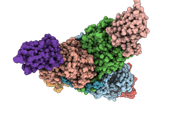 |
Crystal Structure Of The Complex Between Nb474 Mutant R53A,D125A And Trypanosoma Congolense Fructose-1,6-Bisphosphate Aldolase
Organism: Trypanosoma congolense il3000, Vicugna pacos
Method: X-RAY DIFFRACTION Resolution:1.75 Å Release Date: 2025-10-01 Classification: LYASE Ligands: ACT, GOL |
Organism: Trypanosoma congolense il3000, Vicugna pacos
Method: X-RAY DIFFRACTION
Release Date: 2025-10-01
Ligands: ACT, GOL
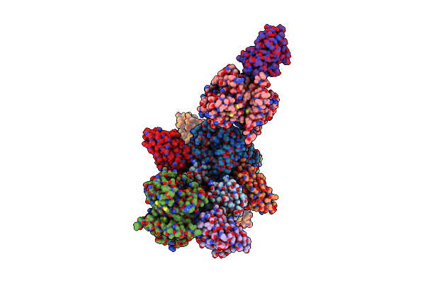 |
Crystal Structure Of Trypanosoma Congolense Pyruvate Kinase In Complex With A Single-Domain Antibody (Tcopyk-Sdab42)
Organism: Trypanosoma congolense, Vicugna pacos
Method: X-RAY DIFFRACTION Resolution:2.80 Å Release Date: 2025-04-16 Classification: TRANSFERASE Ligands: GOL |
Organism: Trypanosoma congolense, Vicugna pacos
Method: X-RAY DIFFRACTION
Release Date: 2025-04-16
Ligands: GOL
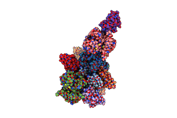 |
Crystal Structure Of Trypanosoma Congolense Pyruvate Kinase In Complex With A Single-Domain Antibody (Tcopyk-Sdab42) In The Presence Of Sulfate
Organism: Trypanosoma congolense, Vicugna pacos
Method: X-RAY DIFFRACTION Resolution:3.19 Å Release Date: 2025-04-16 Classification: TRANSFERASE Ligands: SO4, GOL, GLU |
Organism: Trypanosoma congolense, Vicugna pacos
Method: X-RAY DIFFRACTION
Release Date: 2025-04-16
Ligands: SO4, GOL, GLU
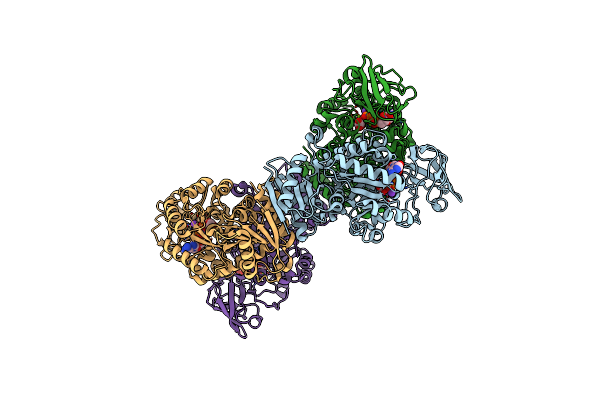 |
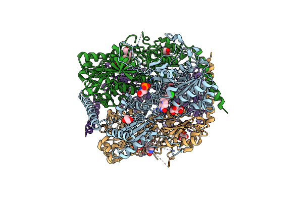
Crystal Structure Of Pyruvate Kinase (Pyk) From Plasmodium Falciparum In Complex With Oxalate And Atp
Organism: Plasmodium falciparum (isolate 3d7)
Method: X-RAY DIFFRACTION Resolution:2.60 Å Release Date: 2020-08-26 Classification: TRANSFERASE Ligands: MG, ATP, K, OXL |
Organism: Plasmodium falciparum (isolate 3d7)
Method: X-RAY DIFFRACTION
Release Date: 2020-08-26
Ligands: MG, ATP, K, OXL
 |
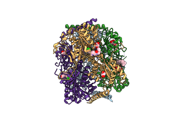
Crystal Structure Of Phosphofructokinase From Trypanosoma Brucei In Complex With An Allosteric Inhibitor Ctcb360
Organism: Trypanosoma brucei brucei
Method: X-RAY DIFFRACTION Resolution:2.35 Å Release Date: 2020-03-18 Classification: TRANSFERASE Ligands: JJ5, CIT, GOL, SEP |
Organism: Trypanosoma brucei brucei
Method: X-RAY DIFFRACTION
Release Date: 2020-03-18
Ligands: JJ5, CIT, GOL, SEP
 |
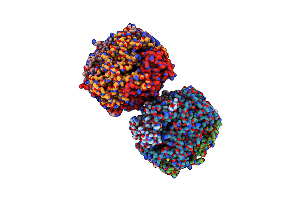
Crystal Structure Of Phosphofructokinase From Trypanosoma Brucei In Complex With An Allosteric Inhibitor Ctcb405
Organism: Trypanosoma brucei brucei
Method: X-RAY DIFFRACTION Resolution:2.75 Å Release Date: 2020-03-18 Classification: TRANSFERASE Ligands: JJ2, CIT, GOL |
Organism: Trypanosoma brucei brucei
Method: X-RAY DIFFRACTION
Release Date: 2020-03-18
Ligands: JJ2, CIT, GOL
 |
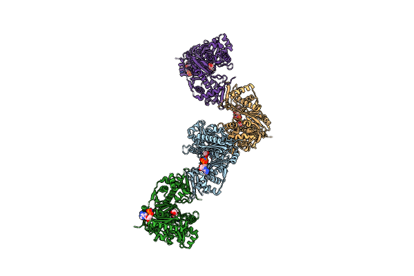
Crystal Structure Of Phosphofructokinase From Trypanosoma Brucei In Complex With An Allosteric Inhibitor Ctcb12
Organism: Trypanosoma brucei brucei
Method: X-RAY DIFFRACTION Resolution:3.40 Å Release Date: 2020-03-18 Classification: TRANSFERASE Ligands: CIT, GOL, JJ8 |
Organism: Trypanosoma brucei brucei
Method: X-RAY DIFFRACTION
Release Date: 2020-03-18
Ligands: CIT, GOL, JJ8
 |
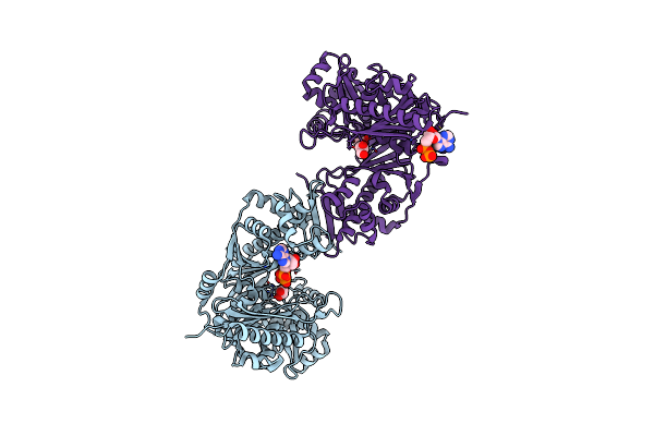
Crystal Structure Of Trypanosoma Brucei Gambiense Glycerol Kinase Complex With Amp-Pnp.
Organism: Trypanosoma brucei gambiense
Method: X-RAY DIFFRACTION Resolution:2.70 Å Release Date: 2020-01-29 Classification: TRANSFERASE Ligands: GOL, ANP |
Organism: Trypanosoma brucei gambiense
Method: X-RAY DIFFRACTION
Release Date: 2020-01-29
Ligands: GOL, ANP
 |
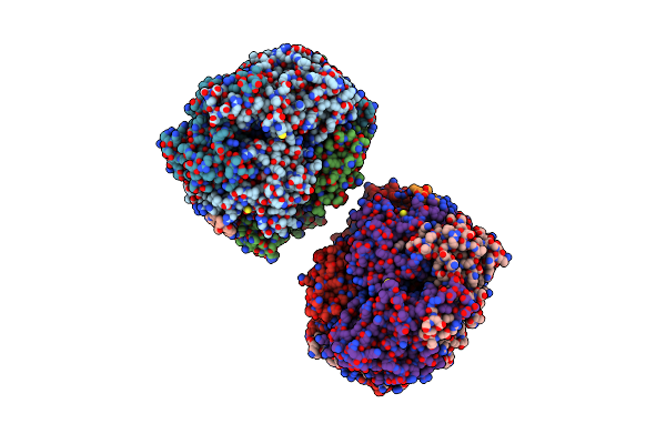
Crystal Structure Of Trypanosoma Brucei Gambiense Glycerol Kinase Complex With Adp.
Organism: Trypanosoma brucei gambiense
Method: X-RAY DIFFRACTION Resolution:2.40 Å Release Date: 2020-01-29 Classification: TRANSFERASE Ligands: ADP, GOL |
Organism: Trypanosoma brucei gambiense
Method: X-RAY DIFFRACTION
Release Date: 2020-01-29
Ligands: ADP, GOL
 |
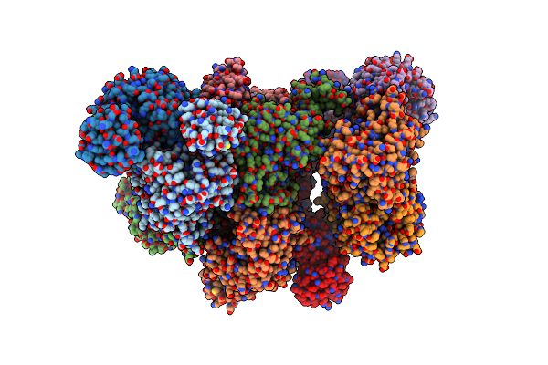
Structure Of Trypanosome Brucei Phosphofructokinase In Complex With Amp.
Organism: Trypanosoma brucei brucei
Method: X-RAY DIFFRACTION Resolution:2.75 Å Release Date: 2019-12-25 Classification: TRANSFERASE Ligands: BNZ, GOL, AMP |
Organism: Trypanosoma brucei brucei
Method: X-RAY DIFFRACTION
Release Date: 2019-12-25
Ligands: BNZ, GOL, AMP
 |
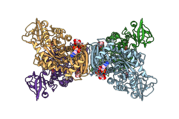
Crystal Structure Of M2 Pyk In Complex With Alanine.
Organism: Homo sapiens
Method: X-RAY DIFFRACTION Resolution:3.72 Å Release Date: 2019-09-04 Classification: TRANSFERASE Ligands: PO4, ALA |
Organism: Homo sapiens
Method: X-RAY DIFFRACTION
Release Date: 2019-09-04
Ligands: PO4, ALA
 |
Crystal Structure Of Pyruvate Kinase (Pyk) From Mycobacterium Tuberculosis In Complex With Oxalate, Amp And Inhibitor Ribose 5-Phosphate
Organism: Mycobacterium tuberculosis (strain atcc 25618 / h37rv)
Method: X-RAY DIFFRACTION Resolution:2.55 Å Release Date: 2019-08-14 Classification: TRANSFERASE Ligands: MG, OXL, R5P, AMP |
Organism: Mycobacterium tuberculosis (strain atcc 25618 / h37rv)
Method: X-RAY DIFFRACTION
Release Date: 2019-08-14
Ligands: MG, OXL, R5P, AMP
 |
Crystal Structure Of M2 Pyk In Complex With Phenyalanine.
Organism: Homo sapiens
Method: X-RAY DIFFRACTION Resolution:2.46 Å Release Date: 2018-05-23 Classification: TRANSFERASE Ligands: PO4, PHE, K |
Organism: Homo sapiens
Method: X-RAY DIFFRACTION
Release Date: 2018-05-23
Ligands: PO4, PHE, K
 |
Crystal Structure Of M2 Pyk In Complex With Tryptophan.
Organism: Homo sapiens
Method: X-RAY DIFFRACTION Resolution:3.20 Å Release Date: 2018-05-23 Classification: TRANSFERASE Ligands: PO4, TRP, K |
Organism: Homo sapiens
Method: X-RAY DIFFRACTION
Release Date: 2018-05-23
Ligands: PO4, TRP, K
 |
Crystal Structure Of M2 Pyk In Complex With Serine.
Organism: Homo sapiens
Method: X-RAY DIFFRACTION Resolution:2.96 Å Release Date: 2018-05-23 Classification: TRANSFERASE Ligands: K, MG, SER, PO4 |
Organism: Homo sapiens
Method: X-RAY DIFFRACTION
Release Date: 2018-05-23
Ligands: K, MG, SER, PO4
 |
Crystal Structure Of Leishmania Major Fructose-1,6-Bisphosphatase In Holo Form.
Organism: Leishmania major
Method: X-RAY DIFFRACTION Resolution:2.80 Å Release Date: 2017-09-20 Classification: HYDROLASE Ligands: PO4, MN, K, CIT |
Organism: Leishmania major
Method: X-RAY DIFFRACTION
Release Date: 2017-09-20
Ligands: PO4, MN, K, CIT
 |
Crystal Structure Of Leishmania Major Fructose-1,6-Bisphosphatase In Apo Form.
Organism: Leishmania major
Method: X-RAY DIFFRACTION Resolution:2.41 Å Release Date: 2017-09-20 Classification: HYDROLASE |
Organism: Leishmania major
Method: X-RAY DIFFRACTION
Release Date: 2017-09-20
 |
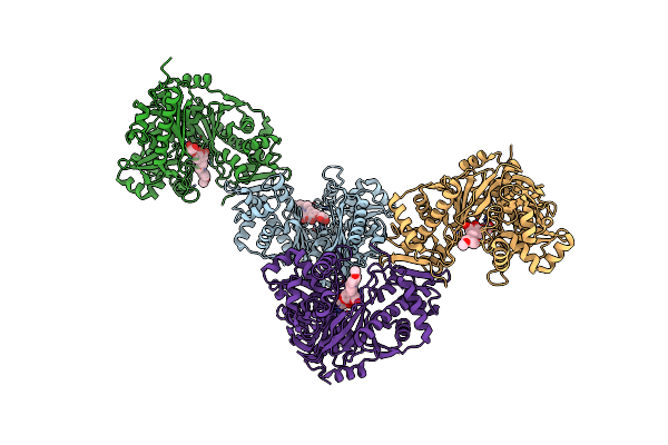
Crystal Structure Of Leishmania Major Fructose-1,6-Bisphosphatase In T-State.
Organism: Leishmania major
Method: X-RAY DIFFRACTION Resolution:2.62 Å Release Date: 2017-09-20 Classification: TRANSFERASE Ligands: AMP, F6P, CL |
Organism: Leishmania major
Method: X-RAY DIFFRACTION
Release Date: 2017-09-20
Ligands: AMP, F6P, CL
 |
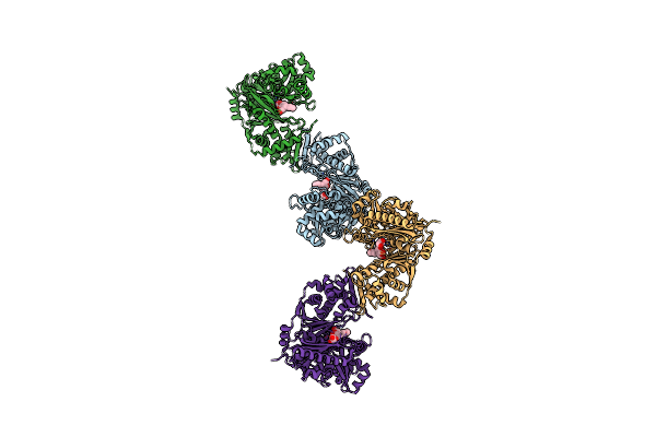
Crystal Structure Of Glycerol Kinase From Trypanosoma Brucei Gambiense Complexed With Cumarin Derivative-17
Organism: Trypanosoma brucei gambiense
Method: X-RAY DIFFRACTION Resolution:2.85 Å Release Date: 2017-07-26 Classification: TRANSFERASE Ligands: 6XZ, GOL |
Organism: Trypanosoma brucei gambiense
Method: X-RAY DIFFRACTION
Release Date: 2017-07-26
Ligands: 6XZ, GOL
 |
Crystal Structure Of Glycerol Kinase From Trypanosoma Brucei Gambiense Complexed With Cumarin Derivative-17B
Organism: Trypanosoma brucei gambiense
Method: X-RAY DIFFRACTION Resolution:2.50 Å Release Date: 2017-07-26 Classification: TRANSFERASE Ligands: 6Y0, GOL |
Organism: Trypanosoma brucei gambiense
Method: X-RAY DIFFRACTION
Release Date: 2017-07-26
Ligands: 6Y0, GOL