

Search Count: 16
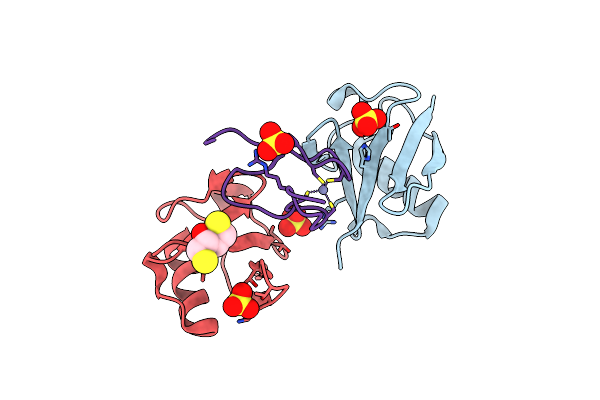 |
Structure Of Tab2 Nzf Domain Bound To K6 / Lys6-Linked Diubiquitin
Organism: Homo sapiens
Method: X-RAY DIFFRACTION Resolution:1.50 Å Release Date: 2024-07-31 Classification: SIGNALING PROTEIN Ligands: SO4, DTT, ZN |
Organism: Homo sapiens
Method: X-RAY DIFFRACTION
Release Date: 2024-07-31
Ligands: SO4, DTT, ZN
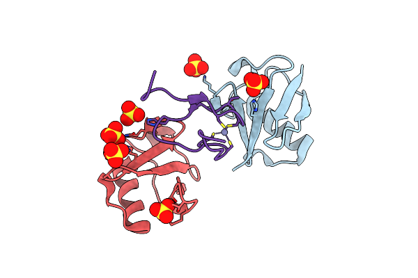 |
Structure Of Tab2 Nzf Domain Bound To K6 / Lys6-Linked Diubiquitin
Organism: Homo sapiens
Method: X-RAY DIFFRACTION Resolution:1.75 Å Release Date: 2024-07-31 Classification: SIGNALING PROTEIN Ligands: SO4, ZN |
Organism: Homo sapiens
Method: X-RAY DIFFRACTION
Release Date: 2024-07-31
Ligands: SO4, ZN
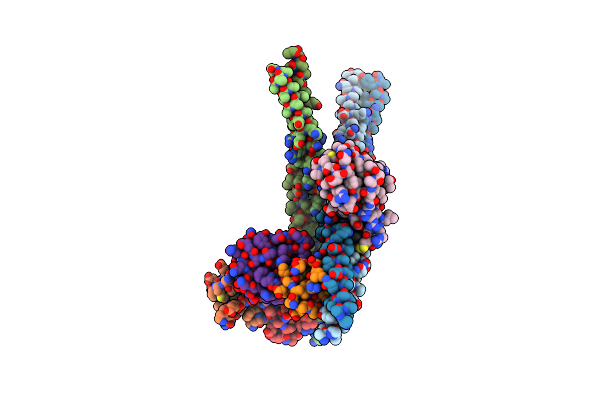 |
Structure Of Ubiquitinated Nemo Uban K285C-Ub G76C Bound To Hoip Nzf1
Organism: Homo sapiens
Method: X-RAY DIFFRACTION Resolution:3.32 Å Release Date: 2024-07-31 Classification: SIGNALING PROTEIN Ligands: ZN |
Organism: Homo sapiens
Method: X-RAY DIFFRACTION
Release Date: 2024-07-31
Ligands: ZN
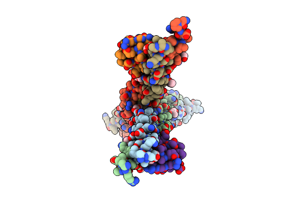 |
Structure Of Optineurin Bound To Hoip Nzf1 Domain
Organism: Homo sapiens
Method: X-RAY DIFFRACTION Resolution:1.70 Å Release Date: 2024-07-31 Classification: SIGNALING PROTEIN Ligands: PGE, PG4, ZN |
Organism: Homo sapiens
Method: X-RAY DIFFRACTION
Release Date: 2024-07-31
Ligands: PGE, PG4, ZN
 |
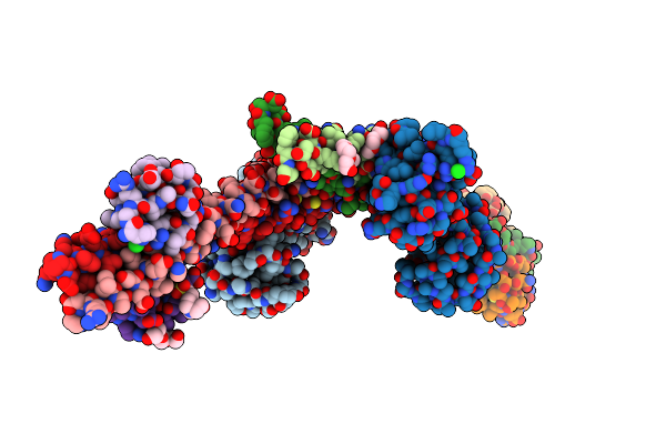
Structure Of Optineurin Bound To Hoip Nzf1 Domain And M1-Linked Diubiquitin, Crystal Form 2
Organism: Homo sapiens
Method: X-RAY DIFFRACTION Resolution:2.41 Å Release Date: 2024-07-31 Classification: SIGNALING PROTEIN Ligands: ZN |
Organism: Homo sapiens
Method: X-RAY DIFFRACTION
Release Date: 2024-07-31
Ligands: ZN
 |
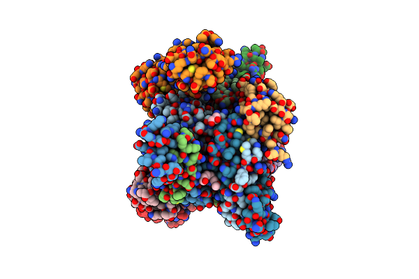
Structure Of Optineurin Bound To Hoip Nzf1 Domain And M1-Linked Diubiquitin, Crystal Form 1
Organism: Homo sapiens
Method: X-RAY DIFFRACTION Resolution:1.81 Å Release Date: 2024-07-31 Classification: SIGNALING PROTEIN Ligands: PG4, PGE, CL, EPE, ZN |
Organism: Homo sapiens
Method: X-RAY DIFFRACTION
Release Date: 2024-07-31
Ligands: PG4, PGE, CL, EPE, ZN
 |
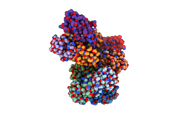
K6-Specific Affimer Bound To K6 Diub
Organism: Synthetic construct, Homo sapiens
Method: X-RAY DIFFRACTION Resolution:2.50 Å Release Date: 2017-10-04 Classification: SIGNALING PROTEIN Ligands: GOL, 15P |
Organism: Synthetic construct, Homo sapiens
Method: X-RAY DIFFRACTION
Release Date: 2017-10-04
Ligands: GOL, 15P
 |
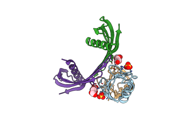
K33-Specific Affimer Bound To K33 Diub
Organism: Homo sapiens, Synthetic construct
Method: X-RAY DIFFRACTION Resolution:3.80 Å Release Date: 2017-10-04 Classification: SIGNALING PROTEIN Ligands: 15P |
Organism: Homo sapiens, Synthetic construct
Method: X-RAY DIFFRACTION
Release Date: 2017-10-04
Ligands: 15P
 |
K33-Specific Affimer Bound To K33 Diub
Organism: Homo sapiens, Synthetic construct
Method: X-RAY DIFFRACTION Resolution:2.80 Å Release Date: 2017-10-04 Classification: SIGNALING PROTEIN Ligands: SO4, GOL |
Organism: Homo sapiens, Synthetic construct
Method: X-RAY DIFFRACTION
Release Date: 2017-10-04
Ligands: SO4, GOL
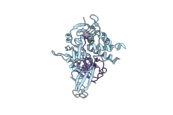 |
Crystal Structure Of Usp30 In Covalent Complex With Ubiquitin Propargylamide (High Resolution)
Organism: Homo sapiens
Method: X-RAY DIFFRACTION Resolution:2.34 Å Release Date: 2017-09-20 Classification: HYDROLASE Ligands: ZN, AYE |
Organism: Homo sapiens
Method: X-RAY DIFFRACTION
Release Date: 2017-09-20
Ligands: ZN, AYE
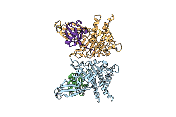 |
Crystal Structure Of Usp30 In Covalent Complex With Ubiquitin Propargylamide (Low Resolution)
Organism: Homo sapiens
Method: X-RAY DIFFRACTION Resolution:3.60 Å Release Date: 2017-09-20 Classification: HYDROLASE Ligands: ZN |
Organism: Homo sapiens
Method: X-RAY DIFFRACTION
Release Date: 2017-09-20
Ligands: ZN
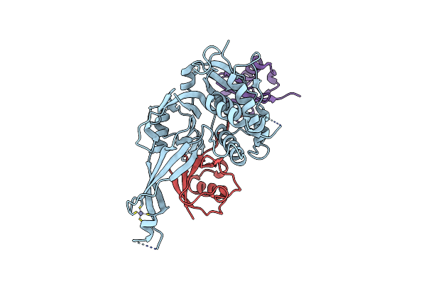 |
Crystal Structure Of Usp30 (C77A) In Complex With Lys6-Linked Diubiquitin
Organism: Homo sapiens
Method: X-RAY DIFFRACTION Resolution:2.80 Å Release Date: 2017-09-20 Classification: HYDROLASE Ligands: ZN |
Organism: Homo sapiens
Method: X-RAY DIFFRACTION
Release Date: 2017-09-20
Ligands: ZN
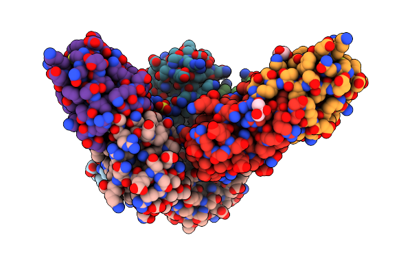 |
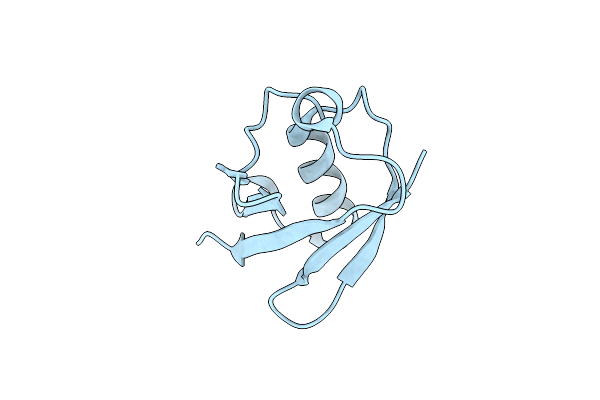
Structure Of Lys33-Linked Diub
Organism: Homo sapiens
Method: X-RAY DIFFRACTION Resolution:1.85 Å Release Date: 2015-03-25 Classification: SIGNALING PROTEIN Ligands: GOL, SO4 |
Organism: Homo sapiens
Method: X-RAY DIFFRACTION
Release Date: 2015-03-25
Ligands: GOL, SO4
 |
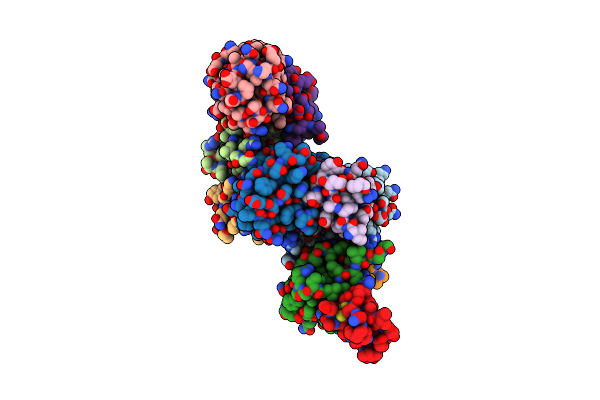
Structure Of Lys33-Linked Triub S.G. P 212121
Organism: Homo sapiens
Method: X-RAY DIFFRACTION Resolution:1.68 Å Release Date: 2015-03-25 Classification: SIGNALING PROTEIN |
Organism: Homo sapiens
Method: X-RAY DIFFRACTION
Release Date: 2015-03-25
 |
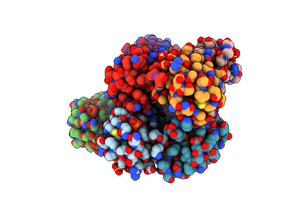
Structure Of Lys33-Linked Diub Bound To Trabid Nzf1
Organism: Homo sapiens
Method: X-RAY DIFFRACTION Resolution:3.40 Å Release Date: 2015-03-25 Classification: SIGNALING PROTEIN Ligands: ZN |
Organism: Homo sapiens
Method: X-RAY DIFFRACTION
Release Date: 2015-03-25
Ligands: ZN
 |
Ser65 Phosphorylated Ubiquitin, Major Conformation
Organism: Homo sapiens
Method: X-RAY DIFFRACTION Resolution:1.90 Å Release Date: 2015-01-14 Classification: SIGNALING PROTEIN Ligands: SO4 |
Organism: Homo sapiens
Method: X-RAY DIFFRACTION
Release Date: 2015-01-14
Ligands: SO4