

Search Count: 23
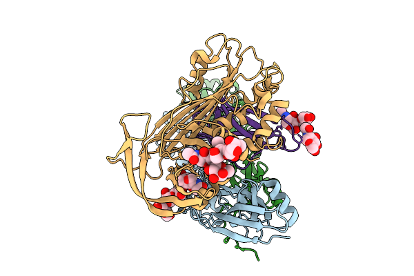 |
Structure Of A16/G9 In Complex With A56/K2 (Vaccinia Virus)
Organism: Vaccinia virus western reserve
Method: ELECTRON MICROSCOPY Release Date: 2025-09-03 Classification: VIRAL PROTEIN Ligands: NAG |
Organism: Vaccinia virus western reserve
Method: ELECTRON MICROSCOPY
Release Date: 2025-09-03
Ligands: NAG
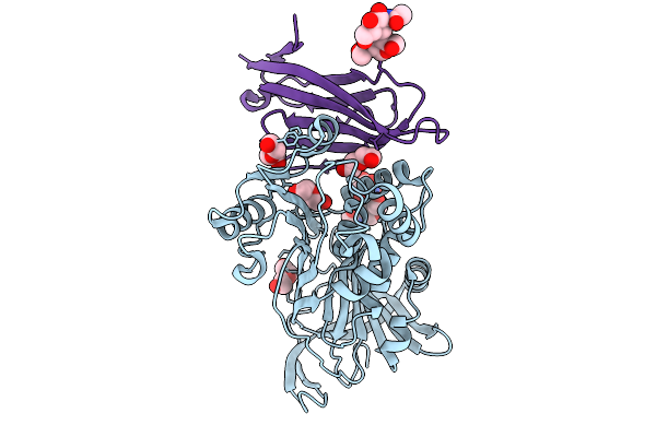 |
Structure Of A56/K2 (Vaccinia Virus)
Organism: Vaccinia virus western reserve
Method: X-RAY DIFFRACTION Resolution:2.80 Å Release Date: 2025-09-03 Classification: VIRAL PROTEIN Ligands: PEG |
Organism: Vaccinia virus western reserve
Method: X-RAY DIFFRACTION
Release Date: 2025-09-03
Ligands: PEG
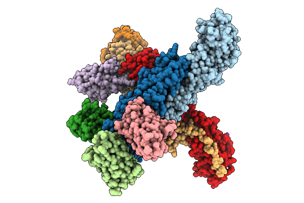 |
Structure Of A16/G9 (Vaccinia Virus) In Complex With Vhh D07, Vhh B01 And Vhh C05
Organism: Vaccinia virus western reserve, Vicugna pacos
Method: X-RAY DIFFRACTION Resolution:3.20 Å Release Date: 2025-09-03 Classification: VIRAL PROTEIN Ligands: K, PO4 |
Organism: Vaccinia virus western reserve, Vicugna pacos
Method: X-RAY DIFFRACTION
Release Date: 2025-09-03
Ligands: K, PO4
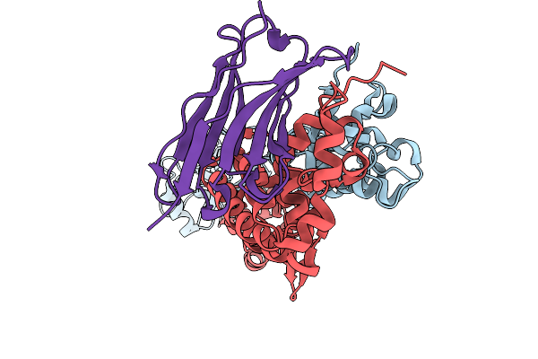 |
Structure Of A16/G9 (G9 Mutant - H44Y) Of Vaccinia Virus In Complex With Vhh D07
Organism: Vaccinia virus western reserve, Vicugna pacos
Method: X-RAY DIFFRACTION Resolution:2.50 Å Release Date: 2025-09-03 Classification: VIRAL PROTEIN |
Organism: Vaccinia virus western reserve, Vicugna pacos
Method: X-RAY DIFFRACTION
Release Date: 2025-09-03
 |
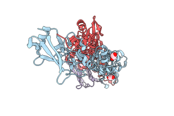
Structure Of A16/G9 (Vaccinia Virus) In Complex With Vhh D07 And Vhh E12
Organism: Vaccinia virus western reserve, Vicugna pacos
Method: X-RAY DIFFRACTION Resolution:3.10 Å Release Date: 2025-09-03 Classification: VIRAL PROTEIN Ligands: PO4, SO4, PEG |
Organism: Vaccinia virus western reserve, Vicugna pacos
Method: X-RAY DIFFRACTION
Release Date: 2025-09-03
Ligands: PO4, SO4, PEG
 |
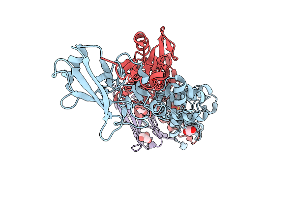
Structure Of A16/G9 (Vaccinia Virus) In Complex With Vhh D07 At Ph 5.7
Organism: Vaccinia virus western reserve, Vicugna pacos
Method: X-RAY DIFFRACTION Resolution:2.45 Å Release Date: 2025-09-03 Classification: VIRAL PROTEIN Ligands: MES, GOL |
Organism: Vaccinia virus western reserve, Vicugna pacos
Method: X-RAY DIFFRACTION
Release Date: 2025-09-03
Ligands: MES, GOL
 |
Structure Of A16/G9 (Vaccinia Virus) In Complex With Vhh D07 At Ph 6.5
Organism: Vaccinia virus western reserve, Vicugna pacos
Method: X-RAY DIFFRACTION Resolution:2.45 Å Release Date: 2025-09-03 Classification: VIRAL PROTEIN Ligands: MES, GOL |
Organism: Vaccinia virus western reserve, Vicugna pacos
Method: X-RAY DIFFRACTION
Release Date: 2025-09-03
Ligands: MES, GOL
 |
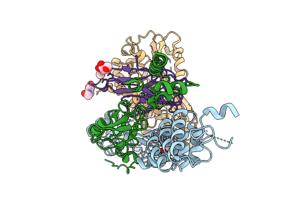
Structure Of A16/G9 (Vaccinia Virus) In Complex With Vhh D07 At Ph 7.2
Organism: Vaccinia virus western reserve, Vicugna pacos
Method: X-RAY DIFFRACTION Resolution:2.45 Å Release Date: 2025-09-03 Classification: VIRAL PROTEIN Ligands: GOL, MES |
Organism: Vaccinia virus western reserve, Vicugna pacos
Method: X-RAY DIFFRACTION
Release Date: 2025-09-03
Ligands: GOL, MES
 |
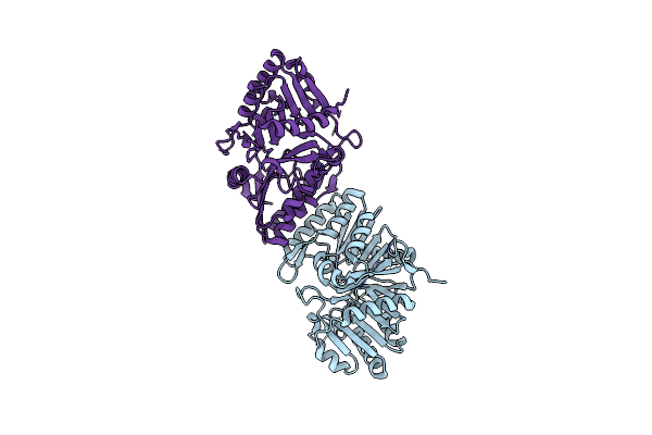
Structure Of A16/G9 In Complex With A56/K2 At Ph 5.5 (Vaccinia Virus)
Organism: Vaccinia virus western reserve
Method: ELECTRON MICROSCOPY Release Date: 2025-09-03 Classification: VIRAL PROTEIN Ligands: NAG |
Organism: Vaccinia virus western reserve
Method: ELECTRON MICROSCOPY
Release Date: 2025-09-03
Ligands: NAG
 |
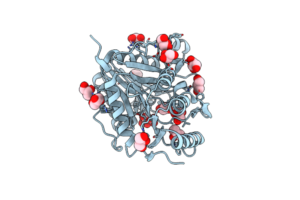
Structure Of The F13 Protein Of Vaccinia Virus (P21 Crystal Form)
Organism: Vaccinia virus western reserve
Method: X-RAY DIFFRACTION Resolution:2.10 Å Release Date: 2025-03-12 Classification: VIRAL PROTEIN |
Organism: Vaccinia virus western reserve
Method: X-RAY DIFFRACTION
Release Date: 2025-03-12
 |
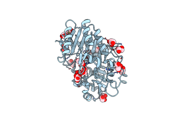
Structure Of The F13 Protein Of Vaccinia Virus (F432 Crystal Form)
Organism: Vaccinia virus western reserve
Method: X-RAY DIFFRACTION Resolution:2.82 Å Release Date: 2025-03-12 Classification: VIRAL PROTEIN Ligands: CIT, GOL |
Organism: Vaccinia virus western reserve
Method: X-RAY DIFFRACTION
Release Date: 2025-03-12
Ligands: CIT, GOL
 |
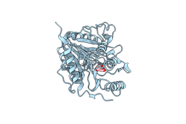
Structure Of The F13 Protein (Mutant A295E) Of Vaccinia Virus
Organism: Vaccinia virus western reserve
Method: X-RAY DIFFRACTION Resolution:2.60 Å Release Date: 2025-03-12 Classification: VIRAL PROTEIN Ligands: CIT, GOL |
Organism: Vaccinia virus western reserve
Method: X-RAY DIFFRACTION
Release Date: 2025-03-12
Ligands: CIT, GOL
 |
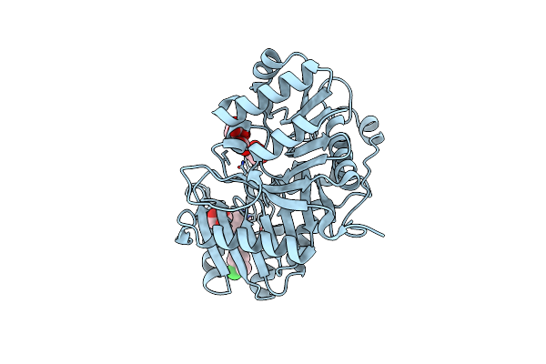
Structure Of The F13 Protein (Mutant G277C) Of Vaccinia Virus
Organism: Vaccinia virus western reserve
Method: X-RAY DIFFRACTION Resolution:4.00 Å Release Date: 2025-03-12 Classification: VIRAL PROTEIN Ligands: GOL |
Organism: Vaccinia virus western reserve
Method: X-RAY DIFFRACTION
Release Date: 2025-03-12
Ligands: GOL
 |
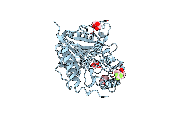
Structure Of The F13 Protein Of Vaccinia Virus In Complex With Imcbh
Organism: Vaccinia virus western reserve
Method: X-RAY DIFFRACTION Resolution:3.80 Å Release Date: 2025-03-12 Classification: VIRAL PROTEIN Ligands: CIT, GOL, A1IDM |
Organism: Vaccinia virus western reserve
Method: X-RAY DIFFRACTION
Release Date: 2025-03-12
Ligands: CIT, GOL, A1IDM
 |
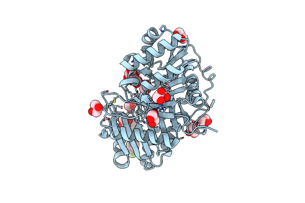
Structure Of The F13 Protein (A295E Mutant) Of Vaccinia Virus In Complex With Tecovirimat
Organism: Vaccinia virus western reserve
Method: X-RAY DIFFRACTION Resolution:3.50 Å Release Date: 2025-03-12 Classification: VIRAL PROTEIN Ligands: GOL, A1ICJ |
Organism: Vaccinia virus western reserve
Method: X-RAY DIFFRACTION
Release Date: 2025-03-12
Ligands: GOL, A1ICJ
 |
Structure Of The F13 Protein Of Vaccinia Virus In Complex With Tecovirimat
Organism: Vaccinia virus western reserve
Method: X-RAY DIFFRACTION Resolution:2.60 Å Release Date: 2025-03-12 Classification: VIRAL PROTEIN Ligands: A1ICJ, CIT, GOL |
Organism: Vaccinia virus western reserve
Method: X-RAY DIFFRACTION
Release Date: 2025-03-12
Ligands: A1ICJ, CIT, GOL
 |
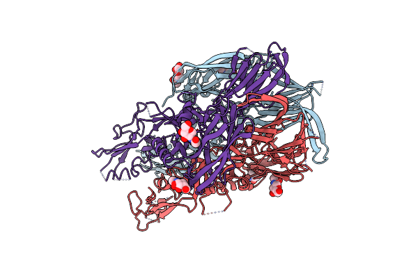
Crystal Structure Of Human Pancreatic Phospholipase A2
Organism: Homo sapiens
Method: X-RAY DIFFRACTION Resolution:1.90 Å Release Date: 2020-04-15 Classification: HYDROLASE Ligands: CL |
Organism: Homo sapiens
Method: X-RAY DIFFRACTION
Release Date: 2020-04-15
Ligands: CL
 |
Crystal Structure Of A C-Terminally Truncated Trimeric Ectodomain Of The Chlamydomonas Reinhardtii Gamete Fusion Protein Hap2
Organism: Chlamydomonas reinhardtii
Method: X-RAY DIFFRACTION Resolution:3.30 Å Release Date: 2017-03-08 Classification: MEMBRANE PROTEIN Ligands: NAG, NGA |
Organism: Chlamydomonas reinhardtii
Method: X-RAY DIFFRACTION
Release Date: 2017-03-08
Ligands: NAG, NGA
 |
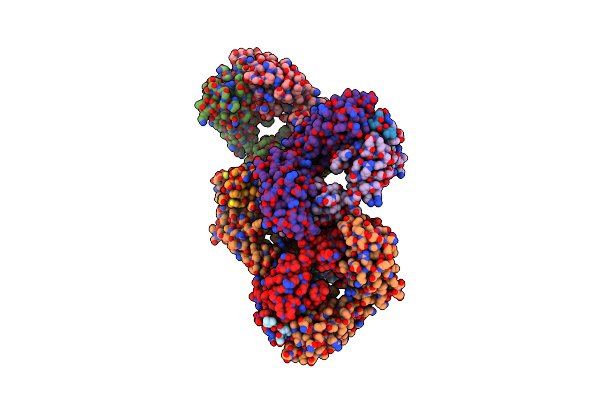
Structure Of The Hepatitis C Virus Envelope Glycoprotein E2 Antigenic Region 412-423 Bound To The Broadly Neutralizing Antibody 3/11, P1 Crystal Form
Organism: Rattus norvegicus, Hepatitis c virus
Method: X-RAY DIFFRACTION Resolution:2.22 Å Release Date: 2014-12-17 Classification: VIRAL PROTEIN |
Organism: Rattus norvegicus, Hepatitis c virus
Method: X-RAY DIFFRACTION
Release Date: 2014-12-17
 |
Structure Of The Hepatitis C Virus Envelope Glycoprotein E2 Antigenic Region 412-423 Bound To The Broadly Neutralizing Antibody 3/11, P21 Crystal Form
Organism: Rattus norvegicus, Hepatitis c virus
Method: X-RAY DIFFRACTION Resolution:2.62 Å Release Date: 2014-12-17 Classification: VIRAL PROTEIN |
Organism: Rattus norvegicus, Hepatitis c virus
Method: X-RAY DIFFRACTION
Release Date: 2014-12-17