

Search Count: 30
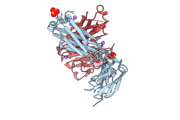 |
Crystal Structure Of 19B Fab Bound To The Third Variable (V3) Loop Peptide From The Hiv-1 Jr-Fl Envelope (Env) Glycoprotein
Organism: Homo sapiens, Human immunodeficiency virus 1
Method: X-RAY DIFFRACTION Resolution:1.78 Å Release Date: 2026-01-21 Classification: IMMUNE SYSTEM Ligands: ACT, GOL, SO4, NA |
Organism: Homo sapiens, Human immunodeficiency virus 1
Method: X-RAY DIFFRACTION
Release Date: 2026-01-21
Ligands: ACT, GOL, SO4, NA
 |
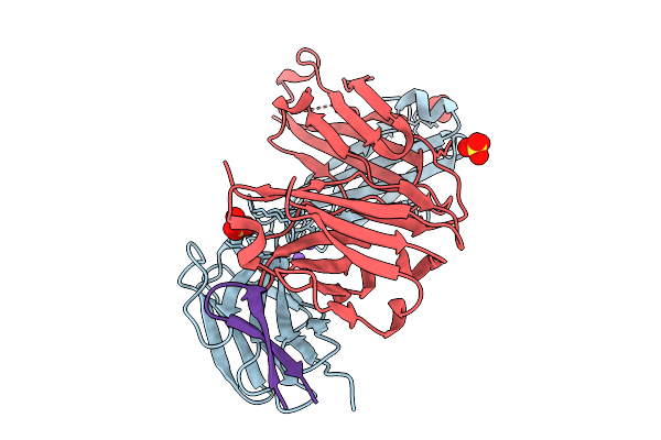
Crystal Structure Of 19B Fab Bound To The Third Variable (V3) Loop Peptide From The Hiv-1 5768-P27 Envelope (Env) Glycoprotein
Organism: Homo sapiens, Human immunodeficiency virus 1
Method: X-RAY DIFFRACTION Resolution:2.50 Å Release Date: 2026-01-21 Classification: IMMUNE SYSTEM/VIRAL PROTEIN Ligands: SO4 |
Organism: Homo sapiens, Human immunodeficiency virus 1
Method: X-RAY DIFFRACTION
Release Date: 2026-01-21
Ligands: SO4
 |
Crystal Structure Of 19B Fab Bound To The Third Variable (V3) Loop Peptide From The Hiv-1 92Br020 Envelope (Env) Glycoprotein
Organism: Homo sapiens, Human immunodeficiency virus 1
Method: X-RAY DIFFRACTION Resolution:1.94 Å Release Date: 2026-01-21 Classification: IMMUNE SYSTEM/VIRAL PROTEIN Ligands: SO4, NA |
Organism: Homo sapiens, Human immunodeficiency virus 1
Method: X-RAY DIFFRACTION
Release Date: 2026-01-21
Ligands: SO4, NA
 |
Crystal Structure Of 19B Fab Bound To The Third Variable (V3) Loop Peptide From The Hiv-1 5768X-P27 Envelope (Env) Glycoprotein
Organism: Homo sapiens, Human immunodeficiency virus 1
Method: X-RAY DIFFRACTION Resolution:1.74 Å Release Date: 2026-01-21 Classification: IMMUNE SYSTEM/VIRAL PROTEIN Ligands: SO4, NA |
Organism: Homo sapiens, Human immunodeficiency virus 1
Method: X-RAY DIFFRACTION
Release Date: 2026-01-21
Ligands: SO4, NA
 |
Crystal Structure Of 19B Fab
Organism: Homo sapiens
Method: X-RAY DIFFRACTION Resolution:2.10 Å Release Date: 2026-01-21 Classification: IMMUNE SYSTEM |
Organism: Homo sapiens
Method: X-RAY DIFFRACTION
Release Date: 2026-01-21
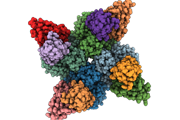 |
Structure Of Fab Hb420 In Complex With Influenza H3N2 A/Moscow/10/1999 Neuraminidase
Organism: Influenza a virus, Homo sapiens
Method: ELECTRON MICROSCOPY Release Date: 2025-10-01 Classification: VIRAL PROTEIN/IMMUNE SYSTEM |
Organism: Influenza a virus, Homo sapiens
Method: ELECTRON MICROSCOPY
Release Date: 2025-10-01
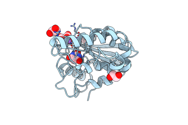 |
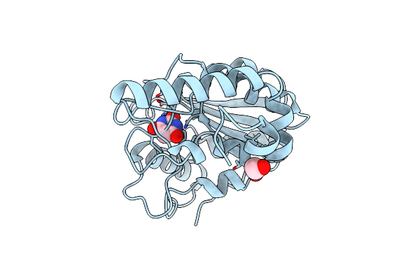
Crystal Structure Of Mycobacterium Tuberculosis Uracil-Dna Glycosylase In Complex With Barbituric Acid And Citric Acid, Form I
Organism: Mycobacterium tuberculosis h37rv
Method: X-RAY DIFFRACTION Resolution:1.24 Å Release Date: 2023-07-12 Classification: HYDROLASE Ligands: BR8, CIT, EDO, NA |
Organism: Mycobacterium tuberculosis h37rv
Method: X-RAY DIFFRACTION
Release Date: 2023-07-12
Ligands: BR8, CIT, EDO, NA
 |
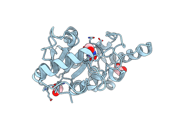
Crystal Structure Of Mycobacterium Tuberculosis Uracil-Dna Glycosylase In Complex With Barbituric Acid, Form I
Organism: Mycobacterium tuberculosis h37rv
Method: X-RAY DIFFRACTION Resolution:1.26 Å Release Date: 2023-07-12 Classification: HYDROLASE/INHIBITOR Ligands: BR8, EDO, CL |
Organism: Mycobacterium tuberculosis h37rv
Method: X-RAY DIFFRACTION
Release Date: 2023-07-12
Ligands: BR8, EDO, CL
 |
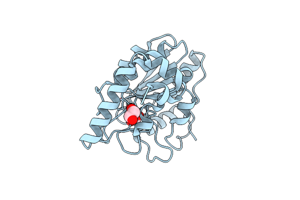
Crystal Structure Of Mycobacterium Tuberculosis Uracil-Dna Glycosylase In Complex With Barbituric Acid, Form Iii
Organism: Mycobacterium tuberculosis h37rv
Method: X-RAY DIFFRACTION Resolution:1.95 Å Release Date: 2023-07-12 Classification: HYDROLASE/INHIBITOR Ligands: BR8, EDO |
Organism: Mycobacterium tuberculosis h37rv
Method: X-RAY DIFFRACTION
Release Date: 2023-07-12
Ligands: BR8, EDO
 |
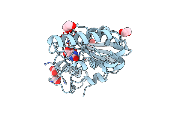
Crystal Structure Of Mycobacterium Tuberculosis Uracil-Dna Glycosylase In Complex With Barbituric Acid, Form Ii
Organism: Mycobacterium tuberculosis h37rv
Method: X-RAY DIFFRACTION Resolution:2.26 Å Release Date: 2023-07-12 Classification: HYDROLASE/INHIBITOR Ligands: BR8, EDO |
Organism: Mycobacterium tuberculosis h37rv
Method: X-RAY DIFFRACTION
Release Date: 2023-07-12
Ligands: BR8, EDO
 |
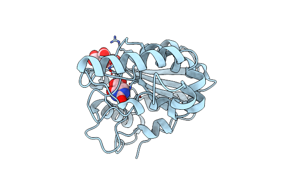
Crystal Structure Of Mycobacterium Tuberculosis Uracil-Dna Glycosylase In Complex With Isoorotic Acid (2,4-Dihydroxypyrimidine-5-Carboxylic Acid), Form I
Organism: Mycobacterium tuberculosis h37rv
Method: X-RAY DIFFRACTION Resolution:1.72 Å Release Date: 2023-07-12 Classification: HYDROLASE/INHIBITOR Ligands: 5CU, EDO |
Organism: Mycobacterium tuberculosis h37rv
Method: X-RAY DIFFRACTION
Release Date: 2023-07-12
Ligands: 5CU, EDO
 |
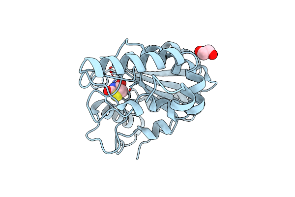
Crystal Structure Of Mycobacterium Tuberculosis Uracil-Dna Glycosylase In Complex With Isoorotic Acid (2,4-Dihydroxypyrimidine-5-Carboxylic Acid) And Citric Acid, Form I
Organism: Mycobacterium tuberculosis h37rv
Method: X-RAY DIFFRACTION Resolution:2.60 Å Release Date: 2023-07-12 Classification: HYDROLASE/INHIBITOR Ligands: 5CU, CIT |
Organism: Mycobacterium tuberculosis h37rv
Method: X-RAY DIFFRACTION
Release Date: 2023-07-12
Ligands: 5CU, CIT
 |
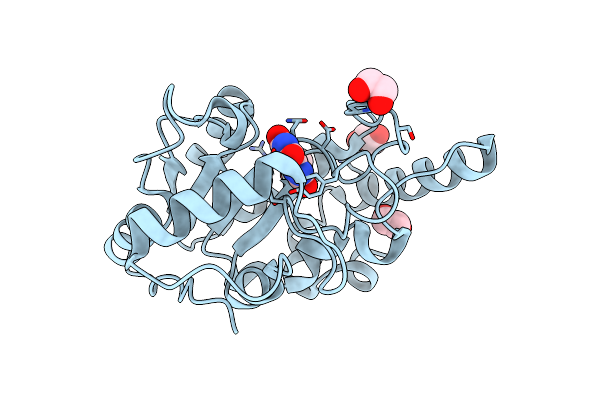
Crystal Structure Of Mycobacterium Tuberculosis Uracil-Dna Glycosylase In Complex With 2,4-Thiazolidinedione, Form I
Organism: Mycobacterium tuberculosis h37rv
Method: X-RAY DIFFRACTION Resolution:1.72 Å Release Date: 2023-07-12 Classification: HYDROLASE/INHIBITOR Ligands: OD3, EDO |
Organism: Mycobacterium tuberculosis h37rv
Method: X-RAY DIFFRACTION
Release Date: 2023-07-12
Ligands: OD3, EDO
 |
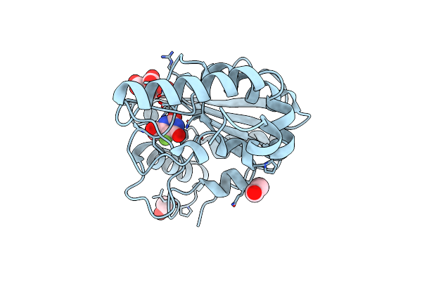
Crystal Structure Of Mycobacterium Tuberculosis Uracil-Dna Glycosylase In Complex With Uric Acid, Form Iii
Organism: Mycobacterium tuberculosis h37rv
Method: X-RAY DIFFRACTION Resolution:1.88 Å Release Date: 2023-07-12 Classification: HYDROLASE/INHIBITOR Ligands: URC, EDO |
Organism: Mycobacterium tuberculosis h37rv
Method: X-RAY DIFFRACTION
Release Date: 2023-07-12
Ligands: URC, EDO
 |
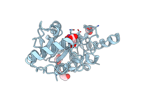
Crystal Structure Of Mycobacterium Tuberculosis Uracil-Dna Glycosylase In Complex With 5-Fluoroorotic Acid And Citric Acid, Form I
Organism: Mycobacterium tuberculosis h37rv
Method: X-RAY DIFFRACTION Resolution:2.00 Å Release Date: 2023-07-12 Classification: HYDROLASE/INHIBITOR Ligands: FOT, CIT, EDO |
Organism: Mycobacterium tuberculosis h37rv
Method: X-RAY DIFFRACTION
Release Date: 2023-07-12
Ligands: FOT, CIT, EDO
 |
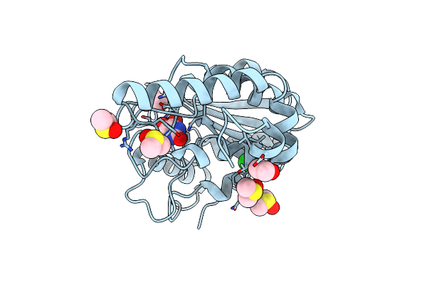
Crystal Structure Of Mycobacterium Tuberculosis Uracil-Dna Glycosylase In Complex With Orotic Acid, Form Iii
Organism: Mycobacterium tuberculosis h37rv
Method: X-RAY DIFFRACTION Resolution:2.00 Å Release Date: 2023-07-12 Classification: HYDROLASE/INHIBITOR Ligands: ORO, EDO |
Organism: Mycobacterium tuberculosis h37rv
Method: X-RAY DIFFRACTION
Release Date: 2023-07-12
Ligands: ORO, EDO
 |
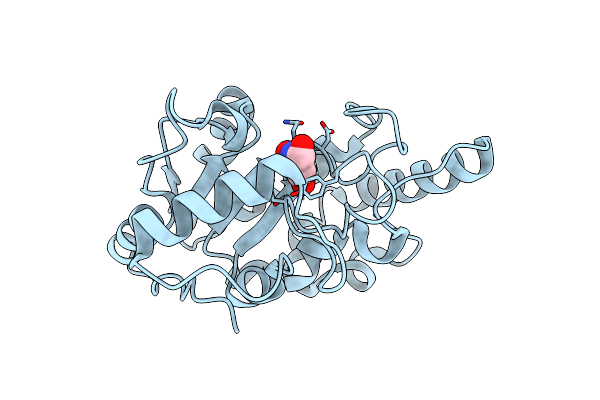
Crystal Structure Of Mycobacterium Tuberculosis Uracil-Dna Glycosylase In Complex With 5-Hydroxy-2,4(1H,3H)-Pyrimidinedione, Form I
Organism: Mycobacterium tuberculosis h37rv
Method: X-RAY DIFFRACTION Resolution:1.60 Å Release Date: 2023-07-12 Classification: HYDROLASE/INHIBITOR Ligands: ODF, EDO, DMS, NA, CL |
Organism: Mycobacterium tuberculosis h37rv
Method: X-RAY DIFFRACTION
Release Date: 2023-07-12
Ligands: ODF, EDO, DMS, NA, CL
 |
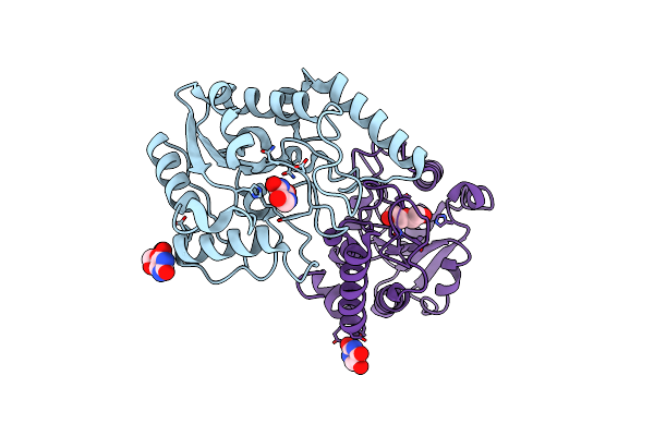
Crystal Structure Of Mycobacterium Tuberculosis Uracil-Dna Glycosylase In Complex With 6-Formyl-Uracil, Form Iii
Organism: Mycobacterium tuberculosis h37rv
Method: X-RAY DIFFRACTION Resolution:2.28 Å Release Date: 2023-07-12 Classification: HYDROLASE/INHIBITOR Ligands: OG6 |
Organism: Mycobacterium tuberculosis h37rv
Method: X-RAY DIFFRACTION
Release Date: 2023-07-12
Ligands: OG6
 |
Crystal Structure Of Mycobacterium Tuberculosis Uracil-Dna Glycosylase In Complex With 5-Hydroxy-2,4(1H,3H)-Pyrimidinedione, Form Vi
Organism: Mycobacterium tuberculosis h37rv
Method: X-RAY DIFFRACTION Resolution:2.40 Å Release Date: 2023-07-12 Classification: HYDROLASE/INHIBITOR Ligands: ODF, EDO, PEG |
Organism: Mycobacterium tuberculosis h37rv
Method: X-RAY DIFFRACTION
Release Date: 2023-07-12
Ligands: ODF, EDO, PEG
 |
Aleuria Aurantia Lectin Aal N224Q Mutant In Complex With Fucalpha1-6Glcnac
Organism: Aleuria aurantia
Method: X-RAY DIFFRACTION Resolution:1.08 Å Release Date: 2019-06-19 Classification: SUGAR BINDING PROTEIN Ligands: FUC, GOL, EDO |
Organism: Aleuria aurantia
Method: X-RAY DIFFRACTION
Release Date: 2019-06-19
Ligands: FUC, GOL, EDO