

Search Count: 62
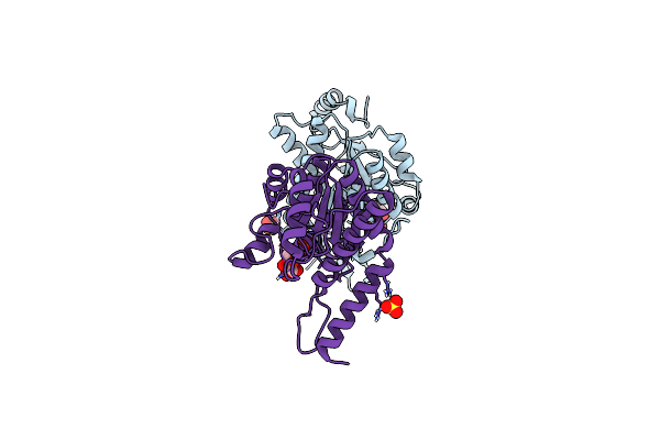 |
N-Acetylglucosamine 6-Phosphate Dehydratase: Inhibited 6-Phosphogluconic Acid State Of Nags
Organism: Streptomyces coelicolor
Method: X-RAY DIFFRACTION Resolution:1.69 Å Release Date: 2025-02-19 Classification: SUGAR BINDING PROTEIN Ligands: SO4, 6PG |
Organism: Streptomyces coelicolor
Method: X-RAY DIFFRACTION
Release Date: 2025-02-19
Ligands: SO4, 6PG
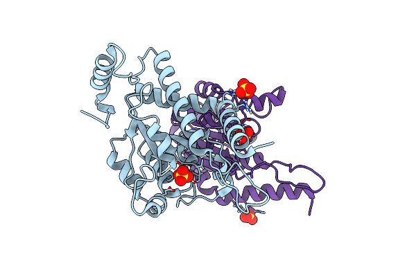 |
N-Acetylglucosamine 6-Phosphate Dehydratase: Apo Form Of Nags
Organism: Streptomyces coelicolor
Method: X-RAY DIFFRACTION Resolution:2.30 Å Release Date: 2025-02-19 Classification: SUGAR BINDING PROTEIN Ligands: SO4 |
Organism: Streptomyces coelicolor
Method: X-RAY DIFFRACTION
Release Date: 2025-02-19
Ligands: SO4
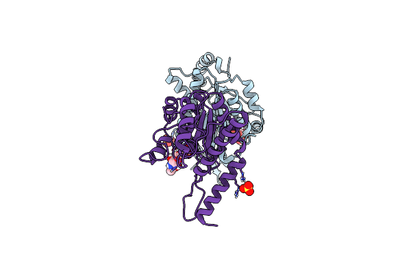 |
N-Acetylglucosamine 6-Phosphate Dehydratase: Glcnac6P Substrate-Bound State Of Nags
Organism: Streptomyces coelicolor
Method: X-RAY DIFFRACTION Resolution:2.59 Å Release Date: 2025-02-19 Classification: SUGAR BINDING PROTEIN Ligands: SO4, 16G |
Organism: Streptomyces coelicolor
Method: X-RAY DIFFRACTION
Release Date: 2025-02-19
Ligands: SO4, 16G
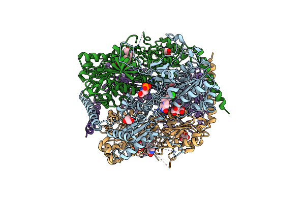 |
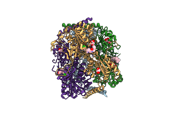
Crystal Structure Of Phosphofructokinase From Trypanosoma Brucei In Complex With An Allosteric Inhibitor Ctcb360
Organism: Trypanosoma brucei brucei
Method: X-RAY DIFFRACTION Resolution:2.35 Å Release Date: 2020-03-18 Classification: TRANSFERASE Ligands: JJ5, CIT, GOL, SEP |
Organism: Trypanosoma brucei brucei
Method: X-RAY DIFFRACTION
Release Date: 2020-03-18
Ligands: JJ5, CIT, GOL, SEP
 |
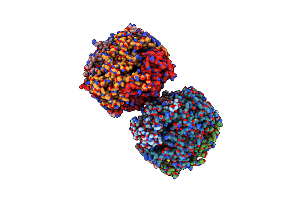
Crystal Structure Of Phosphofructokinase From Trypanosoma Brucei In Complex With An Allosteric Inhibitor Ctcb405
Organism: Trypanosoma brucei brucei
Method: X-RAY DIFFRACTION Resolution:2.75 Å Release Date: 2020-03-18 Classification: TRANSFERASE Ligands: JJ2, CIT, GOL |
Organism: Trypanosoma brucei brucei
Method: X-RAY DIFFRACTION
Release Date: 2020-03-18
Ligands: JJ2, CIT, GOL
 |
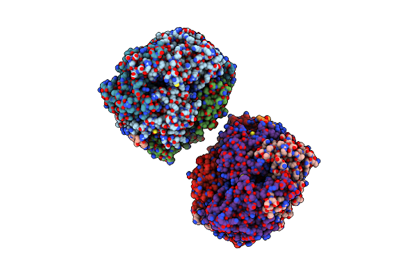
Crystal Structure Of Phosphofructokinase From Trypanosoma Brucei In Complex With An Allosteric Inhibitor Ctcb12
Organism: Trypanosoma brucei brucei
Method: X-RAY DIFFRACTION Resolution:3.40 Å Release Date: 2020-03-18 Classification: TRANSFERASE Ligands: CIT, GOL, JJ8 |
Organism: Trypanosoma brucei brucei
Method: X-RAY DIFFRACTION
Release Date: 2020-03-18
Ligands: CIT, GOL, JJ8
 |
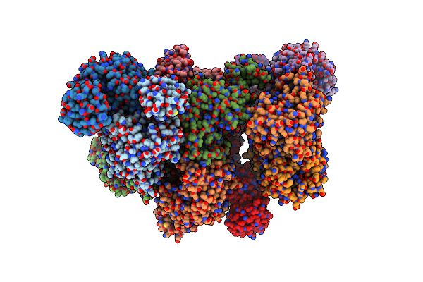
Structure Of Trypanosome Brucei Phosphofructokinase In Complex With Amp.
Organism: Trypanosoma brucei brucei
Method: X-RAY DIFFRACTION Resolution:2.75 Å Release Date: 2019-12-25 Classification: TRANSFERASE Ligands: BNZ, GOL, AMP |
Organism: Trypanosoma brucei brucei
Method: X-RAY DIFFRACTION
Release Date: 2019-12-25
Ligands: BNZ, GOL, AMP
 |
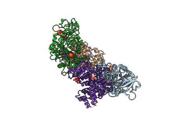
Crystal Structure Of M2 Pyk In Complex With Alanine.
Organism: Homo sapiens
Method: X-RAY DIFFRACTION Resolution:3.72 Å Release Date: 2019-09-04 Classification: TRANSFERASE Ligands: PO4, ALA |
Organism: Homo sapiens
Method: X-RAY DIFFRACTION
Release Date: 2019-09-04
Ligands: PO4, ALA
 |
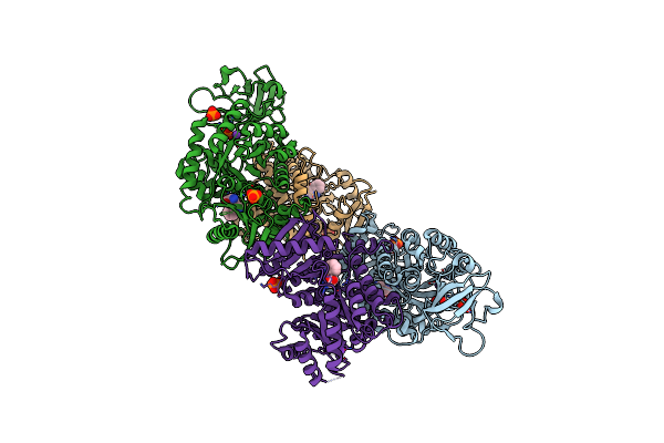
Crystal Structure Of M2 Pyk In Complex With Phenyalanine.
Organism: Homo sapiens
Method: X-RAY DIFFRACTION Resolution:2.46 Å Release Date: 2018-05-23 Classification: TRANSFERASE Ligands: PO4, PHE, K |
Organism: Homo sapiens
Method: X-RAY DIFFRACTION
Release Date: 2018-05-23
Ligands: PO4, PHE, K
 |
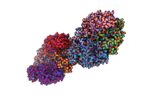
Crystal Structure Of M2 Pyk In Complex With Tryptophan.
Organism: Homo sapiens
Method: X-RAY DIFFRACTION Resolution:3.20 Å Release Date: 2018-05-23 Classification: TRANSFERASE Ligands: PO4, TRP, K |
Organism: Homo sapiens
Method: X-RAY DIFFRACTION
Release Date: 2018-05-23
Ligands: PO4, TRP, K
 |
Crystal Structure Of M2 Pyk In Complex With Serine.
Organism: Homo sapiens
Method: X-RAY DIFFRACTION Resolution:2.96 Å Release Date: 2018-05-23 Classification: TRANSFERASE Ligands: K, MG, SER, PO4 |
Organism: Homo sapiens
Method: X-RAY DIFFRACTION
Release Date: 2018-05-23
Ligands: K, MG, SER, PO4
 |
Crystal Structure Of Leishmania Major Fructose-1,6-Bisphosphatase In Holo Form.
Organism: Leishmania major
Method: X-RAY DIFFRACTION Resolution:2.80 Å Release Date: 2017-09-20 Classification: HYDROLASE Ligands: PO4, MN, K, CIT |
Organism: Leishmania major
Method: X-RAY DIFFRACTION
Release Date: 2017-09-20
Ligands: PO4, MN, K, CIT
 |
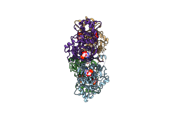
Crystal Structure Of Leishmania Major Fructose-1,6-Bisphosphatase In Apo Form.
Organism: Leishmania major
Method: X-RAY DIFFRACTION Resolution:2.41 Å Release Date: 2017-09-20 Classification: HYDROLASE |
Organism: Leishmania major
Method: X-RAY DIFFRACTION
Release Date: 2017-09-20
 |
Crystal Structure Of Leishmania Major Fructose-1,6-Bisphosphatase In T-State.
Organism: Leishmania major
Method: X-RAY DIFFRACTION Resolution:2.62 Å Release Date: 2017-09-20 Classification: TRANSFERASE Ligands: AMP, F6P, CL |
Organism: Leishmania major
Method: X-RAY DIFFRACTION
Release Date: 2017-09-20
Ligands: AMP, F6P, CL
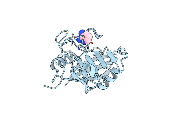 |
Structure Of Cyclophilin A In Complex With 2,3-Diaminopyridine
Organism: Homo sapiens
Method: X-RAY DIFFRACTION Resolution:1.25 Å Release Date: 2017-04-05 Classification: ISOMERASE Ligands: 76X |
Organism: Homo sapiens
Method: X-RAY DIFFRACTION
Release Date: 2017-04-05
Ligands: 76X
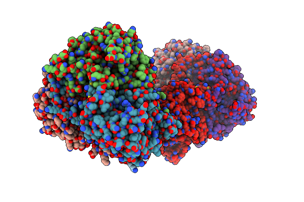 |
Co-Crystal Structure Of Ldh Liganded With Oxamate
Organism: Rattus norvegicus
Method: X-RAY DIFFRACTION Resolution:2.29 Å Release Date: 2016-01-13 Classification: OXIDOREDUCTASE Ligands: OXM |
Organism: Rattus norvegicus
Method: X-RAY DIFFRACTION
Release Date: 2016-01-13
Ligands: OXM
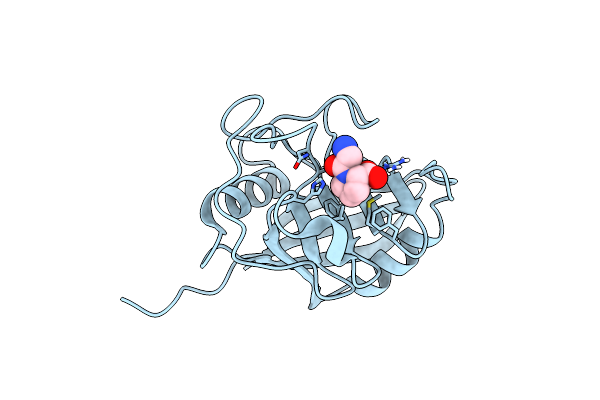 |
Structure Of Cyclophilin A In Complex With Glypro.
Organism: Homo sapiens
Method: X-RAY DIFFRACTION Resolution:1.15 Å Release Date: 2015-08-12 Classification: ISOMERASE Ligands: GLY, PRO |
Organism: Homo sapiens
Method: X-RAY DIFFRACTION
Release Date: 2015-08-12
Ligands: GLY, PRO
 |
Structure Of Cyclophilin A In Complex With Benzamide.
Organism: Homo sapiens
Method: X-RAY DIFFRACTION Resolution:1.50 Å Release Date: 2015-08-12 Classification: ISOMERASE Ligands: UNU |
Organism: Homo sapiens
Method: X-RAY DIFFRACTION
Release Date: 2015-08-12
Ligands: UNU
 |
Structure Of Cyclophilin A In Complex With Saccharin.
Organism: Homo sapiens
Method: X-RAY DIFFRACTION Resolution:1.75 Å Release Date: 2015-08-12 Classification: ISOMERASE Ligands: LSA |
Organism: Homo sapiens
Method: X-RAY DIFFRACTION
Release Date: 2015-08-12
Ligands: LSA
 |
Structure Of Cyclophilin A In Complex With Picolinamide.
Organism: Homo sapiens
Method: X-RAY DIFFRACTION Resolution:1.90 Å Release Date: 2015-08-12 Classification: ISOMERASE Ligands: WM1 |
Organism: Homo sapiens
Method: X-RAY DIFFRACTION
Release Date: 2015-08-12
Ligands: WM1