

Search Count: 101
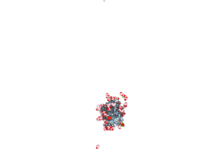 |
Crystal Structure Of Thermomyces Lanuginosa Lipase With Bound 1,3 Diacylglycerol And Fatty Acid Acyl Intermediates: Space Group H32
Organism: Thermomyces lanuginosus
Method: X-RAY DIFFRACTION Resolution:1.30 Å Release Date: 2026-08-05 Classification: LIPID BINDING PROTEIN Ligands: NAG, PG4, PO4, DGA, PEG, LTV, GOL, CA |
Organism: Thermomyces lanuginosus
Method: X-RAY DIFFRACTION
Release Date: 2026-08-05
Ligands: NAG, PG4, PO4, DGA, PEG, LTV, GOL, CA
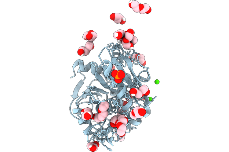 |
Crystal Structure Of Thermomyces Lanuginosa Lipase With Acyl Intermediate: Space Group I222
Organism: Thermomyces lanuginosus
Method: X-RAY DIFFRACTION Resolution:1.45 Å Release Date: 2026-08-05 Classification: LIPID BINDING PROTEIN Ligands: CA, PLM, NAG, PO4, PG4, PEG, CL |
Organism: Thermomyces lanuginosus
Method: X-RAY DIFFRACTION
Release Date: 2026-08-05
Ligands: CA, PLM, NAG, PO4, PG4, PEG, CL
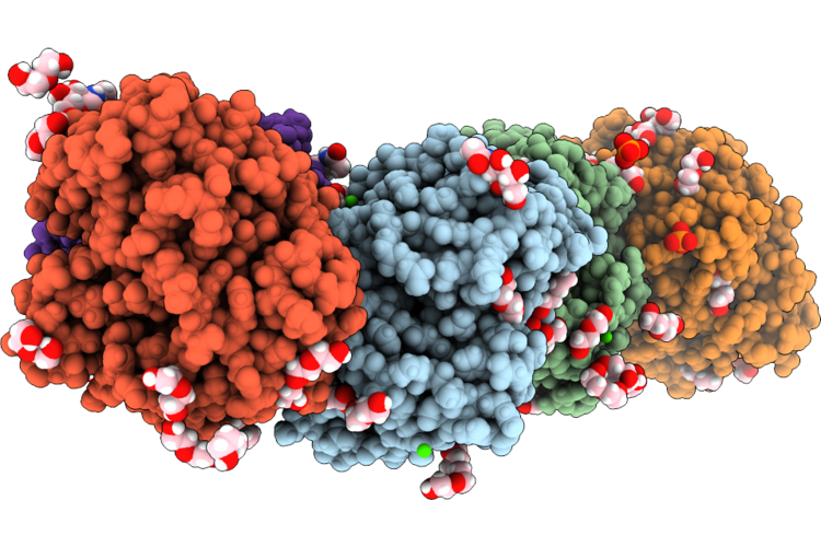 |
Crystal Structure Of Thermomyces Lanuginosa Lipase With Bound 1,3 Diacylglycrol And Fatty Acid Acyl Intermediates: Monoclinic Crystals
Organism: Thermomyces lanuginosus
Method: X-RAY DIFFRACTION Resolution:1.43 Å Release Date: 2026-08-05 Classification: LIPID BINDING PROTEIN Ligands: OCA, ACT, PEG, PG4, GOL, CA, NAG, LTV, PO4, PG5 |
Organism: Thermomyces lanuginosus
Method: X-RAY DIFFRACTION
Release Date: 2026-08-05
Ligands: OCA, ACT, PEG, PG4, GOL, CA, NAG, LTV, PO4, PG5
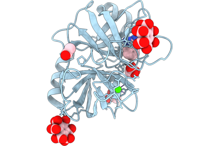 |
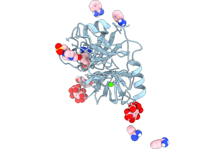
Structure Of Bovine Trypsin Complexed With Mellitic Acid At Room Temperature
Organism: Bos taurus
Method: X-RAY DIFFRACTION Resolution:1.30 Å Release Date: 2026-05-27 Classification: HYDROLASE Ligands: BEN, MLI, ACT, BHC, CA |
Organism: Bos taurus
Method: X-RAY DIFFRACTION
Release Date: 2026-05-27
Ligands: BEN, MLI, ACT, BHC, CA
 |
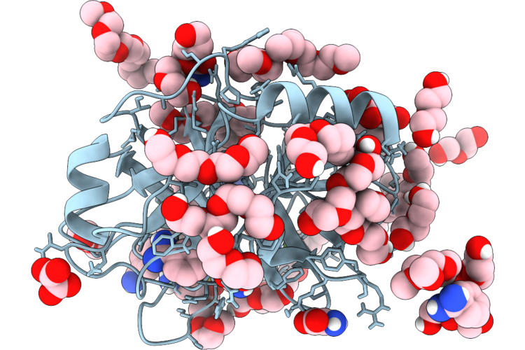
The Structure Of Bovine Trypsin Complexed With Mellitic Acid At 173 Degrees
Organism: Bos taurus
Method: X-RAY DIFFRACTION Resolution:1.50 Å Release Date: 2026-05-27 Classification: HYDROLASE Ligands: EPE, BHC, PG4, CA, BEN, ACT |
Organism: Bos taurus
Method: X-RAY DIFFRACTION
Release Date: 2026-05-27
Ligands: EPE, BHC, PG4, CA, BEN, ACT
 |
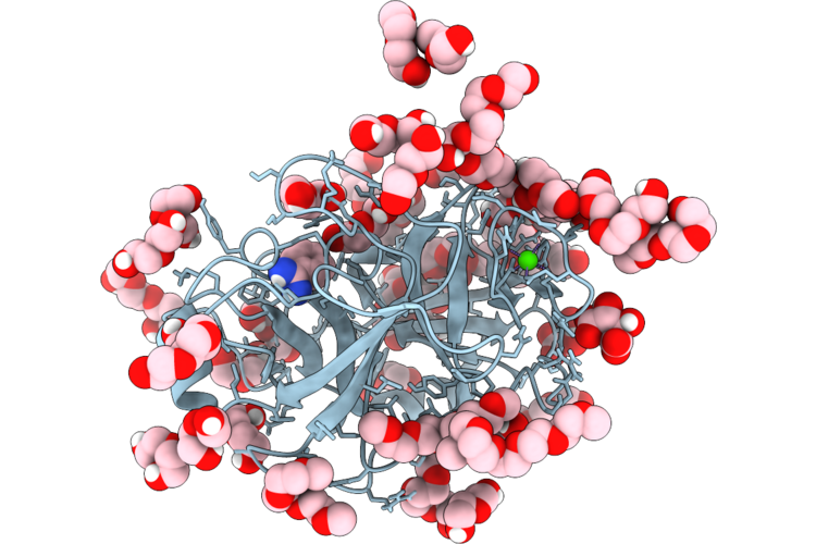
The Structure Of Porcine Trypsin In Complex With Crystallization Additives I
Organism: Sus scrofa
Method: X-RAY DIFFRACTION Resolution:1.28 Å Release Date: 2026-05-27 Classification: HYDROLASE Ligands: CA, BEN, PG5, PEG, PG4, OXL, A1CHW, OXM, PG6 |
Organism: Sus scrofa
Method: X-RAY DIFFRACTION
Release Date: 2026-05-27
Ligands: CA, BEN, PG5, PEG, PG4, OXL, A1CHW, OXM, PG6
 |
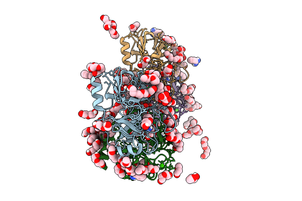
Porcine Trypsin Grown From Peg And Complexed With Crystallization Additives Ii
Organism: Sus scrofa
Method: X-RAY DIFFRACTION Resolution:1.27 Å Release Date: 2026-05-27 Classification: HYDROLASE Ligands: CA, TLA, PEG, PG4, PG6, PG5, BEN |
Organism: Sus scrofa
Method: X-RAY DIFFRACTION
Release Date: 2026-05-27
Ligands: CA, TLA, PEG, PG4, PG6, PG5, BEN
 |
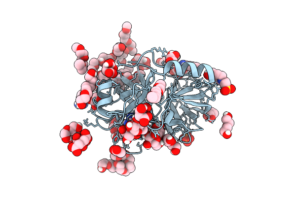
Structure Of Porcine Trypsin Crystals Grown From Peg And Complexed With Crystallization Additives Iv
Organism: Sus scrofa
Method: X-RAY DIFFRACTION Resolution:1.18 Å Release Date: 2025-09-24 Classification: PEPTIDE BINDING PROTEIN Ligands: BEN, PG5, PG6, PEG, CA, MLI, PG4, PMA, EPE, CL |
Organism: Sus scrofa
Method: X-RAY DIFFRACTION
Release Date: 2025-09-24
Ligands: BEN, PG5, PG6, PEG, CA, MLI, PG4, PMA, EPE, CL
 |
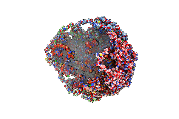
Structure Of Porcine Trypsin Crystals Grown From Peg Complexed With Crystallization Additives Iii
Organism: Sus scrofa
Method: X-RAY DIFFRACTION Resolution:1.41 Å Release Date: 2025-09-17 Classification: PEPTIDE BINDING PROTEIN Ligands: CA, BEN, FLC, PG4, PEG, GOL, PG5, MLT, PG6, EPE, ACT, PO4, TAR |
 |
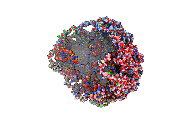
Crystallographic Structure Of The Cubic Crystal Form Of Stmv (77.9 Degree Rotation) Grown From Nacl
Organism: Satellite tobacco mosaic virus
Method: X-RAY DIFFRACTION Resolution:2.94 Å Release Date: 2021-12-15 Classification: VIRUS/RNA Ligands: CL, MG |
Organism: Satellite tobacco mosaic virus
Method: X-RAY DIFFRACTION
Release Date: 2021-12-15
Ligands: CL, MG
 |
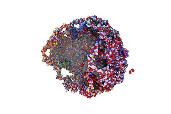
Crystallographic Structure Of A Cubic Crystal Form Of Stmv (84.5 Degree Rotation) Grown From Chloride
Organism: Satellite tobacco mosaic virus
Method: X-RAY DIFFRACTION Resolution:3.00 Å Release Date: 2021-12-15 Classification: VIRUS Ligands: CL, MG |
Organism: Satellite tobacco mosaic virus
Method: X-RAY DIFFRACTION
Release Date: 2021-12-15
Ligands: CL, MG
 |
Crystallographic Structure Of A Cubic Form Of Stmv Grown From Nitrate
Organism: Satellite tobacco mosaic virus
Method: X-RAY DIFFRACTION Resolution:3.19 Å Release Date: 2021-12-15 Classification: VIRUS Ligands: NO3, MG |
Organism: Satellite tobacco mosaic virus
Method: X-RAY DIFFRACTION
Release Date: 2021-12-15
Ligands: NO3, MG
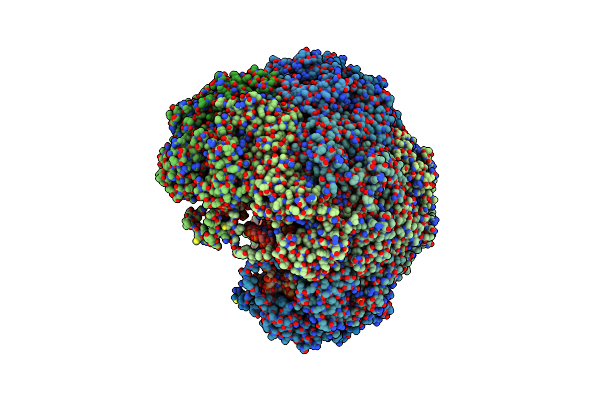 |
Crystallographic Structure Of The Monoclinic Form Of Satellite Tobacco Mosaic Virus
Organism: Satellite tobacco mosaic virus
Method: X-RAY DIFFRACTION Resolution:2.71 Å Release Date: 2021-12-15 Classification: VIRUS/RNA Ligands: MG, PO4, SO4 |
Organism: Satellite tobacco mosaic virus
Method: X-RAY DIFFRACTION
Release Date: 2021-12-15
Ligands: MG, PO4, SO4
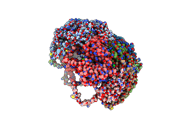 |
Crystallographic Structure Of The Rhombohedral Crystal Form Of Stmv Grown From Chloride
Organism: Satellite tobacco mosaic virus
Method: X-RAY DIFFRACTION Resolution:1.80 Å Release Date: 2021-12-15 Classification: VIRUS/RNA Ligands: CL, MG, PO4 |
Organism: Satellite tobacco mosaic virus
Method: X-RAY DIFFRACTION
Release Date: 2021-12-15
Ligands: CL, MG, PO4
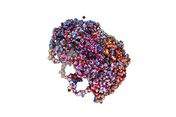 |
Crystallographic Structure Of The Rhombohedral Crystal Form Of Stmv Grown From Bromide
Organism: Satellite tobacco mosaic virus
Method: X-RAY DIFFRACTION Resolution:2.10 Å Release Date: 2021-12-15 Classification: VIRUS Ligands: BR, MG |
Organism: Satellite tobacco mosaic virus
Method: X-RAY DIFFRACTION
Release Date: 2021-12-15
Ligands: BR, MG
 |
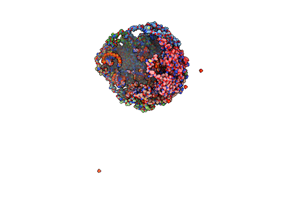
Crystallographic Structure Of A Cubic Crystal Of Stmv (80.7 Degree Rotation About 111) Grown From Chloride
Organism: Satellite tobacco mosaic virus
Method: X-RAY DIFFRACTION Resolution:3.20 Å Release Date: 2021-12-15 Classification: VIRUS/RNA Ligands: PO4, CL, MG |
Organism: Satellite tobacco mosaic virus
Method: X-RAY DIFFRACTION
Release Date: 2021-12-15
Ligands: PO4, CL, MG
 |
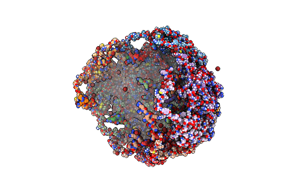
Crystallographic Structure Of A Cubic Crystal Form Of Stmv Grown From Ammonium Sulfate
Organism: Satellite tobacco mosaic virus
Method: X-RAY DIFFRACTION Resolution:2.31 Å Release Date: 2021-12-15 Classification: VIRUS Ligands: PO4, MG, CL, SO4 |
Organism: Satellite tobacco mosaic virus
Method: X-RAY DIFFRACTION
Release Date: 2021-12-15
Ligands: PO4, MG, CL, SO4
 |
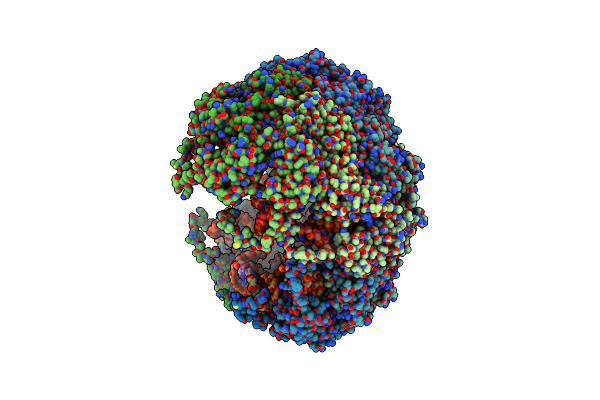
Crystallographic Structure Of A Cubic Crystal Form Of Stmv Grown From Bromide
Organism: Satellite tobacco mosaic virus
Method: X-RAY DIFFRACTION Resolution:3.80 Å Release Date: 2021-12-15 Classification: VIRUS Ligands: CA, BR, MG, CL |
Organism: Satellite tobacco mosaic virus
Method: X-RAY DIFFRACTION
Release Date: 2021-12-15
Ligands: CA, BR, MG, CL
 |
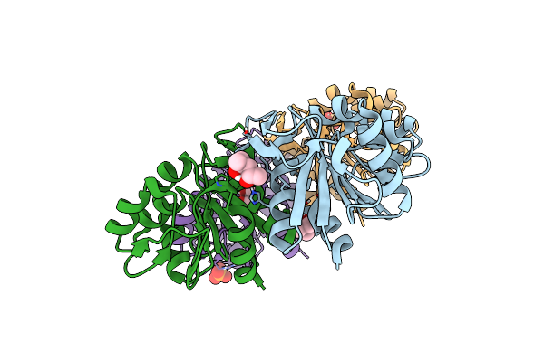
Crystallographic Structure Of A Primitive Orthorhombic Crystal Form Of Stmv
Organism: Satellite tobacco mosaic virus
Method: X-RAY DIFFRACTION Resolution:4.00 Å Release Date: 2021-12-15 Classification: VIRUS/RNA Ligands: PO4 |
Organism: Satellite tobacco mosaic virus
Method: X-RAY DIFFRACTION
Release Date: 2021-12-15
Ligands: PO4
 |
Structure Of Pfp1 Protease From Thermococcus Thioreducens: Large Unit Cell At 1.44 A Resolution
Organism: Thermococcus thioreducens
Method: X-RAY DIFFRACTION Resolution:1.44 Å Release Date: 2021-09-29 Classification: HYDROLASE Ligands: MPD, PO4 |
Organism: Thermococcus thioreducens
Method: X-RAY DIFFRACTION
Release Date: 2021-09-29
Ligands: MPD, PO4