

Search Count: 16
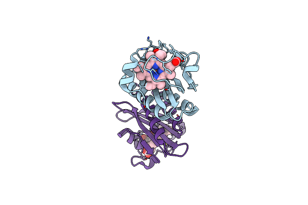 |
Doss Gafa Domain Reduced Nitric Oxide Bound Crystal Structure
Organism: Mycobacterium tuberculosis (strain cdc 1551 / oshkosh)
Method: X-RAY DIFFRACTION Resolution:1.90 Å Release Date: 2016-03-23 Classification: OXIDOREDUCTASE Ligands: HEM, NO |
Organism: Mycobacterium tuberculosis (strain cdc 1551 / oshkosh)
Method: X-RAY DIFFRACTION
Release Date: 2016-03-23
Ligands: HEM, NO
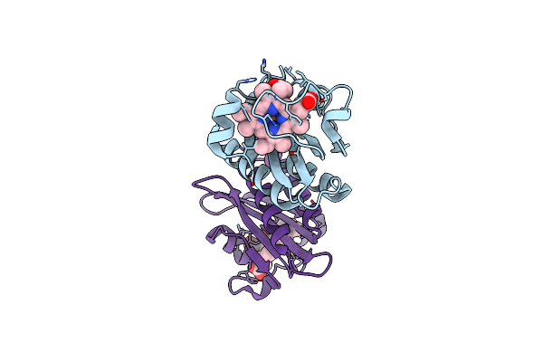 |
Doss Gafa Domain Reduced Co Bound Crystal Structure
Organism: Mycobacterium tuberculosis (strain cdc 1551 / oshkosh)
Method: X-RAY DIFFRACTION Resolution:1.92 Å Release Date: 2016-02-24 Classification: OXIDOREDUCTASE Ligands: HEM, CMO |
Organism: Mycobacterium tuberculosis (strain cdc 1551 / oshkosh)
Method: X-RAY DIFFRACTION
Release Date: 2016-02-24
Ligands: HEM, CMO
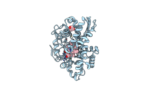 |
Crystal Structure Of Cyp119 From Sulfolobus Acidocaldarius, Complexed With 4-(4-Bromophenyl)-1H Imidazole
Organism: Sulfolobus acidocaldarius
Method: X-RAY DIFFRACTION Resolution:2.18 Å Release Date: 2015-02-18 Classification: OXIDOREDUCTASE Ligands: HEM, 36Y, GOL |
Organism: Sulfolobus acidocaldarius
Method: X-RAY DIFFRACTION
Release Date: 2015-02-18
Ligands: HEM, 36Y, GOL
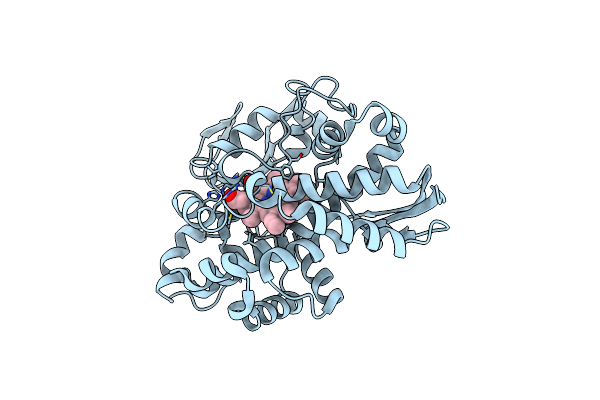 |
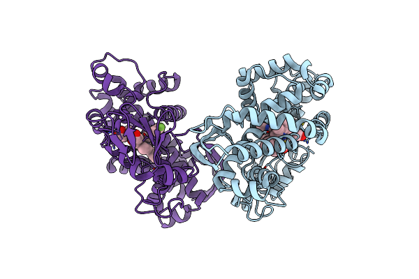
X-Ray Crystal Structure Of Cyp119 From Sulfolobus Acidocaldarius, Complexed With 4-(4-Chlorophenyl)Imidazole
Organism: Sulfolobus acidocaldarius
Method: X-RAY DIFFRACTION Resolution:2.50 Å Release Date: 2015-02-18 Classification: OXIDOREDUCTASE Ligands: HEM, CPZ |
Organism: Sulfolobus acidocaldarius
Method: X-RAY DIFFRACTION
Release Date: 2015-02-18
Ligands: HEM, CPZ
 |
X-Ray Crystal Structure Of Cyp119 Complexed With 4-(4-Flourophenyl)-1H-Imidazole
Organism: Sulfolobus acidocaldarius
Method: X-RAY DIFFRACTION Resolution:2.00 Å Release Date: 2015-02-18 Classification: OXIDOREDUCTASE Ligands: 3SQ, HEM |
Organism: Sulfolobus acidocaldarius
Method: X-RAY DIFFRACTION
Release Date: 2015-02-18
Ligands: 3SQ, HEM
 |
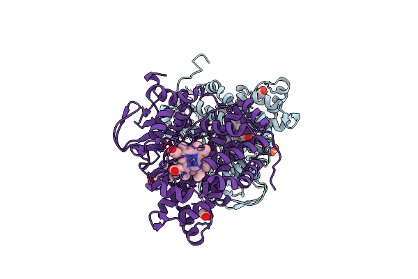
Crystal Structure Of Cyp119 From Sulfolobus Acidocaldarius, Collected At 298K And Complexed With 4-(4-Bromophenyl)-1H Imidazole
Organism: Sulfolobus acidocaldarius
Method: X-RAY DIFFRACTION Resolution:2.70 Å Release Date: 2015-02-18 Classification: OXIDOREDUCTASE Ligands: HEM, 36Y, GOL |
Organism: Sulfolobus acidocaldarius
Method: X-RAY DIFFRACTION
Release Date: 2015-02-18
Ligands: HEM, 36Y, GOL
 |
X-Ray Crystal Structure Of Cyp142A2 From Mycobacterium Smegmatis, Complexed With Cholesterol Sulfate.
Organism: Mycobacterium smegmatis
Method: X-RAY DIFFRACTION Resolution:2.00 Å Release Date: 2014-09-17 Classification: OXIDOREDUCTASE Ligands: HEM, C3S, GOL |
Organism: Mycobacterium smegmatis
Method: X-RAY DIFFRACTION
Release Date: 2014-09-17
Ligands: HEM, C3S, GOL
 |
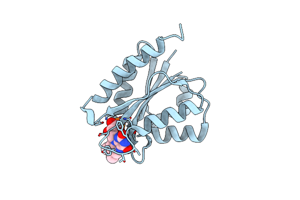
X-Ray Crystal Structure Of Ligand Free Cyp142A2 From Mycobacterium Smegmatis
Organism: Mycobacterium smegmatis
Method: X-RAY DIFFRACTION Resolution:1.78 Å Release Date: 2014-09-17 Classification: OXIDOREDUCTASE Ligands: HEM |
Organism: Mycobacterium smegmatis
Method: X-RAY DIFFRACTION
Release Date: 2014-09-17
Ligands: HEM
 |
Crystal Structure Of Cindoxin, Surface Entropy Reduction Mutant
Organism: Citrobacter braakii
Method: X-RAY DIFFRACTION Resolution:1.21 Å Release Date: 2014-04-16 Classification: ELECTRON TRANSPORT Ligands: FMN |
Organism: Citrobacter braakii
Method: X-RAY DIFFRACTION
Release Date: 2014-04-16
Ligands: FMN
 |
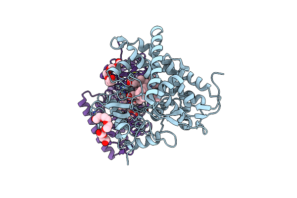
Crystal Structure Of P450Cin Y81F Mutant, Crystallized In 7 Mm 1,8-Cineole
Organism: Citrobacter braakii
Method: X-RAY DIFFRACTION Resolution:1.37 Å Release Date: 2013-07-24 Classification: OXIDOREDUCTASE Ligands: HEM, CNL, SO4, PGE |
Organism: Citrobacter braakii
Method: X-RAY DIFFRACTION
Release Date: 2013-07-24
Ligands: HEM, CNL, SO4, PGE
 |
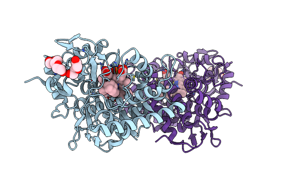
P450Cin Active Site Water: Implications For Substrate Binding And Solvent Accessibility
Organism: Citrobacter braakii
Method: X-RAY DIFFRACTION Resolution:1.38 Å Release Date: 2013-07-24 Classification: OXIDOREDUCTASE Ligands: HEM, CNL, PG4, PEG, SO4 |
Organism: Citrobacter braakii
Method: X-RAY DIFFRACTION
Release Date: 2013-07-24
Ligands: HEM, CNL, PG4, PEG, SO4
 |
Crystal Structure Of P450Cin Y81F Mutant, Crystallized In 3 Mm 1,8-Cineole
Organism: Citrobacter braakii
Method: X-RAY DIFFRACTION Resolution:2.14 Å Release Date: 2013-07-24 Classification: OXIDOREDUCTASE Ligands: HEM, CNL, PEG, PGE, SO4, CL, PG4 |
Organism: Citrobacter braakii
Method: X-RAY DIFFRACTION
Release Date: 2013-07-24
Ligands: HEM, CNL, PEG, PGE, SO4, CL, PG4
 |
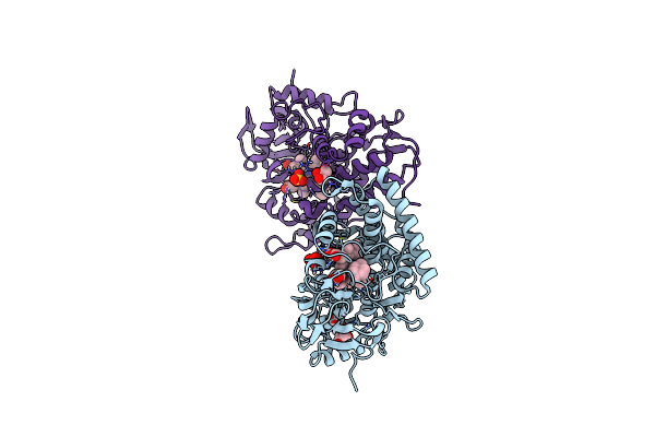
Crystal Structure Of Nitrosyl Cytochrome P450Cam
Organism: Pseudomonas putida
Method: X-RAY DIFFRACTION Resolution:2.20 Å Release Date: 2012-08-01 Classification: OXIDOREDUCTASE Ligands: HEM, CAM, K, NO |
Organism: Pseudomonas putida
Method: X-RAY DIFFRACTION
Release Date: 2012-08-01
Ligands: HEM, CAM, K, NO
 |
Crystal Structure Of Substrate-Bound P450Cin
Organism: Citrobacter braakii
Method: X-RAY DIFFRACTION Resolution:1.55 Å Release Date: 2012-07-25 Classification: OXIDOREDUCTASE Ligands: HEM, CNL, SO4, GOL |
Organism: Citrobacter braakii
Method: X-RAY DIFFRACTION
Release Date: 2012-07-25
Ligands: HEM, CNL, SO4, GOL
 |
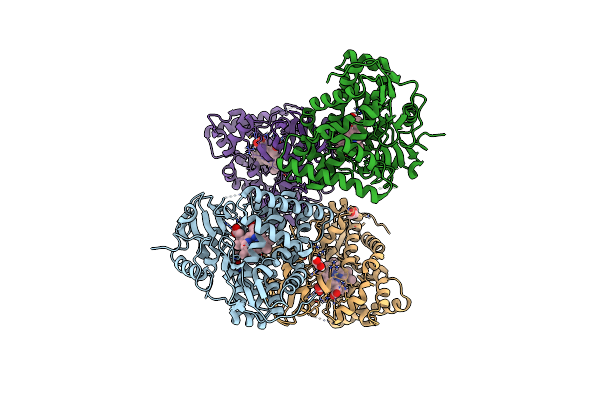
Crystal Structure Of Nitrosyl Cytochrome P450Cin
Organism: Citrobacter braakii
Method: X-RAY DIFFRACTION Resolution:2.32 Å Release Date: 2012-07-25 Classification: OXIDOREDUCTASE Ligands: HEM, CNL, NO, PG4, PEG |
Organism: Citrobacter braakii
Method: X-RAY DIFFRACTION
Release Date: 2012-07-25
Ligands: HEM, CNL, NO, PG4, PEG
 |
Crystal Structure Of Substrate-Free P450Cin
Organism: Citrobacter braakii
Method: X-RAY DIFFRACTION Resolution:1.37 Å Release Date: 2012-07-18 Classification: OXIDOREDUCTASE Ligands: HEM, EDO, CL |
Organism: Citrobacter braakii
Method: X-RAY DIFFRACTION
Release Date: 2012-07-18
Ligands: HEM, EDO, CL