

Search Count: 19
 |
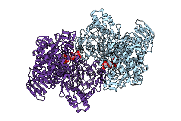
Gh161 Phosphorylase In Complex With Beta-1,3 Glucooligosaccharide
Organism: Gut metagenome
Method: ELECTRON MICROSCOPY Release Date: 2025-12-10 Classification: CYTOSOLIC PROTEIN |
Organism: Gut metagenome
Method: ELECTRON MICROSCOPY
Release Date: 2025-12-10
 |
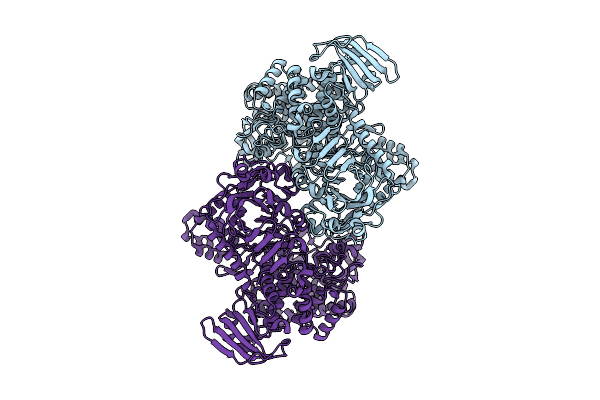
Catalytically Active Gh161A Phosphorylase Refined In C1
Organism: Gut metagenome
Method: ELECTRON MICROSCOPY Release Date: 2025-12-10 Classification: HYDROLASE Ligands: PO4, BGC |
Organism: Gut metagenome
Method: ELECTRON MICROSCOPY
Release Date: 2025-12-10
Ligands: PO4, BGC
 |
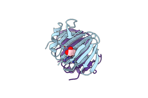
The Cryo-Em Structure Of Bacterial Beta-1,3-Glucan Phosphorylase From Family Gh161
Organism: Gut metagenome
Method: ELECTRON MICROSCOPY Release Date: 2025-11-12 Classification: SUGAR BINDING PROTEIN |
Organism: Gut metagenome
Method: ELECTRON MICROSCOPY
Release Date: 2025-11-12
 |
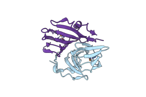
Crystal Structure Of Cellulose Oxidative Enzyme In Acidic Ph With Glycerol
Organism: Metagenome
Method: X-RAY DIFFRACTION Resolution:2.30 Å Release Date: 2024-12-11 Classification: OXIDOREDUCTASE Ligands: GOL, CU |
Organism: Metagenome
Method: X-RAY DIFFRACTION
Release Date: 2024-12-11
Ligands: GOL, CU
 |
Crystal Structure Of Cellulose Oxidative Enzyme Without Ligand
Organism: Metagenome
Method: X-RAY DIFFRACTION Resolution:2.20 Å Release Date: 2024-12-04 Classification: OXIDOREDUCTASE Ligands: CU |
Organism: Metagenome
Method: X-RAY DIFFRACTION
Release Date: 2024-12-04
Ligands: CU
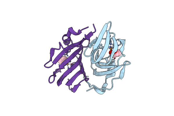 |
Crystal Structure Of Cellulose Oxidative Enzyme With Glycerol
Organism: Metagenome
Method: X-RAY DIFFRACTION Resolution:1.65 Å Release Date: 2024-12-04 Classification: OXIDOREDUCTASE Ligands: GOL, CU |
Organism: Metagenome
Method: X-RAY DIFFRACTION
Release Date: 2024-12-04
Ligands: GOL, CU
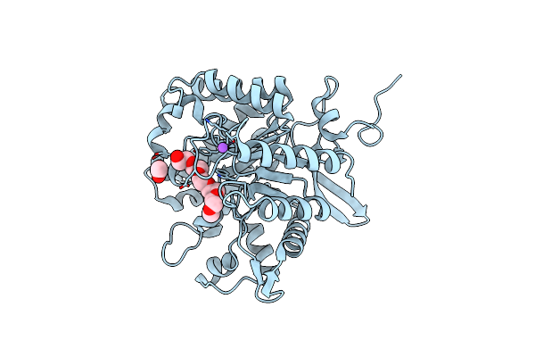 |
Crystal Structure Of A Novel Gh5 Enzyme Retrieved From Capybara Gut Metagenome
Organism: Metagenome
Method: X-RAY DIFFRACTION Resolution:1.60 Å Release Date: 2022-11-02 Classification: HYDROLASE Ligands: 1PG, ACT, NA, CL |
Organism: Metagenome
Method: X-RAY DIFFRACTION
Release Date: 2022-11-02
Ligands: 1PG, ACT, NA, CL
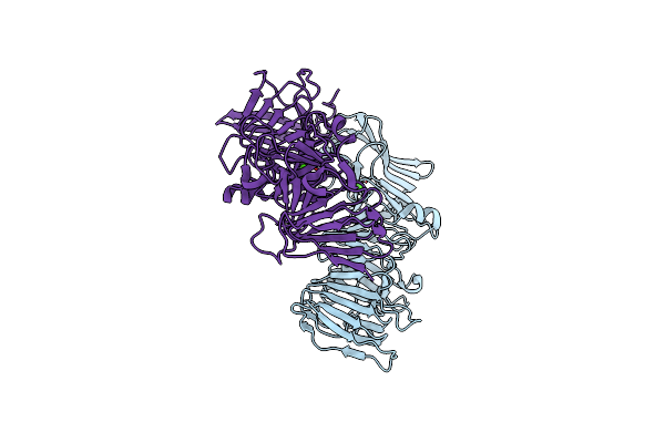 |
Crystal Structure Of A Beta-Helix Domain Retrieved From Capybara Gut Metagenome
Organism: Metagenome
Method: X-RAY DIFFRACTION Resolution:1.85 Å Release Date: 2021-09-08 Classification: UNKNOWN FUNCTION Ligands: CA |
Organism: Metagenome
Method: X-RAY DIFFRACTION
Release Date: 2021-09-08
Ligands: CA
 |
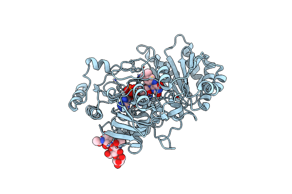
Structure Of Fgceldh7C
Organism: Gibberella zeae (strain ph-1 / atcc mya-4620 / fgsc 9075 / nrrl 31084)
Method: X-RAY DIFFRACTION Resolution:1.64 Å Release Date: 2021-03-03 Classification: OXIDOREDUCTASE Ligands: FAD, NAG, SCN, 1PE, CL, PG4, PEG |
Organism: Gibberella zeae (strain ph-1 / atcc mya-4620 / fgsc 9075 / nrrl 31084)
Method: X-RAY DIFFRACTION
Release Date: 2021-03-03
Ligands: FAD, NAG, SCN, 1PE, CL, PG4, PEG
 |
Structure Of Fgchi7B
Organism: Gibberella zeae (strain ph-1 / atcc mya-4620 / fgsc 9075 / nrrl 31084)
Method: X-RAY DIFFRACTION Resolution:2.38 Å Release Date: 2021-03-03 Classification: OXIDOREDUCTASE Ligands: FAD |
Organism: Gibberella zeae (strain ph-1 / atcc mya-4620 / fgsc 9075 / nrrl 31084)
Method: X-RAY DIFFRACTION
Release Date: 2021-03-03
Ligands: FAD
 |
Iodide Structure Of Trichoderma Reesei Carbohydrate-Active Enzymes Family Aa12
Organism: Hypocrea jecorina (strain atcc 56765 / bcrc 32924 / nrrl 11460 / rut c-30)
Method: X-RAY DIFFRACTION Resolution:1.99 Å Release Date: 2019-11-13 Classification: OXIDOREDUCTASE Ligands: NAG, PGE, CA, IOD |
Organism: Hypocrea jecorina (strain atcc 56765 / bcrc 32924 / nrrl 11460 / rut c-30)
Method: X-RAY DIFFRACTION
Release Date: 2019-11-13
Ligands: NAG, PGE, CA, IOD
 |
Calcium Structure Of Trichoderma Reesei Carbohydrate-Active Enzymes Family Aa12
Organism: Hypocrea jecorina (strain atcc 56765 / bcrc 32924 / nrrl 11460 / rut c-30)
Method: X-RAY DIFFRACTION Resolution:1.80 Å Release Date: 2019-11-13 Classification: OXIDOREDUCTASE Ligands: NAG, PGE, CA |
Organism: Hypocrea jecorina (strain atcc 56765 / bcrc 32924 / nrrl 11460 / rut c-30)
Method: X-RAY DIFFRACTION
Release Date: 2019-11-13
Ligands: NAG, PGE, CA
 |
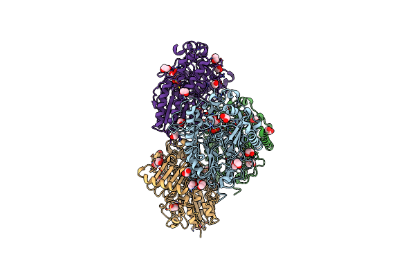
Native Structure Of Trichoderma Reesei Carbohydrate-Active Enzymes Family Aa12
Organism: Hypocrea jecorina (strain atcc 56765 / bcrc 32924 / nrrl 11460 / rut c-30)
Method: X-RAY DIFFRACTION Resolution:2.10 Å Release Date: 2019-08-14 Classification: OXIDOREDUCTASE Ligands: PGE, CA, NAG |
Organism: Hypocrea jecorina (strain atcc 56765 / bcrc 32924 / nrrl 11460 / rut c-30)
Method: X-RAY DIFFRACTION
Release Date: 2019-08-14
Ligands: PGE, CA, NAG
 |
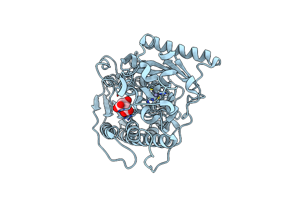
Bt1596 In Complex With Its Substrate 4,5 Unsaturated Uronic Acid Alpha 1,4 D-Glucosamine-2-N, 6-O-Disulfate
Organism: Bacteroides thetaiotaomicron
Method: X-RAY DIFFRACTION Resolution:1.90 Å Release Date: 2017-05-24 Classification: HYDROLASE Ligands: ZN, EDO, CL, UAP |
Organism: Bacteroides thetaiotaomicron
Method: X-RAY DIFFRACTION
Release Date: 2017-05-24
Ligands: ZN, EDO, CL, UAP
 |
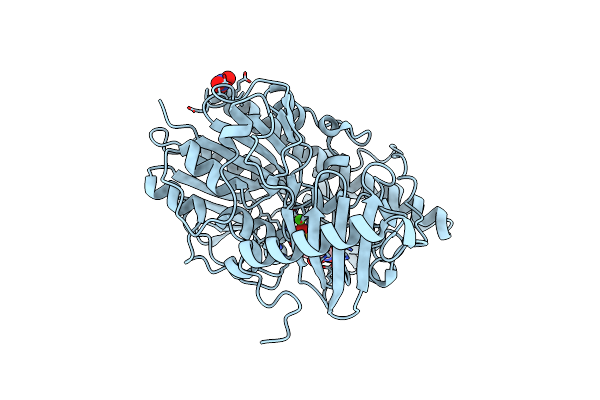
Structure Of Bt1596,A 2-O Gag Sulfatase
Organism: Bacteroides thetaiotaomicron
Method: X-RAY DIFFRACTION Resolution:1.43 Å Release Date: 2017-05-24 Classification: HYDROLASE Ligands: ZN, CIT |
Organism: Bacteroides thetaiotaomicron
Method: X-RAY DIFFRACTION
Release Date: 2017-05-24
Ligands: ZN, CIT
 |
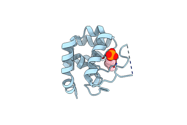
Structure Of Bt4656 In Complex With Its Substrate D-Glucosamine-2-N, 6-O-Disulfate.
Organism: Bacteroides thetaiotaomicron
Method: X-RAY DIFFRACTION Resolution:1.39 Å Release Date: 2017-05-24 Classification: HYDROLASE Ligands: CA, SGN, NO3 |
Organism: Bacteroides thetaiotaomicron
Method: X-RAY DIFFRACTION
Release Date: 2017-05-24
Ligands: CA, SGN, NO3
 |
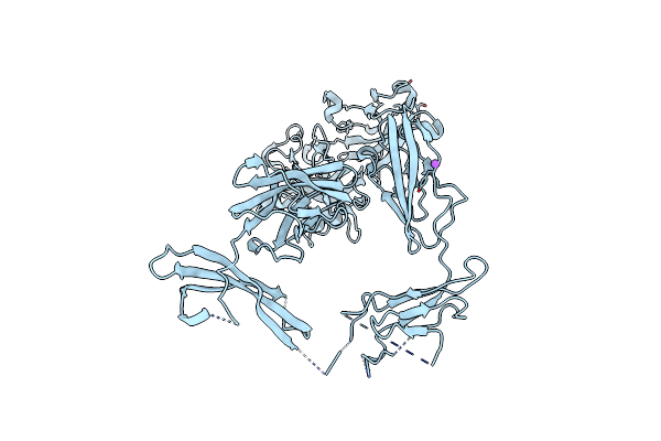
Crystal Structure Of The Endo-Beta-N-Acetylglucosaminidase From Thermotoga Maritima
Organism: Thermotoga maritima
Method: X-RAY DIFFRACTION Resolution:1.70 Å Release Date: 2014-11-12 Classification: HYDROLASE Ligands: PO4, MPD |
Organism: Thermotoga maritima
Method: X-RAY DIFFRACTION
Release Date: 2014-11-12
Ligands: PO4, MPD
 |
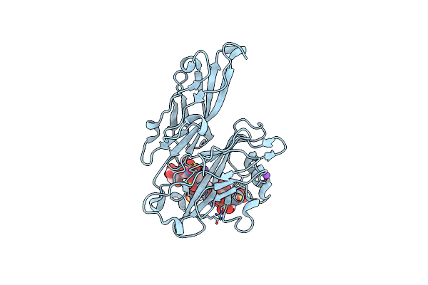
Structure Of Bt4661, A Suse-Like Surface Located Polysaccharide Binding Protein From The Bacteroides Thetaiotaomicron Heparin Utilisation Locus
Organism: Bacteroides thetaiotaomicron
Method: X-RAY DIFFRACTION Resolution:1.95 Å Release Date: 2013-03-06 Classification: HEPARIN-BINDING PROTEIN |
Organism: Bacteroides thetaiotaomicron
Method: X-RAY DIFFRACTION
Release Date: 2013-03-06
 |
Structure Of Bt4661, A Suse-Like Surface Located Polysaccharide Binding Protein From The Bacteroides Thetaiotaomicron Heparin Utilisation Locus
Organism: Bacteroides thetaiotaomicron
Method: X-RAY DIFFRACTION Resolution:1.35 Å Release Date: 2013-03-06 Classification: HEPARIN-BINDING PROTEIN |
Organism: Bacteroides thetaiotaomicron
Method: X-RAY DIFFRACTION
Release Date: 2013-03-06