

Search Count: 34
 |
Sheep Galectin-11 (Lgals11) Complex With Glycerol
Organism: Ovis aries
Method: X-RAY DIFFRACTION Resolution:2.00 Å Release Date: 2020-05-20 Classification: SUGAR BINDING PROTEIN Ligands: GOL |
Organism: Ovis aries
Method: X-RAY DIFFRACTION
Release Date: 2020-05-20
Ligands: GOL
 |
Crystal Structure Of Hiv Protease Complexed With Methyl N-[(1S)-1-Benzhydryl-2-(3-Morpholin-4-Ium-2-Ylpropylamino)-2-Oxo-Ethyl]Carbamate
Organism: Human immunodeficiency virus 1
Method: X-RAY DIFFRACTION Resolution:1.57 Å Release Date: 2016-05-18 Classification: HYDROLASE/INHIBITOR Ligands: 6EH, CL |
Organism: Human immunodeficiency virus 1
Method: X-RAY DIFFRACTION
Release Date: 2016-05-18
Ligands: 6EH, CL
 |
Crystal Structure Of Hiv Protease Complexed With Methyl N-[(1S)-1-[[2-[(3S)-3-[(4-Aminophenyl)Methylamino]-4-Hydroxy-Butyl]Phenyl]Carbamoyl]-2,2-Diphenyl-Ethyl]Carbamate
Organism: Human immunodeficiency virus 1
Method: X-RAY DIFFRACTION Resolution:1.50 Å Release Date: 2016-05-18 Classification: HYDROLASE/INHIBITOR Ligands: CL, 6EG |
Organism: Human immunodeficiency virus 1
Method: X-RAY DIFFRACTION
Release Date: 2016-05-18
Ligands: CL, 6EG
 |
Crystal Structure Of Hiv Protease Complexed With Methyl N-[(1S)-1-Benzhydryl-2-[2-[2-[(2R,5S)-5-(Benzylcarbamoyloxymethyl)Morpholin-2-Yl]Ethyl]Anilino]-2-Oxo-Ethyl]Carbamate
Organism: Human immunodeficiency virus 1
Method: X-RAY DIFFRACTION Resolution:1.46 Å Release Date: 2016-05-18 Classification: HYDROLASE/INHIBITOR Ligands: CL, 6EF |
Organism: Human immunodeficiency virus 1
Method: X-RAY DIFFRACTION
Release Date: 2016-05-18
Ligands: CL, 6EF
 |
Crystal Structure Of Hiv Protease Complexed With [(1S)-1-[(S)-(4-Chlorophenyl)-(3,5-Difluorophenyl)Methyl]-2-[[5-Fluoro-4-[2-[(2R,5S)-5-(2,2,2-Trifluoroethylcarbamoyloxymethyl)Morpholin-4-Ium-2-Yl]Ethyl]Pyridin-1-Ium-3-Yl]Amino]-2-Oxo-Ethyl]Ammonium
Organism: Human immunodeficiency virus 1
Method: X-RAY DIFFRACTION Resolution:1.15 Å Release Date: 2016-05-18 Classification: HYDROLASE/INHIBITOR Ligands: ACT, 6EE, CL |
Organism: Human immunodeficiency virus 1
Method: X-RAY DIFFRACTION
Release Date: 2016-05-18
Ligands: ACT, 6EE, CL
 |
Jc Virus Large T-Antigen Origin Binding Domain F258L Mutant
Organism: Jc polyomavirus
Method: X-RAY DIFFRACTION Resolution:2.70 Å Release Date: 2015-12-16 Classification: viral protein, dna binding protein Ligands: EDO |
Organism: Jc polyomavirus
Method: X-RAY DIFFRACTION
Release Date: 2015-12-16
Ligands: EDO
 |
Sv40 Large T Antigen Origin Binding Domain Bound To Artificial Dna Fork
Organism: Simian virus 40, Synthetic construct
Method: X-RAY DIFFRACTION Resolution:1.70 Å Release Date: 2015-09-09 Classification: replication/dna Ligands: EDO, CA, ACT Modified Residue(s): CME |
Organism: Simian virus 40, Synthetic construct
Method: X-RAY DIFFRACTION
Release Date: 2015-09-09
Ligands: EDO, CA, ACT
 |
Crystal Structure Of The Jcv Large T-Antigen Origin Binding Domain
Organism: Jc polyomavirus
Method: X-RAY DIFFRACTION Resolution:1.50 Å Release Date: 2014-04-02 Classification: HYDROLASE Ligands: GOL, FLC |
Organism: Jc polyomavirus
Method: X-RAY DIFFRACTION
Release Date: 2014-04-02
Ligands: GOL, FLC
 |
Crystal Structure Of The Jcv Large T-Antigen Origin Binding Domain
Organism: Jc polyomavirus
Method: X-RAY DIFFRACTION Resolution:2.60 Å Release Date: 2014-03-05 Classification: Hydrolase, dna binding protein Ligands: SO4 |
Organism: Jc polyomavirus
Method: X-RAY DIFFRACTION
Release Date: 2014-03-05
Ligands: SO4
 |
Crystal Structure Of The Jcv Large T-Antigen Origin Binding Domain
Organism: Jc polyomavirus
Method: X-RAY DIFFRACTION Resolution:1.32 Å Release Date: 2014-03-05 Classification: HYDROLASE Ligands: TLA |
Organism: Jc polyomavirus
Method: X-RAY DIFFRACTION
Release Date: 2014-03-05
Ligands: TLA
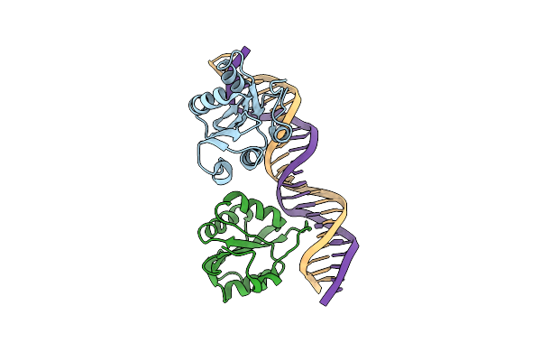 |
Crystal Structure Of The Sv40 Large T-Antigen Origin Bining Domain Bound To Site I Dna
Organism: Simian virus 40
Method: X-RAY DIFFRACTION Resolution:3.20 Å Release Date: 2013-01-23 Classification: DNA BINDING PROTEIN/DNA |
Organism: Simian virus 40
Method: X-RAY DIFFRACTION
Release Date: 2013-01-23
 |
Crystal Structure Of Hiv-1 Reverse Transcriptase (Wild Type) In Complex With Inhibitor M05
Organism: Hiv-1 m:b_hxb2r
Method: X-RAY DIFFRACTION Resolution:2.60 Å Release Date: 2011-10-26 Classification: TRANSFERASE/TRANSFERASE INHIBITOR Ligands: 5MA |
Organism: Hiv-1 m:b_hxb2r
Method: X-RAY DIFFRACTION
Release Date: 2011-10-26
Ligands: 5MA
 |
Crystal Structure Of Hiv-1 Reverse Transcriptase (K103N Mutant) In Complex With Inhibitor M05
Organism: Hiv-1 m:b_hxb2r
Method: X-RAY DIFFRACTION Resolution:2.40 Å Release Date: 2011-10-26 Classification: TRANSFERASE/TRANSFERASE INHIBITOR Ligands: 5MA |
Organism: Hiv-1 m:b_hxb2r
Method: X-RAY DIFFRACTION
Release Date: 2011-10-26
Ligands: 5MA
 |
Crystal Structure Of Hiv-1 Reverse Transcriptase (K103N Mutant) In Complex With Inhibitor M06
Organism: Hiv-1 m:b_hxb2r
Method: X-RAY DIFFRACTION Resolution:2.51 Å Release Date: 2011-10-26 Classification: Transferase/transferase Inhibitor Ligands: M06 |
Organism: Hiv-1 m:b_hxb2r
Method: X-RAY DIFFRACTION
Release Date: 2011-10-26
Ligands: M06
 |
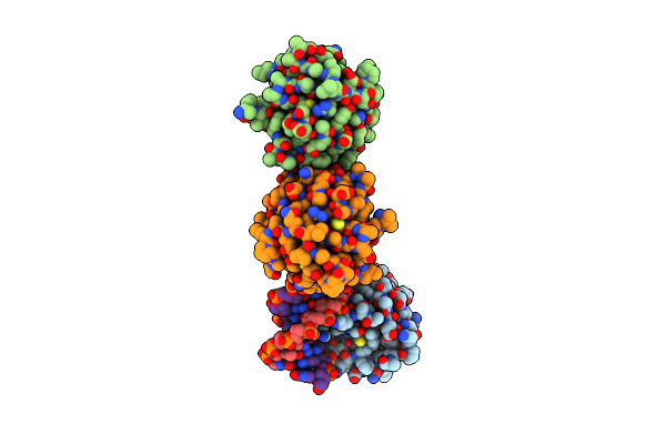
Structure-Based Design Of A Disulfide-Linked Oligomeric Form Of The Simian Virus 40 (Sv40) Large T Antigen Dna Binding Domain
Organism: Simian virus 40
Method: X-RAY DIFFRACTION Resolution:1.66 Å Release Date: 2011-06-01 Classification: HYDROLASE Ligands: FLC |
Organism: Simian virus 40
Method: X-RAY DIFFRACTION
Release Date: 2011-06-01
Ligands: FLC
 |
Asymmetric Assembly Of Merkel Cell Polyomavirus Large T-Antigen Origin Binding Domains At The Viral Origin
Organism: Merkel cell polyomavirus
Method: X-RAY DIFFRACTION Resolution:2.90 Å Release Date: 2011-04-27 Classification: DNA BINDING PROTEIN/DNA |
Organism: Merkel cell polyomavirus
Method: X-RAY DIFFRACTION
Release Date: 2011-04-27
 |
Structure-Based Analysis Of The Interaction Between The Simian Virus 40 T-Antigen Origin Binding Domain And Single-Stranded Dna
Organism: Simian virus 40
Method: X-RAY DIFFRACTION Resolution:1.64 Å Release Date: 2011-02-23 Classification: DNA BINDING PROTEIN Ligands: SCN Modified Residue(s): CME |
Organism: Simian virus 40
Method: X-RAY DIFFRACTION
Release Date: 2011-02-23
Ligands: SCN
 |
Crystal Structure Of A Complex Between Dok7 Ph-Ptb And The Musk Juxtamembrane Region
Organism: Mus musculus
Method: X-RAY DIFFRACTION Resolution:2.60 Å Release Date: 2010-07-14 Classification: SIGNALING PROTEIN Modified Residue(s): MSE, PTR |
Organism: Mus musculus
Method: X-RAY DIFFRACTION
Release Date: 2010-07-14
 |
Crystal Structure Of Hiv Reverse Transcriptase In Complex With Inhibitor 3
Organism: Hiv-1 m:b_hxb2r
Method: X-RAY DIFFRACTION Resolution:2.98 Å Release Date: 2009-08-25 Classification: TRANSFERASE Ligands: RT3 |
Organism: Hiv-1 m:b_hxb2r
Method: X-RAY DIFFRACTION
Release Date: 2009-08-25
Ligands: RT3
 |
Crystal Structure Of Hiv Reverse Transcriptase In Complex With Inhibitor 7
Organism: Hiv-1 m:b_hxb2r
Method: X-RAY DIFFRACTION Resolution:2.70 Å Release Date: 2009-08-25 Classification: TRANSFERASE Ligands: RT7 |
Organism: Hiv-1 m:b_hxb2r
Method: X-RAY DIFFRACTION
Release Date: 2009-08-25
Ligands: RT7