

Search Count: 34
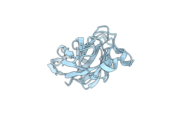 |
Crystal Structure Of The Ucad Lectin-Binding Domain
Organism: Proteus mirabilis
Method: X-RAY DIFFRACTION Resolution:1.62 Å Release Date: 2022-07-06 Classification: SUGAR BINDING PROTEIN Ligands: CL |
Organism: Proteus mirabilis
Method: X-RAY DIFFRACTION
Release Date: 2022-07-06
Ligands: CL
 |
Crystal Structure Of The Ucld Lectin-Binding Domain
Organism: Escherichia coli
Method: X-RAY DIFFRACTION Resolution:2.20 Å Release Date: 2022-07-06 Classification: SUGAR BINDING PROTEIN Ligands: IOD |
Organism: Escherichia coli
Method: X-RAY DIFFRACTION
Release Date: 2022-07-06
Ligands: IOD
 |
Crystal Structure Of The Ucad Lectin-Binding Domain In Complex With Fucose
Organism: Proteus mirabilis
Method: X-RAY DIFFRACTION Resolution:1.50 Å Release Date: 2022-07-06 Classification: SUGAR BINDING PROTEIN Ligands: FUL, CL |
Organism: Proteus mirabilis
Method: X-RAY DIFFRACTION
Release Date: 2022-07-06
Ligands: FUL, CL
 |
Crystal Structure Of The Ucad Lectin-Binding Domain In Complex With Glucose
Organism: Proteus mirabilis
Method: X-RAY DIFFRACTION Resolution:1.78 Å Release Date: 2022-07-06 Classification: SUGAR BINDING PROTEIN Ligands: BGC, CL |
Organism: Proteus mirabilis
Method: X-RAY DIFFRACTION
Release Date: 2022-07-06
Ligands: BGC, CL
 |
Crystal Structure Of The Ucad Lectin-Binding Domain In Complex With Galactose
Organism: Proteus mirabilis
Method: X-RAY DIFFRACTION Resolution:1.72 Å Release Date: 2022-07-06 Classification: SUGAR BINDING PROTEIN Ligands: GLA, CL |
Organism: Proteus mirabilis
Method: X-RAY DIFFRACTION
Release Date: 2022-07-06
Ligands: GLA, CL
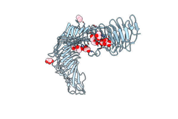 |
Crystal Structure Of Antigen 43 From Uropathogenic Escherichia Coli Uti89
Organism: Escherichia coli
Method: X-RAY DIFFRACTION Resolution:2.43 Å Release Date: 2022-03-09 Classification: MICROBIAL PROTEIN Ligands: GOL, IPA, CIT, PEG |
Organism: Escherichia coli
Method: X-RAY DIFFRACTION
Release Date: 2022-03-09
Ligands: GOL, IPA, CIT, PEG
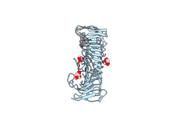 |
Crystal Structure Of Antigen 43B From Escherichia Coli Cft073
Organism: Escherichia coli
Method: X-RAY DIFFRACTION Resolution:2.08 Å Release Date: 2022-03-09 Classification: MICROBIAL PROTEIN Ligands: GOL |
Organism: Escherichia coli
Method: X-RAY DIFFRACTION
Release Date: 2022-03-09
Ligands: GOL
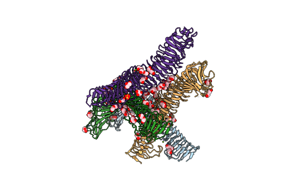 |
Crystal Structure Of Antigen 43 From Escherichia Coli Edl933
Organism: Escherichia coli o157:h7
Method: X-RAY DIFFRACTION Resolution:2.98 Å Release Date: 2022-03-09 Classification: MICROBIAL PROTEIN Ligands: PEG, P6G, GOL, MG, CL, SO4 |
Organism: Escherichia coli o157:h7
Method: X-RAY DIFFRACTION
Release Date: 2022-03-09
Ligands: PEG, P6G, GOL, MG, CL, SO4
 |
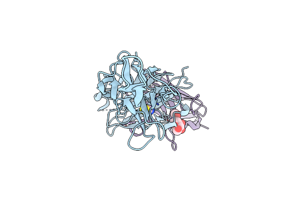
Heterodimeric Complex Of Pf12 And Pf41 Of Plasmodium Falciparum
Organism: Plasmodium falciparum
Method: X-RAY DIFFRACTION Resolution:2.85 Å Release Date: 2022-03-02 Classification: UNKNOWN FUNCTION Ligands: NAG, CD |
Organism: Plasmodium falciparum
Method: X-RAY DIFFRACTION
Release Date: 2022-03-02
Ligands: NAG, CD
 |
Plasmodium Falciparum Protein Pf12 Bound To Nanobody G7
Organism: Plasmodium falciparum 3d7, Vicugna pacos
Method: X-RAY DIFFRACTION Resolution:1.77 Å Release Date: 2022-03-02 Classification: UNKNOWN FUNCTION Ligands: NAG, SCN, NA |
Organism: Plasmodium falciparum 3d7, Vicugna pacos
Method: X-RAY DIFFRACTION
Release Date: 2022-03-02
Ligands: NAG, SCN, NA
 |
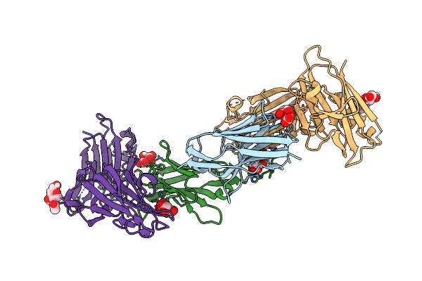
Plasmodium Falciparum Protein Pf12P
Organism: Plasmodium falciparum
Method: X-RAY DIFFRACTION Resolution:2.80 Å Release Date: 2021-02-03 Classification: UNKNOWN FUNCTION |
Organism: Plasmodium falciparum
Method: X-RAY DIFFRACTION
Release Date: 2021-02-03
 |
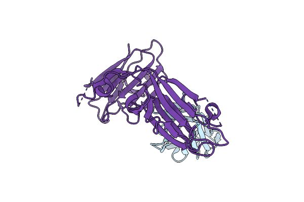
Plasmodium Falciparum Protein Pf12P Bound To Nanobody B9
Organism: Vicugna pacos, Plasmodium falciparum
Method: X-RAY DIFFRACTION Resolution:2.00 Å Release Date: 2021-02-03 Classification: UNKNOWN FUNCTION Ligands: FLC, NAG |
Organism: Vicugna pacos, Plasmodium falciparum
Method: X-RAY DIFFRACTION
Release Date: 2021-02-03
Ligands: FLC, NAG
 |
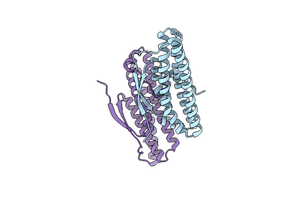
Plasmodium Falciparum Protein Pf12P Bound To Nanobody D9
Organism: Vicugna pacos, Plasmodium falciparum
Method: X-RAY DIFFRACTION Resolution:3.25 Å Release Date: 2021-02-03 Classification: UNKNOWN FUNCTION |
Organism: Vicugna pacos, Plasmodium falciparum
Method: X-RAY DIFFRACTION
Release Date: 2021-02-03
 |
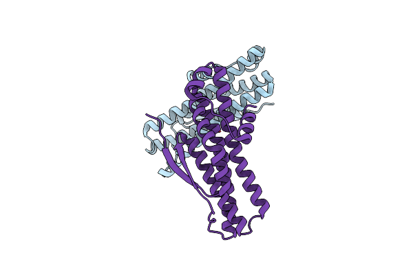
Crystal Structure Of Selenomethionine-Containing Acab From Uropathogenic E. Coli
Organism: Escherichia coli
Method: X-RAY DIFFRACTION Resolution:3.40 Å Release Date: 2020-07-15 Classification: TRANSCRIPTION Ligands: CL |
Organism: Escherichia coli
Method: X-RAY DIFFRACTION
Release Date: 2020-07-15
Ligands: CL
 |
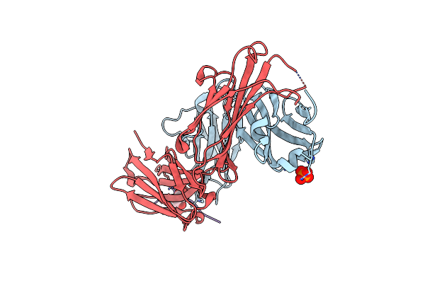
Crystal Structure Of Transcription Regulator Acab From Uropathogenic E. Coli
Organism: Escherichia coli
Method: X-RAY DIFFRACTION Resolution:2.94 Å Release Date: 2020-07-15 Classification: TRANSCRIPTION Ligands: CA |
Organism: Escherichia coli
Method: X-RAY DIFFRACTION
Release Date: 2020-07-15
Ligands: CA
 |
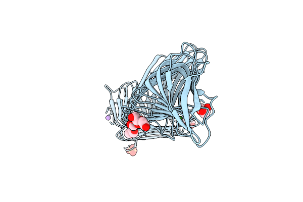
Fab Fragment Complexed With C-Mannosylated Tryptophan Peptide
Organism: Homo sapiens, Mus musculus
Method: X-RAY DIFFRACTION Resolution:1.60 Å Release Date: 2020-07-08 Classification: IMMUNE SYSTEM Ligands: PO4, MAN |
Organism: Homo sapiens, Mus musculus
Method: X-RAY DIFFRACTION
Release Date: 2020-07-08
Ligands: PO4, MAN
 |
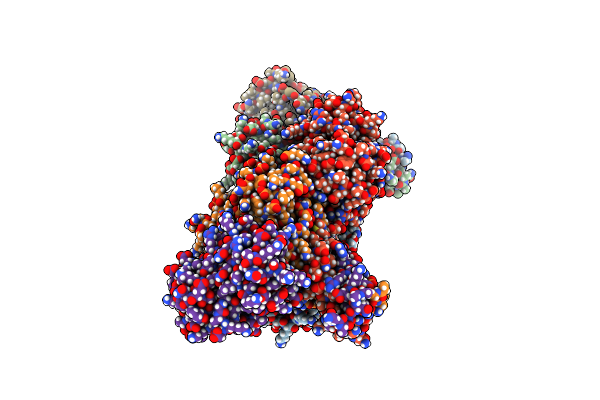
Crystal Structure Of The Autotransporter Upab From E. Coli Strain Cft073
Organism: Escherichia coli o6:h1
Method: X-RAY DIFFRACTION Resolution:1.97 Å Release Date: 2019-04-10 Classification: PROTEIN BINDING Ligands: GOL, P6G, SO4, CA, CL, NA |
Organism: Escherichia coli o6:h1
Method: X-RAY DIFFRACTION
Release Date: 2019-04-10
Ligands: GOL, P6G, SO4, CA, CL, NA
 |
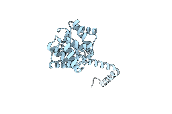
Crystal Structure Of Proteus Mirabilis Scsc In A Transitional Conformation
Organism: Proteus mirabilis atcc 29906
Method: X-RAY DIFFRACTION Resolution:2.56 Å Release Date: 2017-08-02 Classification: ISOMERASE |
Organism: Proteus mirabilis atcc 29906
Method: X-RAY DIFFRACTION
Release Date: 2017-08-02
 |
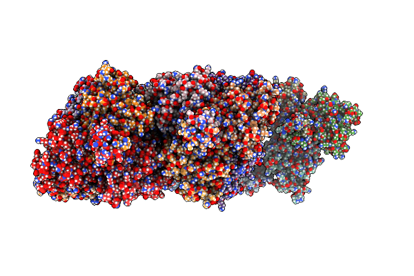
Crystal Structure Of Proteus Mirabilis Scsc In An Extended Conformation
Organism: Proteus mirabilis atcc 29906
Method: X-RAY DIFFRACTION Resolution:2.92 Å Release Date: 2017-07-26 Classification: ISOMERASE |
Organism: Proteus mirabilis atcc 29906
Method: X-RAY DIFFRACTION
Release Date: 2017-07-26
 |
Crystal Structure Of Proteus Mirabilis Scsc In A Compact Conformation
Organism: Proteus mirabilis atcc 29906
Method: X-RAY DIFFRACTION Resolution:2.60 Å Release Date: 2016-06-08 Classification: ISOMERASE |
Organism: Proteus mirabilis atcc 29906
Method: X-RAY DIFFRACTION
Release Date: 2016-06-08