
Search Count: 1,325
All
Selected
 |
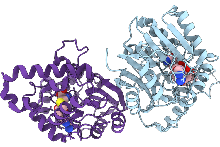
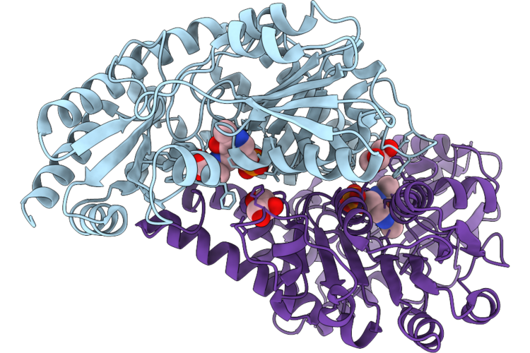
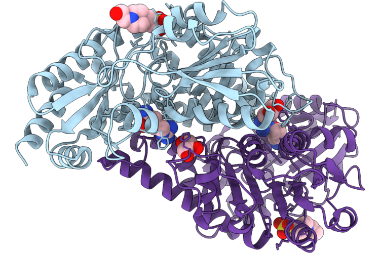
Organism: Streptomyces caespitosus
Method: X-RAY DIFFRACTION Resolution:3.20 Å Release Date: 2026-05-20 Classification: TRANSFERASE Ligands: SAH, A1E3R |
 |
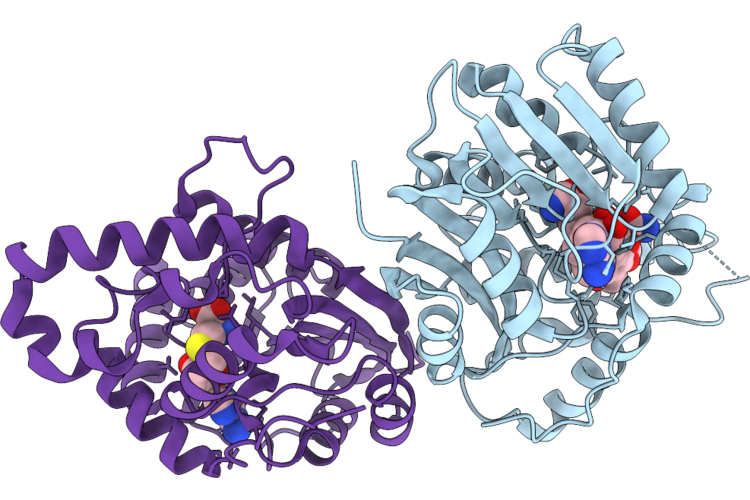
Organism: Streptomyces caespitosus
Method: X-RAY DIFFRACTION Resolution:2.99 Å Release Date: 2026-05-20 Classification: TRANSFERASE Ligands: SAH, A1E3X, MG |
 |
Organism: Streptomyces caespitosus
Method: X-RAY DIFFRACTION Resolution:2.99 Å Release Date: 2026-05-20 Classification: TRANSFERASE Ligands: SAH, A1E3V |
 |
Organism: Streptomyces caespitosus
Method: X-RAY DIFFRACTION Resolution:3.02 Å Release Date: 2026-05-20 Classification: TRANSFERASE Ligands: SAH, A1E3Z |
 |
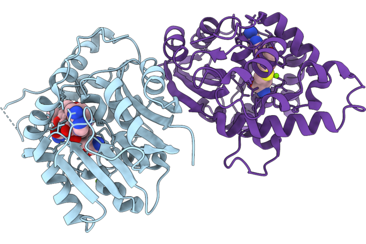
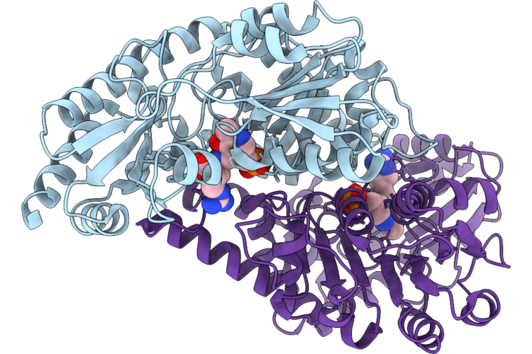
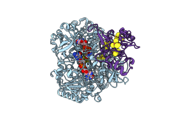
The Costructure Of Mitm And Trans-1-Hydroxy-7-Methoxy-2-Dimethylaminomitosene With Sah
Organism: Streptomyces caespitosus
Method: X-RAY DIFFRACTION Resolution:2.80 Å Release Date: 2026-05-20 Classification: TRANSFERASE Ligands: SAH, A1E30 |
 |
Organism: Gibberella zeae (strain atcc mya-4620 / cbs 123657 / fgsc 9075 / nrrl 31084 / ph-1)
Method: X-RAY DIFFRACTION Resolution:1.67 Å Release Date: 2026-05-20 Classification: BIOSYNTHETIC PROTEIN |
 |
Organism: Gibberella zeae (strain atcc mya-4620 / cbs 123657 / fgsc 9075 / nrrl 31084 / ph-1)
Method: X-RAY DIFFRACTION Resolution:1.90 Å Release Date: 2026-05-20 Classification: BIOSYNTHETIC PROTEIN Ligands: EQJ |
 |
Organism: Fusarium graminearum ph-1
Method: X-RAY DIFFRACTION Resolution:1.86 Å Release Date: 2026-05-20 Classification: BIOSYNTHETIC PROTEIN Ligands: EQJ, GOL |
 |
Organism: Fusarium graminearum ph-1
Method: X-RAY DIFFRACTION Resolution:2.10 Å Release Date: 2026-05-20 Classification: BIOSYNTHETIC PROTEIN Ligands: GOL, 0JO |
 |
Organism: Fusarium graminearum ph-1
Method: X-RAY DIFFRACTION Resolution:1.48 Å Release Date: 2026-05-20 Classification: BIOSYNTHETIC PROTEIN Ligands: GOL, PLP, WYK |
 |
Organism: Fusarium graminearum ph-1
Method: X-RAY DIFFRACTION Resolution:2.18 Å Release Date: 2026-05-20 Classification: BIOSYNTHETIC PROTEIN Ligands: ARG, GOL, EPE |
 |
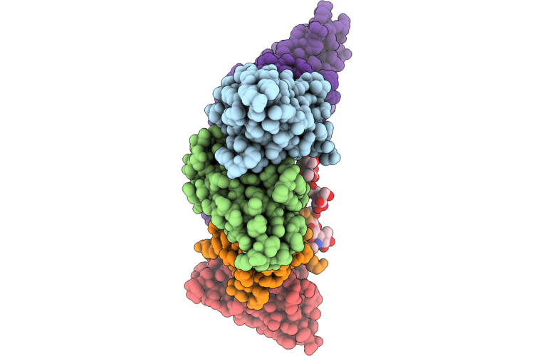
Epitope And Functional Classification Of Human Neutralizing Antibodies Against Sftsv Gn
Organism: Homo sapiens
Method: ELECTRON MICROSCOPY Resolution:2.85 Å Release Date: 2026-05-06 Classification: VIRAL PROTEIN |
 |
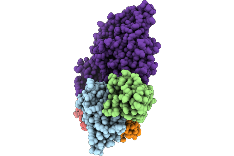
Epitope And Functional Classification Of Human Neutralizing Antibodies Against Sftsv Gn
Organism: Homo sapiens, Severe fever with thrombocytopenia syndrome virus
Method: ELECTRON MICROSCOPY Resolution:2.97 Å Release Date: 2026-04-29 Classification: VIRAL PROTEIN |
 |
Epitope And Functional Classification Of Human Neutralizing Antibodies Against Sftsv Gn
Organism: Homo sapiens, Severe fever with thrombocytopenia syndrome virus
Method: ELECTRON MICROSCOPY Resolution:2.69 Å Release Date: 2026-04-29 Classification: VIRAL PROTEIN |
 |
Epitope And Functional Classification Of Human Neutralizing Antibodies Against Sftsv Gn
Organism: Homo sapiens, Severe fever with thrombocytopenia syndrome virus
Method: ELECTRON MICROSCOPY Resolution:2.69 Å Release Date: 2026-04-29 Classification: VIRAL PROTEIN |
 |
Cryo-Em Structure Of Formate Dehydrogenase From Shewanella Oneidensis Mr-1 (Sofdhab)
Organism: Shewanella oneidensis mr-1
Method: ELECTRON MICROSCOPY Release Date: 2026-04-08 Classification: OXIDOREDUCTASE Ligands: MGD, SF4, H2S, W |
 |
Phosphorylation Dependent Recognition Of Ripk1 By Phosphatidylinositol 3,4,5-Trisphosphate 5-Phosphatase 1
|
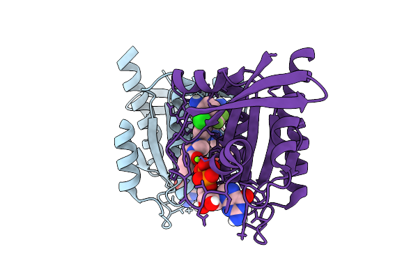 |
Organism: Homo sapiens
Method: X-RAY DIFFRACTION Resolution:1.84 Å Release Date: 2026-03-11 Classification: ONCOPROTEIN Ligands: A1CQ2, GDP, MG |
 |
Organism: Homo sapiens
Method: X-RAY DIFFRACTION Resolution:2.17 Å Release Date: 2026-03-11 Classification: ONCOPROTEIN Ligands: GDP, MG, DMS, EDO, A1AWR, PEG |
 |
Organism: Adeno-associated virus
Method: ELECTRON MICROSCOPY Release Date: 2026-03-11 Classification: VIRUS |