

Search Count: 12
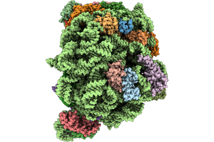 |
Pseudomonas Aeruginosa 50S Ribosome Bound To Rsfs (L1 Stalk In 'Mid' Conformation)
Organism: Pseudomonas aeruginosa pao1
Method: ELECTRON MICROSCOPY Resolution:2.29 Å Release Date: 2026-08-26 Classification: RIBOSOME Ligands: MG, ZN |
Organism: Pseudomonas aeruginosa pao1
Method: ELECTRON MICROSCOPY
Release Date: 2026-08-26
Ligands: MG, ZN
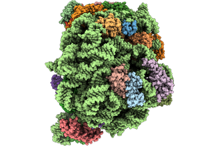 |
Pseudomonas Aeruginosa 50S Ribosome Bound To Rsfs (L1 Stalk 'In' Conformation)
Organism: Pseudomonas aeruginosa pao1
Method: ELECTRON MICROSCOPY Resolution:2.29 Å Release Date: 2026-08-26 Classification: RIBOSOME Ligands: MG, ZN |
Organism: Pseudomonas aeruginosa pao1
Method: ELECTRON MICROSCOPY
Release Date: 2026-08-26
Ligands: MG, ZN
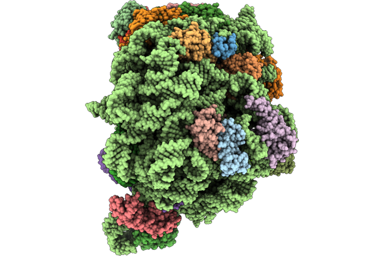 |
Pseudomonas Aeruginosa 50S Ribosome Bound To Rsfs (L1 Stalk In 'Out' Conformation)
Organism: Pseudomonas aeruginosa pao1
Method: ELECTRON MICROSCOPY Resolution:2.29 Å Release Date: 2026-08-26 Classification: RIBOSOME Ligands: MG, ZN |
Organism: Pseudomonas aeruginosa pao1
Method: ELECTRON MICROSCOPY
Release Date: 2026-08-26
Ligands: MG, ZN
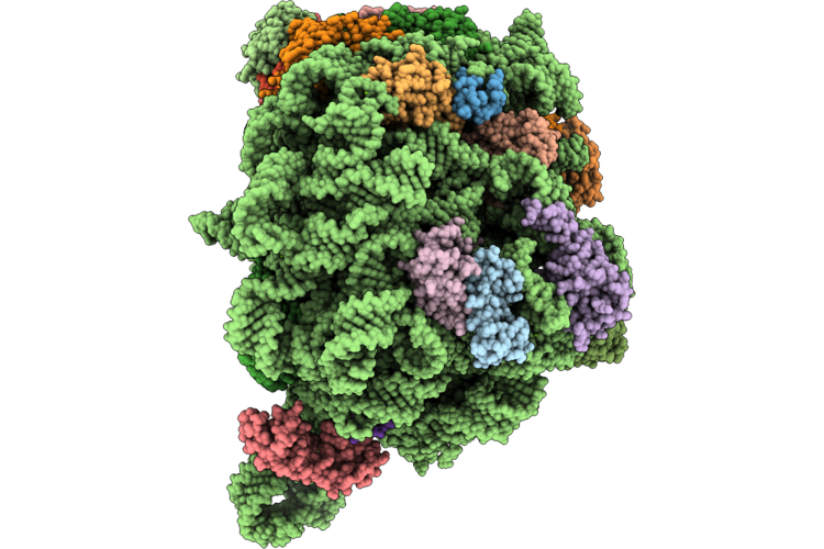 |
Pseudomonas Aeruginosa 50S Ribosome Bound To Rsfs (L1 Stalk In 'Out' Conformation And Missing Ul1 And Trna)
Organism: Pseudomonas aeruginosa pao1
Method: ELECTRON MICROSCOPY Resolution:2.29 Å Release Date: 2026-08-26 Classification: RIBOSOME Ligands: MG, ZN |
Organism: Pseudomonas aeruginosa pao1
Method: ELECTRON MICROSCOPY
Release Date: 2026-08-26
Ligands: MG, ZN
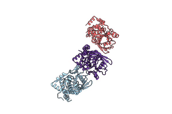 |
Crystal Structure Of Tcptp Catalytic Domain
Organism: Homo sapiens
Method: X-RAY DIFFRACTION Resolution:1.93 Å Release Date: 2021-12-29 Classification: HYDROLASE |
Organism: Homo sapiens
Method: X-RAY DIFFRACTION
Release Date: 2021-12-29
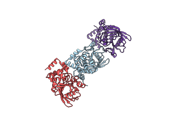 |
Crystal Structure Of Ptpn2 Catalytic Domain
Organism: Homo sapiens
Method: X-RAY DIFFRACTION Resolution:1.70 Å Release Date: 2021-12-29 Classification: HYDROLASE Ligands: IOD |
Organism: Homo sapiens
Method: X-RAY DIFFRACTION
Release Date: 2021-12-29
Ligands: IOD
 |
Orexin Receptor 2 (Ox2R) In Complex With G Protein And Natural Peptide-Agonist Orexin B (Oxb)
Organism: Homo sapiens, Mus musculus
Method: ELECTRON MICROSCOPY Release Date: 2021-02-10 Classification: MEMBRANE PROTEIN |
Organism: Homo sapiens, Mus musculus
Method: ELECTRON MICROSCOPY
Release Date: 2021-02-10
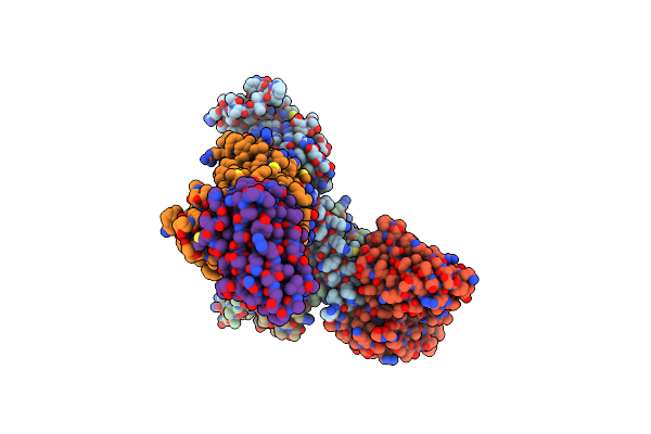 |
Orexin Receptor 2 (Ox2R) In Complex With G Protein And Small-Molecule Agonist Compound 1
Organism: Homo sapiens, Mus musculus, Lama glama
Method: ELECTRON MICROSCOPY Release Date: 2021-02-10 Classification: MEMBRANE PROTEIN Ligands: XGD |
Organism: Homo sapiens, Mus musculus, Lama glama
Method: ELECTRON MICROSCOPY
Release Date: 2021-02-10
Ligands: XGD
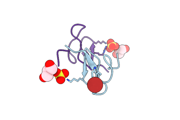 |
Crystal Structure Of Jztx-V
Organism: Chilobrachys guangxiensis
Method: X-RAY DIFFRACTION Resolution:1.05 Å Release Date: 2019-03-06 Classification: TOXIN Ligands: BR, SO4, GOL |
Organism: Chilobrachys guangxiensis
Method: X-RAY DIFFRACTION
Release Date: 2019-03-06
Ligands: BR, SO4, GOL
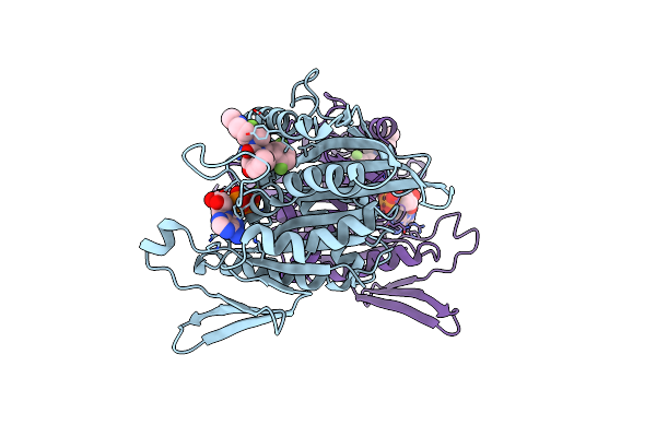 |
Crystal Structure Of Ksp In Complex With Inhibitor 30
Organism: Homo sapiens
Method: X-RAY DIFFRACTION Resolution:2.28 Å Release Date: 2008-07-01 Classification: MOTOR PROTEIN Ligands: MG, ADP, K30 |
Organism: Homo sapiens
Method: X-RAY DIFFRACTION
Release Date: 2008-07-01
Ligands: MG, ADP, K30
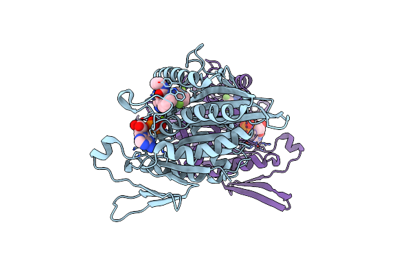 |
Crystal Structure Of Ksp In Complex With Inhibitor 9H
Organism: Homo sapiens
Method: X-RAY DIFFRACTION Resolution:2.51 Å Release Date: 2006-10-03 Classification: CELL CYCLE Ligands: MG, ADP, N9H |
Organism: Homo sapiens
Method: X-RAY DIFFRACTION
Release Date: 2006-10-03
Ligands: MG, ADP, N9H
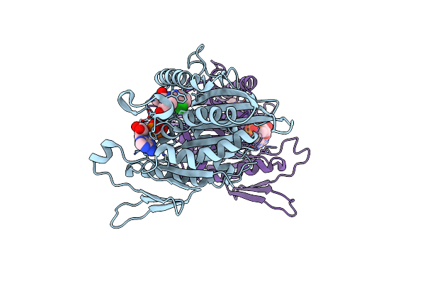 |
Crystal Structure Of Ksp In Complex With Inhibitor 1
Organism: Homo sapiens
Method: X-RAY DIFFRACTION Resolution:2.50 Å Release Date: 2005-04-12 Classification: CELL CYCLE Ligands: MG, ADP, L47 |
Organism: Homo sapiens
Method: X-RAY DIFFRACTION
Release Date: 2005-04-12
Ligands: MG, ADP, L47