

Search Count: 34
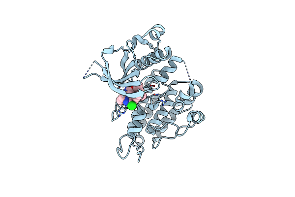 |
Structure Of Human Anaplastic Lymphoma Kinase (Alk) Harboring The G1202R/L1196M Compound Mutation In Complex With Nvl-655
Organism: Homo sapiens
Method: X-RAY DIFFRACTION Resolution:1.58 Å Release Date: 2024-09-18 Classification: ONCOPROTEIN Ligands: A1IJ8 |
Organism: Homo sapiens
Method: X-RAY DIFFRACTION
Release Date: 2024-09-18
Ligands: A1IJ8
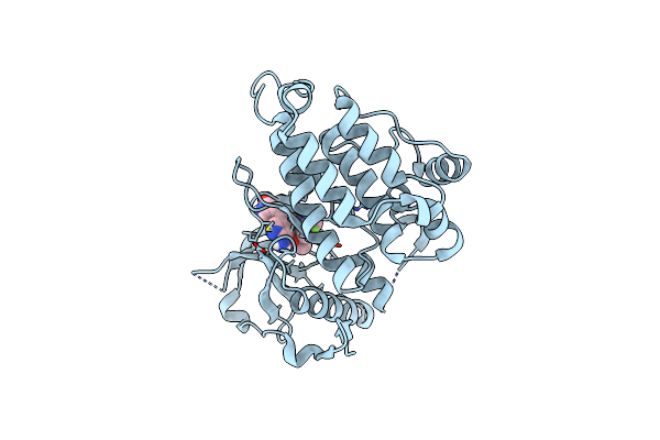 |
Structure Of Human Anaplastic Lymphoma Kinase Domain In Complex With (4-[6-Amino-5-[(1~{R})-1-[5-Fluoro-2-(Triazol-2-Yl)Phenyl]Ethoxy]-3-Pyridyl]Isoindolin-1-One)
Organism: Homo sapiens
Method: X-RAY DIFFRACTION Resolution:1.83 Å Release Date: 2022-06-29 Classification: TRANSFERASE Ligands: 25J |
Organism: Homo sapiens
Method: X-RAY DIFFRACTION
Release Date: 2022-06-29
Ligands: 25J
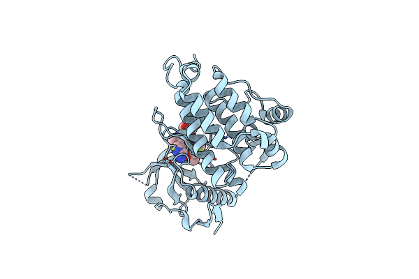 |
Structure Of Human Anaplastic Lymphoma Kinase Domain In Complex With ((2~{R})-2-[5-[6-Amino-5-[(1~{R})-1-[5-Fluoro-2-(Triazol-2-Yl)Phenyl]Ethoxy]-3-Pyridyl]-4-Methyl-Thiazol-2-Yl]Propane-1,2-Diol)
Organism: Homo sapiens
Method: X-RAY DIFFRACTION Resolution:1.94 Å Release Date: 2022-06-29 Classification: TRANSFERASE Ligands: AWJ |
Organism: Homo sapiens
Method: X-RAY DIFFRACTION
Release Date: 2022-06-29
Ligands: AWJ
 |
Crystal Structure Of Aldehyde Dehydrogenase C (Aldc) Mutant (C291A) From Pseudomonas Syringae In Complexed With Nad+ And Octanal
Organism: Pseudomonas syringae pv. tomato (strain atcc baa-871 / dc3000)
Method: X-RAY DIFFRACTION Resolution:2.52 Å Release Date: 2020-09-09 Classification: OXIDOREDUCTASE Ligands: OYA, NAD |
Organism: Pseudomonas syringae pv. tomato (strain atcc baa-871 / dc3000)
Method: X-RAY DIFFRACTION
Release Date: 2020-09-09
Ligands: OYA, NAD
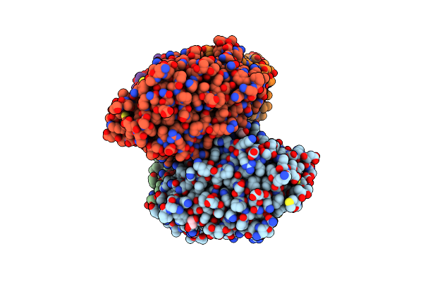 |
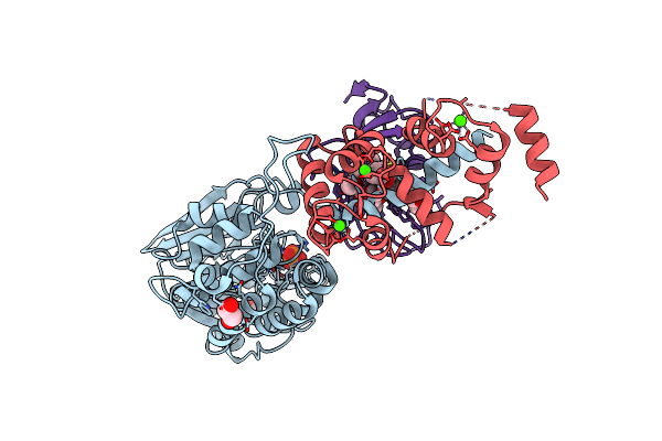
Crystal Structure Of Candida Albicans Calcineurin A, Calcineurin B, Fkbp12 And Fk506 (Tacrolimus)
Organism: Candida albicans (strain wo-1), Candida albicans (strain sc5314 / atcc mya-2876)
Method: X-RAY DIFFRACTION Resolution:2.55 Å Release Date: 2019-09-18 Classification: Hydrolase/Isomerase/Calcium Binding Ligands: PO4, ZN, FE, EDO, CA, FK5 |
Organism: Candida albicans (strain wo-1), Candida albicans (strain sc5314 / atcc mya-2876)
Method: X-RAY DIFFRACTION
Release Date: 2019-09-18
Ligands: PO4, ZN, FE, EDO, CA, FK5
 |
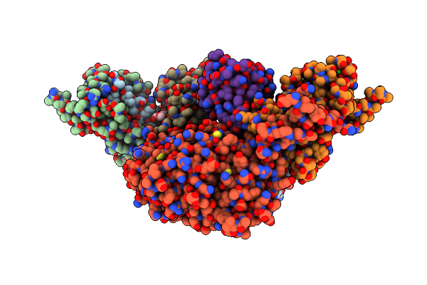
Crystal Structure Of Aspergillus Fumigatus Calcineurin A, Calcineurin B, Fkbp12 And Fk506 (Tacrolimus)
Organism: Neosartorya fumigata (strain atcc mya-4609 / af293 / cbs 101355 / fgsc a1100)
Method: X-RAY DIFFRACTION Resolution:2.50 Å Release Date: 2019-09-18 Classification: Hydrolase/Isomerase/Calcium Binding Ligands: ZN, FE, PO4, EDO, CA, FK5 |
Organism: Neosartorya fumigata (strain atcc mya-4609 / af293 / cbs 101355 / fgsc a1100)
Method: X-RAY DIFFRACTION
Release Date: 2019-09-18
Ligands: ZN, FE, PO4, EDO, CA, FK5
 |
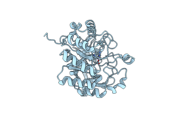
Crystal Structure Of Cryptococcus Neoformans Calceineurin A, Calcineurin B, And Fkbp12 With Fk-506
Organism: Cryptococcus neoformans var. grubii serotype a (strain h99 / atcc 208821 / cbs 10515 / fgsc 9487)
Method: X-RAY DIFFRACTION Resolution:3.30 Å Release Date: 2019-09-18 Classification: Hydrolase/Isomerase/Calcium Binding Ligands: SO4, FE, ZN, CA, FK5 |
Organism: Cryptococcus neoformans var. grubii serotype a (strain h99 / atcc 208821 / cbs 10515 / fgsc 9487)
Method: X-RAY DIFFRACTION
Release Date: 2019-09-18
Ligands: SO4, FE, ZN, CA, FK5
 |
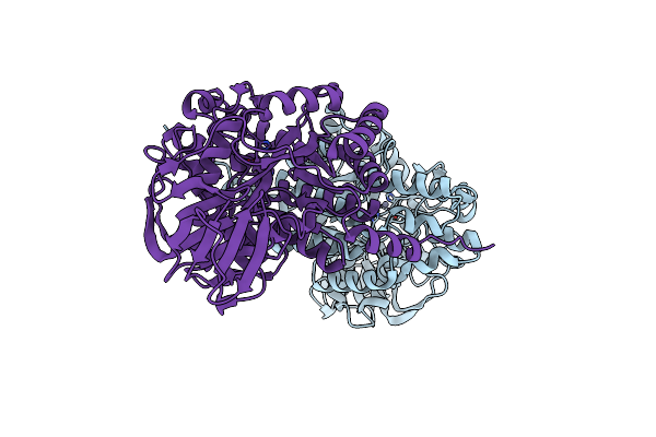
Crystal Structure Of The Dihydroorotase Domain (K1556A) Of Human Cad
Organism: Homo sapiens
Method: X-RAY DIFFRACTION Resolution:2.77 Å Release Date: 2018-10-24 Classification: HYDROLASE Ligands: ZN |
Organism: Homo sapiens
Method: X-RAY DIFFRACTION
Release Date: 2018-10-24
Ligands: ZN
 |
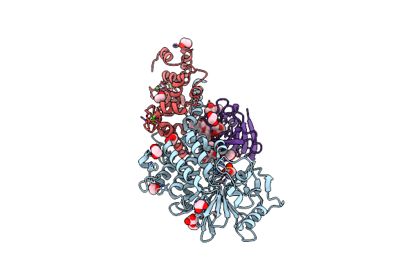
Crystal Structure Of A Monometallic Dihydropyrimidinase From Pseudomonas Aeruginosa Pao1 Reveals No Lysine Carbamylation Within The Active Site
Organism: Pseudomonas aeruginosa (strain atcc 15692 / dsm 22644 / cip 104116 / jcm 14847 / lmg 12228 / 1c / prs 101 / pao1)
Method: X-RAY DIFFRACTION Resolution:2.22 Å Release Date: 2018-10-24 Classification: HYDROLASE Ligands: ZN |
 |
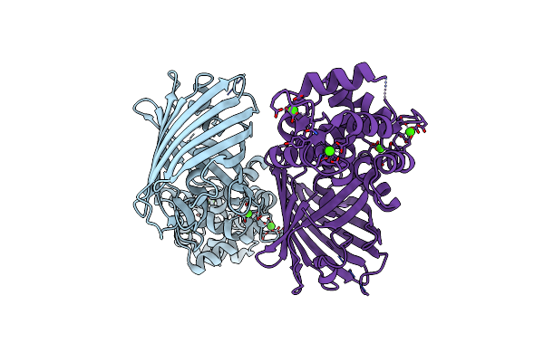
Crystal Structure Of Calcineurin A And Calcineurin B In Complex With Fkbp12 And Fk506 From Coccidioides Immitis Rs
Organism: Coccidioides immitis (strain rs)
Method: X-RAY DIFFRACTION Resolution:1.85 Å Release Date: 2015-05-20 Classification: HYDROLASE Ligands: ZN, FE, MES, EDO, CA, FK5 |
Organism: Coccidioides immitis (strain rs)
Method: X-RAY DIFFRACTION
Release Date: 2015-05-20
Ligands: ZN, FE, MES, EDO, CA, FK5
 |
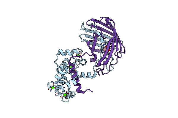
Crystal Structure Of The Genetically Encoded Calcium Indicator Rgeco1
Organism: Gallus gallus, Discosoma sp., Rattus norvegicus
Method: X-RAY DIFFRACTION Resolution:2.20 Å Release Date: 2013-07-24 Classification: FLUORESCENT PROTEIN Ligands: CA |
Organism: Gallus gallus, Discosoma sp., Rattus norvegicus
Method: X-RAY DIFFRACTION
Release Date: 2013-07-24
Ligands: CA
 |
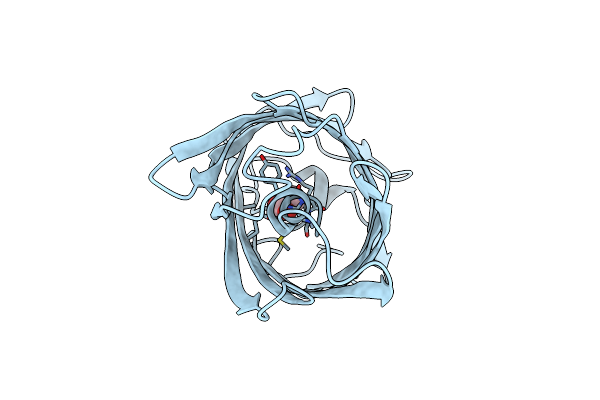
Crystal Structure Of The Genetically Encoded Calcium Indicator Rcamp
Organism: Entacmaea quadricolor
Method: X-RAY DIFFRACTION Resolution:2.10 Å Release Date: 2012-10-03 Classification: FLUORESCENT PROTEIN Ligands: ACT, CA |
Organism: Entacmaea quadricolor
Method: X-RAY DIFFRACTION
Release Date: 2012-10-03
Ligands: ACT, CA
 |
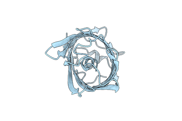
Crystal Structure Of The Engineered Fluorescent Protein Mruby, Crystal Form 1, Ph 4.5
Organism: Entacmaea quadricolor
Method: X-RAY DIFFRACTION Resolution:1.25 Å Release Date: 2012-10-03 Classification: FLUORESCENT PROTEIN Ligands: ACT |
Organism: Entacmaea quadricolor
Method: X-RAY DIFFRACTION
Release Date: 2012-10-03
Ligands: ACT
 |
Crystal Structure Of The Engineered Fluorescent Protein Mruby, Crystal Form 1, Ph 8.5
Organism: Entacmaea quadricolor
Method: X-RAY DIFFRACTION Resolution:1.65 Å Release Date: 2012-10-03 Classification: FLUORESCENT PROTEIN |
Organism: Entacmaea quadricolor
Method: X-RAY DIFFRACTION
Release Date: 2012-10-03
 |
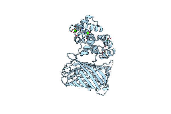
Crystal Structure Of The Engineered Fluorescent Protein Mruby, Crystal Form 2
Organism: Entacmaea quadricolor
Method: X-RAY DIFFRACTION Resolution:1.60 Å Release Date: 2012-10-03 Classification: FLUORESCENT PROTEIN Ligands: SO4 |
Organism: Entacmaea quadricolor
Method: X-RAY DIFFRACTION
Release Date: 2012-10-03
Ligands: SO4
 |
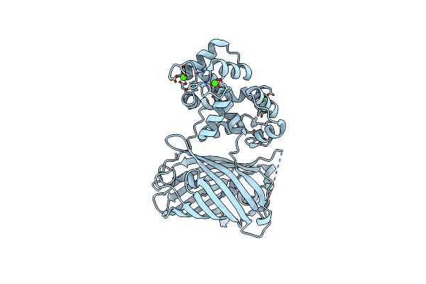
Crystal Structure Of Gcamp2-T116V,D381Y
Organism: Gallus gallus, Aequorea victoria, Rattus norvegicus
Method: X-RAY DIFFRACTION Resolution:2.00 Å Release Date: 2012-06-20 Classification: FLUORESCENT PROTEIN Ligands: CA |
Organism: Gallus gallus, Aequorea victoria, Rattus norvegicus
Method: X-RAY DIFFRACTION
Release Date: 2012-06-20
Ligands: CA
 |
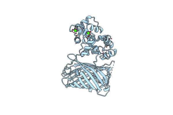
Crystal Structure Of Gcamp3-D380Y
Organism: Gallus gallus, Aequorea victoria, Rattus norvegicus
Method: X-RAY DIFFRACTION Resolution:2.10 Å Release Date: 2012-06-20 Classification: FLUORESCENT PROTEIN Ligands: CA |
Organism: Gallus gallus, Aequorea victoria, Rattus norvegicus
Method: X-RAY DIFFRACTION
Release Date: 2012-06-20
Ligands: CA
 |
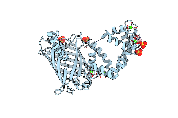
Crystal Structure Of Gcamp3-D380Y, Lp(Linker 2)
Organism: Gallus gallus, Aequorea victoria, Rattus norvegicus
Method: X-RAY DIFFRACTION Resolution:2.40 Å Release Date: 2012-06-20 Classification: FLUORESCENT PROTEIN Ligands: CA |
Organism: Gallus gallus, Aequorea victoria, Rattus norvegicus
Method: X-RAY DIFFRACTION
Release Date: 2012-06-20
Ligands: CA
 |
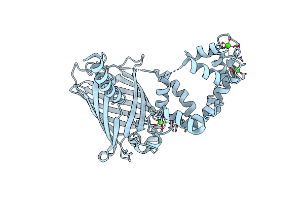
Crystal Structure Of Dimeric Gcamp3-D380Y, Qp(Linker 1), Lp(Linker 2)
Organism: Gallus gallus, Aequorea victoria, Rattus norvegicus
Method: X-RAY DIFFRACTION Resolution:1.90 Å Release Date: 2012-06-20 Classification: FLUORESCENT PROTEIN Ligands: CA, SO4, GOL |
Organism: Gallus gallus, Aequorea victoria, Rattus norvegicus
Method: X-RAY DIFFRACTION
Release Date: 2012-06-20
Ligands: CA, SO4, GOL
 |
Crystal Structure Of Dimeric Gcamp2-Lia(Linker 1)
Organism: Gallus gallus, Aequorea victoria, Rattus norvegicus
Method: X-RAY DIFFRACTION Resolution:1.70 Å Release Date: 2012-06-20 Classification: FLUORESCENT PROTEIN Ligands: CA |
Organism: Gallus gallus, Aequorea victoria, Rattus norvegicus
Method: X-RAY DIFFRACTION
Release Date: 2012-06-20
Ligands: CA