

Search Count: 28
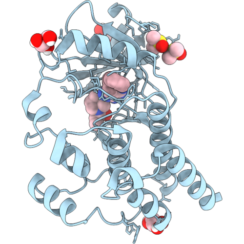 |
Crystal Structure Of Btk Kinase Domain Bound To Ibrutinib
Organism: Mus musculus
Method: X-RAY DIFFRACTION Resolution:1.37 Å Release Date: 2026-09-09 Classification: TRANSFERASE Ligands: 8E8, GOL, DMS |
Organism: Mus musculus
Method: X-RAY DIFFRACTION
Release Date: 2026-09-09
Ligands: 8E8, GOL, DMS
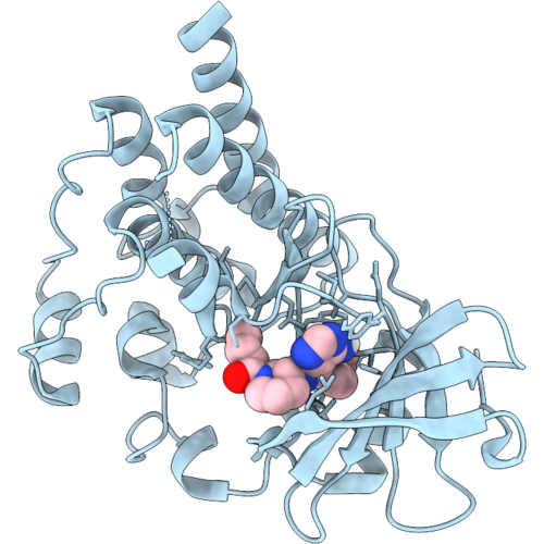 |
Crystal Structure Of Btk Kinase Domain Bound To An Ibrutinib Analog With A 2-Butynamide Warhead
Organism: Mus musculus
Method: X-RAY DIFFRACTION Resolution:2.48 Å Release Date: 2026-09-09 Classification: TRANSFERASE Ligands: A1CSA |
Organism: Mus musculus
Method: X-RAY DIFFRACTION
Release Date: 2026-09-09
Ligands: A1CSA
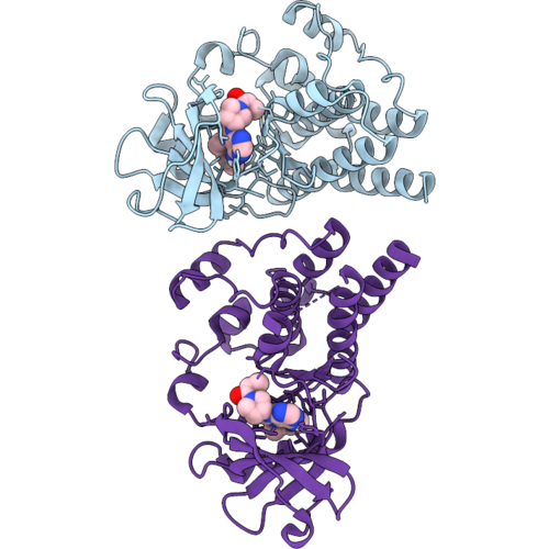 |
Crystal Structure Of Btk Kinase Domain Bound To A Tirabrutinib Analog With An Acrylamide Warhead
Organism: Mus musculus
Method: X-RAY DIFFRACTION Resolution:2.48 Å Release Date: 2026-09-09 Classification: TRANSFERASE Ligands: A1CSB |
Organism: Mus musculus
Method: X-RAY DIFFRACTION
Release Date: 2026-09-09
Ligands: A1CSB
 |
Crystal Structure Of Btk Kinase Domain Bound To Compound Ys1
Organism: Mus musculus
Method: X-RAY DIFFRACTION Resolution:1.60 Å Release Date: 2026-02-25 Classification: TRANSFERASE/TRANSFERASE INHIBITOR Ligands: A1C20, IMD |
Organism: Mus musculus
Method: X-RAY DIFFRACTION
Release Date: 2026-02-25
Ligands: A1C20, IMD
 |
Crystal Structure Of Btk Kinase Domain Bound To Compound Ys2
Organism: Mus musculus
Method: X-RAY DIFFRACTION Resolution:1.27 Å Release Date: 2026-02-25 Classification: TRANSFERASE/TRANSFERASE INHIBITOR Ligands: A1C2Z, IMD |
Organism: Mus musculus
Method: X-RAY DIFFRACTION
Release Date: 2026-02-25
Ligands: A1C2Z, IMD
 |
Bruton'S Tyrosine Kinase In Complex With Compound Pti55
Organism: Mus musculus
Method: X-RAY DIFFRACTION Resolution:2.03 Å Release Date: 2025-08-13 Classification: TRANSFERASE/TRANSFERASE INHIBITOR Ligands: A1BIZ |
Organism: Mus musculus
Method: X-RAY DIFFRACTION
Release Date: 2025-08-13
Ligands: A1BIZ
 |
Bruton'S Tyrosine Kinase In Complex With Compound Pti52
Organism: Mus musculus
Method: X-RAY DIFFRACTION Resolution:2.10 Å Release Date: 2025-08-13 Classification: TRANSFERASE/TRANSFERASE INHIBITOR Ligands: A1BI0 |
Organism: Mus musculus
Method: X-RAY DIFFRACTION
Release Date: 2025-08-13
Ligands: A1BI0
 |
Bruton'S Tyrosine Kinase With Mutations In The Activation Loop In Complex With Compound Pti52
Organism: Mus musculus
Method: X-RAY DIFFRACTION Resolution:2.26 Å Release Date: 2025-08-13 Classification: TRANSFERASE/TRANSFERASE INHIBITOR Ligands: A1BI0, DMS |
Organism: Mus musculus
Method: X-RAY DIFFRACTION
Release Date: 2025-08-13
Ligands: A1BI0, DMS
 |
Bruton'S Tyrosine Kinase With Mutations In The Activation Loop In Complex With Compound Pti42
Organism: Mus musculus
Method: X-RAY DIFFRACTION Resolution:2.86 Å Release Date: 2025-08-13 Classification: TRANSFERASE/TRANSFERASE INHIBITOR Ligands: A1BI1 |
Organism: Mus musculus
Method: X-RAY DIFFRACTION
Release Date: 2025-08-13
Ligands: A1BI1
 |
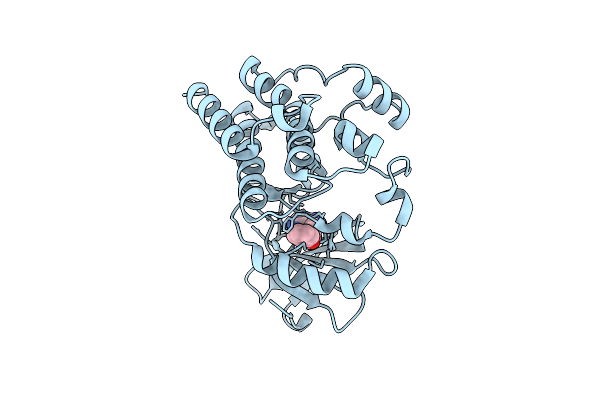
Bruton'S Tyrosine Kinase With Mutations In The Activation Loop In Complex With Compound A110162
Organism: Mus musculus
Method: X-RAY DIFFRACTION Resolution:3.38 Å Release Date: 2025-08-13 Classification: TRANSFERASE/TRANSFERASE INHIBITOR Ligands: A1BKW |
Organism: Mus musculus
Method: X-RAY DIFFRACTION
Release Date: 2025-08-13
Ligands: A1BKW
 |
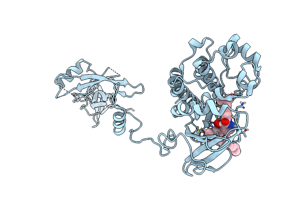
Bruton'S Tyrosine Kinase With Mutations In The Activation Loop In Complex With Compound P301390
Organism: Mus musculus
Method: X-RAY DIFFRACTION Resolution:3.05 Å Release Date: 2025-08-13 Classification: TRANSFERASE/TRANSFERASE INHIBITOR Ligands: A1BJE |
Organism: Mus musculus
Method: X-RAY DIFFRACTION
Release Date: 2025-08-13
Ligands: A1BJE
 |
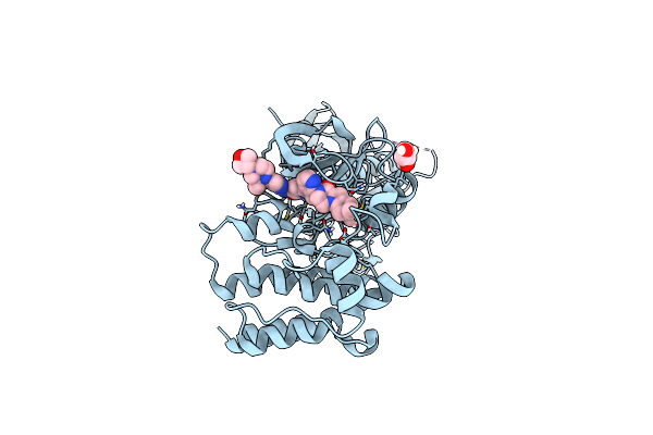
Crystal Structure Of The Full-Length Bruton'S Tyrosine Kinase (Ph-Th Domain Not Visible)
Organism: Mus musculus
Method: X-RAY DIFFRACTION Resolution:3.40 Å Release Date: 2023-08-16 Classification: TRANSFERASE Ligands: 9AJ |
Organism: Mus musculus
Method: X-RAY DIFFRACTION
Release Date: 2023-08-16
Ligands: 9AJ
 |
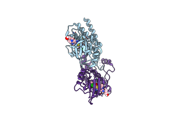
Crystal Structure Of The Ph-Th/Kinase Complex Of Bruton'S Tyrosine Kinase
Organism: Mus musculus
Method: X-RAY DIFFRACTION Resolution:2.10 Å Release Date: 2023-08-16 Classification: TRANSFERASE Ligands: 9AJ, ZN, GOL |
Organism: Mus musculus
Method: X-RAY DIFFRACTION
Release Date: 2023-08-16
Ligands: 9AJ, ZN, GOL
 |
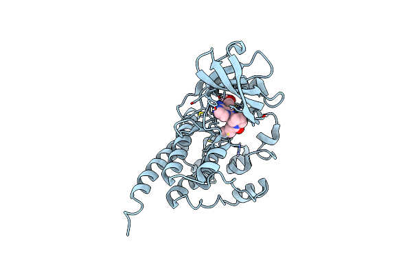
Crystal Structure Of The Kinase Domain Of Bruton'S Tyrosine Kinase Bound To Dasatinib
Organism: Mus musculus
Method: X-RAY DIFFRACTION Resolution:2.60 Å Release Date: 2023-08-16 Classification: TRANSFERASE Ligands: 1N1 |
Organism: Mus musculus
Method: X-RAY DIFFRACTION
Release Date: 2023-08-16
Ligands: 1N1
 |
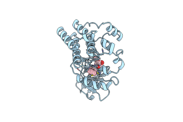
Structure Of Btk Kinase Domain With The Second-Generation Inhibitor Acalabrutinib
Organism: Mus musculus
Method: X-RAY DIFFRACTION Resolution:1.70 Å Release Date: 2023-07-05 Classification: TRANSFERASE/TRANSFERASE INHIBITOR Ligands: XQQ, BR |
Organism: Mus musculus
Method: X-RAY DIFFRACTION
Release Date: 2023-07-05
Ligands: XQQ, BR
 |
Structure Of Btk Kinase Domain With The Second-Generation Inhibitor Tirabrutinib
Organism: Mus musculus
Method: X-RAY DIFFRACTION Resolution:2.60 Å Release Date: 2023-07-05 Classification: TRANSFERASE/TRANSFERASE INHIBITOR Ligands: 7GB |
Organism: Mus musculus
Method: X-RAY DIFFRACTION
Release Date: 2023-07-05
Ligands: 7GB
 |
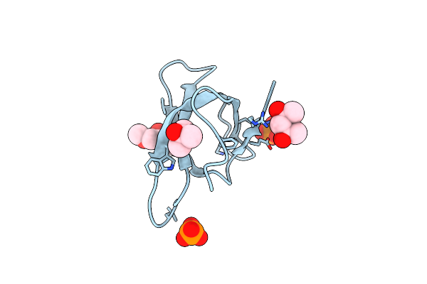
Crystal Structure Of Trimethoprim-Resistant Type Ii Dihydrofolate Reductase In Complex With A Bisbenzimidazole Inhibitor
Organism: Escherichia coli
Method: X-RAY DIFFRACTION Resolution:1.75 Å Release Date: 2019-05-29 Classification: OXIDOREDUCTASE Ligands: D49, MRD, PO4 |
Organism: Escherichia coli
Method: X-RAY DIFFRACTION
Release Date: 2019-05-29
Ligands: D49, MRD, PO4
 |
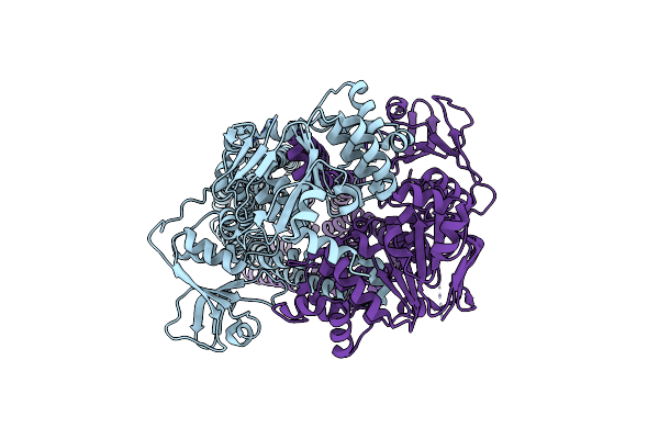
Crystal Structure Of Trimethoprim-Resistant Type Ii Dihydrofolate Reductase In Complex With A Bisbenzimidazole Inhibitor
Organism: Escherichia coli
Method: X-RAY DIFFRACTION Resolution:1.40 Å Release Date: 2019-05-29 Classification: OXIDOREDUCTASE Ligands: LBA, MRD, PO4 |
Organism: Escherichia coli
Method: X-RAY DIFFRACTION
Release Date: 2019-05-29
Ligands: LBA, MRD, PO4
 |
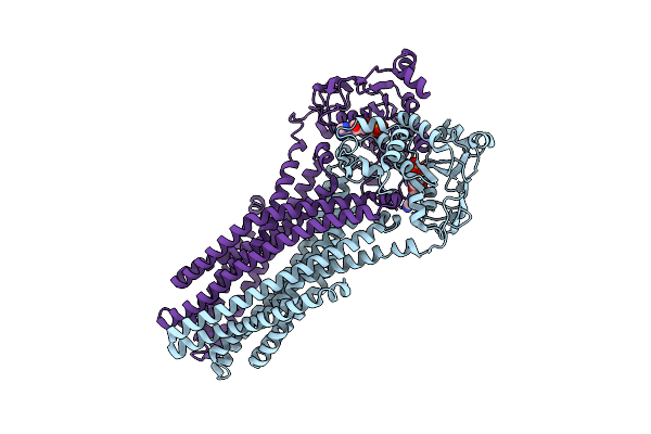
Crystal Structure Of The Peptidase-Containing Abc Transporter Pcat1
Organism: Ruminiclostridium thermocellum atcc 27405
Method: X-RAY DIFFRACTION Resolution:3.61 Å Release Date: 2015-07-22 Classification: TRANSPORT PROTEIN/HYDROLASE |
Organism: Ruminiclostridium thermocellum atcc 27405
Method: X-RAY DIFFRACTION
Release Date: 2015-07-22
 |
Crystal Structure Of The Peptidase-Containing Abc Transporter Pcat1 E648Q Mutant Complexed With Atpgs In An Occluded Conformation
Organism: Ruminiclostridium thermocellum atcc 27405
Method: X-RAY DIFFRACTION Resolution:5.52 Å Release Date: 2015-07-22 Classification: TRANSPORT PROTEIN/HYDROLASE Ligands: AGS |
Organism: Ruminiclostridium thermocellum atcc 27405
Method: X-RAY DIFFRACTION
Release Date: 2015-07-22
Ligands: AGS