

Search Count: 15
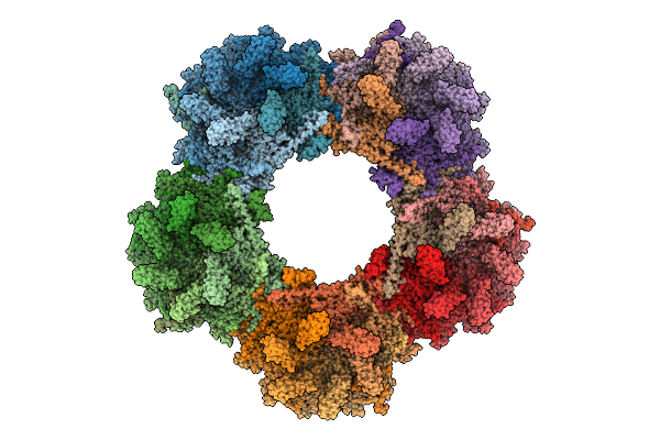 |
Cryo-Em Structure Of Mycobacteriophage Douge Genome-Packed Vertex (Gp8 And Gp113)
Organism: Mycolicibacterium smegmatis mc2 155
Method: ELECTRON MICROSCOPY Release Date: 2025-07-16 Classification: VIRAL PROTEIN |
Organism: Mycolicibacterium smegmatis mc2 155
Method: ELECTRON MICROSCOPY
Release Date: 2025-07-16
 |
Cryo-Em Structure Of Mycobacteriophage Douge Genome-Free Vertex (Gp8)
Organism: Mycolicibacterium smegmatis mc2 155
Method: ELECTRON MICROSCOPY Release Date: 2025-07-16 Classification: VIRAL PROTEIN |
Organism: Mycolicibacterium smegmatis mc2 155
Method: ELECTRON MICROSCOPY
Release Date: 2025-07-16
 |
Cryo-Em Structure Of Mycobacteriophage Douge Genome-Packed Capsid (Gp8 And Gp113)
Organism: Mycolicibacterium smegmatis mc2 155
Method: ELECTRON MICROSCOPY Release Date: 2025-06-25 Classification: VIRUS |
Organism: Mycolicibacterium smegmatis mc2 155
Method: ELECTRON MICROSCOPY
Release Date: 2025-06-25
 |
Cryo-Em Structure Of Mycobacteriophage Douge Genome-Free Capsid (Gp8)
Organism: Mycolicibacterium smegmatis mc2 155
Method: ELECTRON MICROSCOPY Release Date: 2025-06-25 Classification: VIRUS |
Organism: Mycolicibacterium smegmatis mc2 155
Method: ELECTRON MICROSCOPY
Release Date: 2025-06-25
 |
Cryo-Em Structure Of Mycobacteriophage Douge Genome-Packed Connector (Gp5, Gp9, Gp10, Gp12 And Gp13)
Organism: Mycolicibacterium smegmatis mc2 155
Method: ELECTRON MICROSCOPY Release Date: 2025-06-25 Classification: VIRAL PROTEIN |
Organism: Mycolicibacterium smegmatis mc2 155
Method: ELECTRON MICROSCOPY
Release Date: 2025-06-25
 |
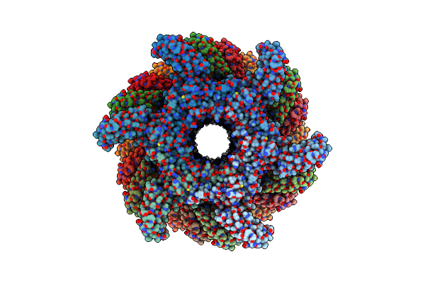
Cryo-Em Structure Of Mycobacteriophage Douge Genome-Free Connector (Gp5, Gp9, Gp10, Gp12 And Gp13)
Organism: Mycolicibacterium smegmatis mc2 155
Method: ELECTRON MICROSCOPY Release Date: 2025-06-25 Classification: VIRAL PROTEIN |
Organism: Mycolicibacterium smegmatis mc2 155
Method: ELECTRON MICROSCOPY
Release Date: 2025-06-25
 |
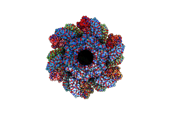
Cryo-Em Structure Of Mycobacteriophage Douge Genome-Free Tail Tube (Gp13)
Organism: Mycolicibacterium smegmatis mc2 155
Method: ELECTRON MICROSCOPY Release Date: 2025-06-25 Classification: VIRAL PROTEIN |
Organism: Mycolicibacterium smegmatis mc2 155
Method: ELECTRON MICROSCOPY
Release Date: 2025-06-25
 |
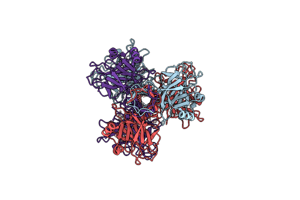
Cryo-Em Structure Of Mycobacteriophage Douge Baseplate (Gp13, Gp17, Gp23, Gp16, Gp18 And Gp20)
Organism: Mycolicibacterium smegmatis mc2 155
Method: ELECTRON MICROSCOPY Release Date: 2025-06-25 Classification: VIRAL PROTEIN |
Organism: Mycolicibacterium smegmatis mc2 155
Method: ELECTRON MICROSCOPY
Release Date: 2025-06-25
 |
Cryo-Em Structure Of Mycobacteriophage Douge Central Fiber (Gp20)
Organism: Mycolicibacterium smegmatis mc2 155
Method: ELECTRON MICROSCOPY Release Date: 2025-06-25 Classification: VIRAL PROTEIN |
Organism: Mycolicibacterium smegmatis mc2 155
Method: ELECTRON MICROSCOPY
Release Date: 2025-06-25
 |
Cryo-Em Structure Of Mycobacteriophage Douge Complete Baseplate (Gp13, Gp17, Gp23, Gp16, Gp18 And Gp20)
Organism: Mycolicibacterium smegmatis mc2 155
Method: ELECTRON MICROSCOPY Release Date: 2025-06-25 Classification: VIRAL PROTEIN |
Organism: Mycolicibacterium smegmatis mc2 155
Method: ELECTRON MICROSCOPY
Release Date: 2025-06-25
 |
Cryo-Em Structure Of Mycobacteriophage Douge Genome-Packed Tail Tube (Gp13)
Organism: Mycolicibacterium smegmatis mc2 155
Method: ELECTRON MICROSCOPY Release Date: 2025-06-25 Classification: VIRAL PROTEIN |
Organism: Mycolicibacterium smegmatis mc2 155
Method: ELECTRON MICROSCOPY
Release Date: 2025-06-25
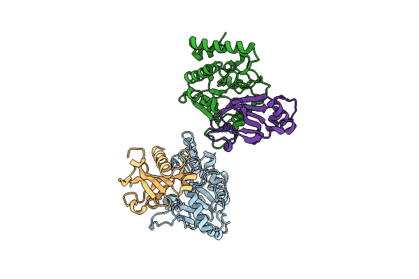 |
The Crystal Structure Of Saugi/Ebvudg Complex
Organism: Epstein-barr virus (strain gd1), Staphylococcus aureus
Method: X-RAY DIFFRACTION Resolution:2.10 Å Release Date: 2020-06-24 Classification: HYDROLASE |
Organism: Epstein-barr virus (strain gd1), Staphylococcus aureus
Method: X-RAY DIFFRACTION
Release Date: 2020-06-24
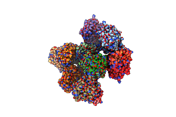 |
The Crystal Structure Of Saugi/Kshvudg Complex
Organism: Human herpesvirus 8, Staphylococcus aureus
Method: X-RAY DIFFRACTION Resolution:2.70 Å Release Date: 2020-06-24 Classification: HYDROLASE |
Organism: Human herpesvirus 8, Staphylococcus aureus
Method: X-RAY DIFFRACTION
Release Date: 2020-06-24
 |
The Crystal Structure Of Phytoplasmal Effector Causing Phyllody Symptoms 1 (Phyl1)
Organism: 'echinacea purpurea' witches'-broom phytoplasma
Method: X-RAY DIFFRACTION Resolution:2.34 Å Release Date: 2019-07-31 Classification: GENE REGULATION Ligands: CD |
Organism: 'echinacea purpurea' witches'-broom phytoplasma
Method: X-RAY DIFFRACTION
Release Date: 2019-07-31
Ligands: CD
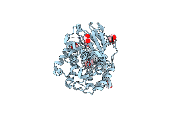 |
The Crystal Structure Of Vaccinia Virus A26 (Residues 1-397)
Organism: Vaccinia virus (strain western reserve)
Method: X-RAY DIFFRACTION Resolution:1.18 Å Release Date: 2019-06-12 Classification: VIRAL PROTEIN Ligands: EDO |
Organism: Vaccinia virus (strain western reserve)
Method: X-RAY DIFFRACTION
Release Date: 2019-06-12
Ligands: EDO