

Search Count: 18
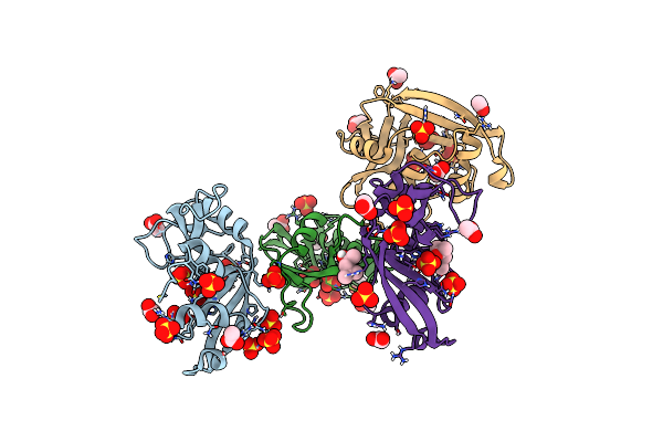 |
Crystal Structure Of A Resurrected Ancestor (Ancrnase) Of The Pancreatic-Type Rnases 2 And 3 Sub-Families
Organism: Synthetic construct
Method: X-RAY DIFFRACTION Resolution:2.03 Å Release Date: 2024-02-28 Classification: HYDROLASE Ligands: SO4, ACT, MPD |
Organism: Synthetic construct
Method: X-RAY DIFFRACTION
Release Date: 2024-02-28
Ligands: SO4, ACT, MPD
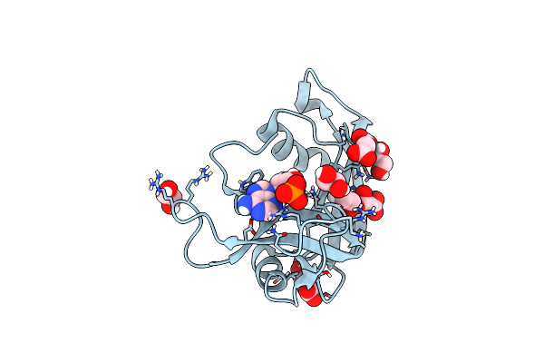 |
Crystal Structure Of Human Eosinophil-Derived Neurotoxin (Edn, Ribonuclease 2) In Complex With 5'-Adenosine Monophosphate (Amp)
Organism: Homo sapiens
Method: X-RAY DIFFRACTION Resolution:1.70 Å Release Date: 2023-11-29 Classification: HYDROLASE Ligands: SIN, EDO, AMP |
Organism: Homo sapiens
Method: X-RAY DIFFRACTION
Release Date: 2023-11-29
Ligands: SIN, EDO, AMP
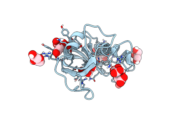 |
Crystal Structure Of Apo Eosinophil Cationic Protein (Ribonuclease 3) From Macaca Fascicularis (Mfecp)
Organism: Macaca fascicularis
Method: X-RAY DIFFRACTION Resolution:1.80 Å Release Date: 2023-08-16 Classification: HYDROLASE Ligands: GOL, CIT, PEG |
Organism: Macaca fascicularis
Method: X-RAY DIFFRACTION
Release Date: 2023-08-16
Ligands: GOL, CIT, PEG
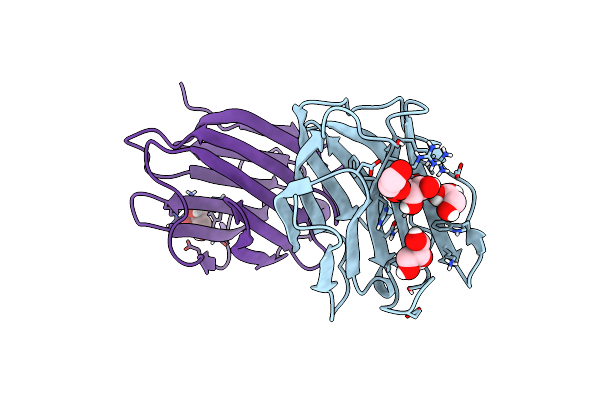 |
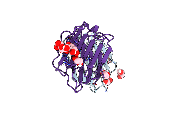
Crystal Structure Of R20A Human Galectin-7 Mutant
Organism: Homo sapiens
Method: X-RAY DIFFRACTION Resolution:2.05 Å Release Date: 2023-01-25 Classification: SUGAR BINDING PROTEIN Ligands: EDO, GOL |
Organism: Homo sapiens
Method: X-RAY DIFFRACTION
Release Date: 2023-01-25
Ligands: EDO, GOL
 |
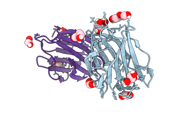
Crystal Structure Of R20A Human Galectin-7 Mutant In Presence Of Lactose
Organism: Homo sapiens
Method: X-RAY DIFFRACTION Resolution:1.83 Å Release Date: 2023-01-25 Classification: SUGAR BINDING PROTEIN Ligands: GOL, EDO |
Organism: Homo sapiens
Method: X-RAY DIFFRACTION
Release Date: 2023-01-25
Ligands: GOL, EDO
 |
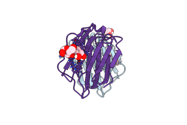
Crystal Structure Of R22A Human Galectin-7 Mutant
Organism: Homo sapiens
Method: X-RAY DIFFRACTION Resolution:2.10 Å Release Date: 2023-01-25 Classification: SUGAR BINDING PROTEIN Ligands: GOL, TRS, EDO, PEG |
Organism: Homo sapiens
Method: X-RAY DIFFRACTION
Release Date: 2023-01-25
Ligands: GOL, TRS, EDO, PEG
 |
Crystal Structure Of R22A Human Galectin-7 Mutant In Presence Of Lactose
Organism: Homo sapiens
Method: X-RAY DIFFRACTION Resolution:2.49 Å Release Date: 2023-01-25 Classification: SUGAR BINDING PROTEIN Ligands: EDO |
Organism: Homo sapiens
Method: X-RAY DIFFRACTION
Release Date: 2023-01-25
Ligands: EDO
 |
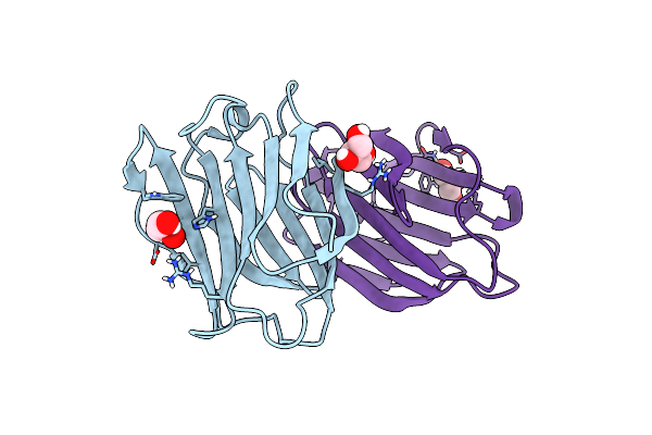
Crystal Structure Of R20A-R22A Human Galectin-7 Mutant
Organism: Homo sapiens
Method: X-RAY DIFFRACTION Resolution:1.95 Å Release Date: 2023-01-25 Classification: SUGAR BINDING PROTEIN Ligands: EDO, TRS, GOL |
Organism: Homo sapiens
Method: X-RAY DIFFRACTION
Release Date: 2023-01-25
Ligands: EDO, TRS, GOL
 |
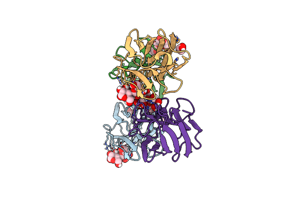
Crystal Structure Of D103A Human Galectin-7 Mutant
Organism: Homo sapiens
Method: X-RAY DIFFRACTION Resolution:2.38 Å Release Date: 2023-01-25 Classification: SUGAR BINDING PROTEIN Ligands: GOL |
Organism: Homo sapiens
Method: X-RAY DIFFRACTION
Release Date: 2023-01-25
Ligands: GOL
 |
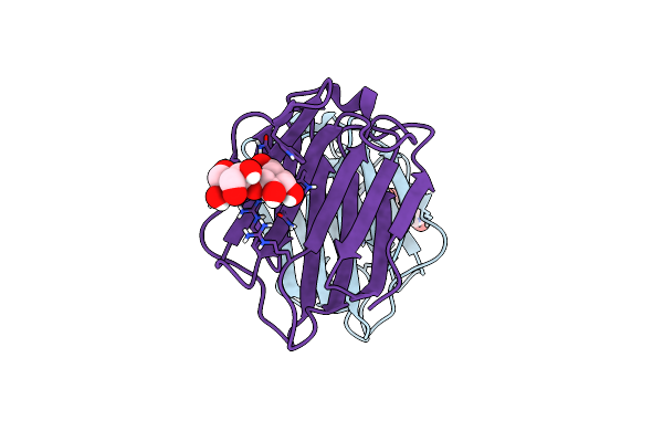
Crystal Structure Of D103A Human Galectin-7 Mutant In Presence Of Lactose
Organism: Homo sapiens
Method: X-RAY DIFFRACTION Resolution:3.00 Å Release Date: 2023-01-25 Classification: SUGAR BINDING PROTEIN Ligands: EDO |
Organism: Homo sapiens
Method: X-RAY DIFFRACTION
Release Date: 2023-01-25
Ligands: EDO
 |
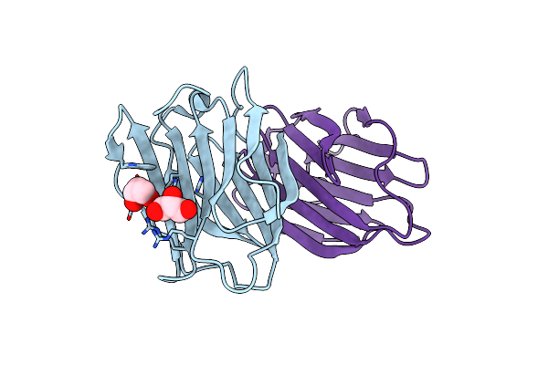
Crystal Structure Of Human Galectin-7 In Complex With 4-O-Beta-D-Galactopyranosyl-D-Glucose
Organism: Homo sapiens
Method: X-RAY DIFFRACTION Resolution:1.69 Å Release Date: 2021-08-25 Classification: SUGAR BINDING PROTEIN Ligands: LBL |
Organism: Homo sapiens
Method: X-RAY DIFFRACTION
Release Date: 2021-08-25
Ligands: LBL
 |
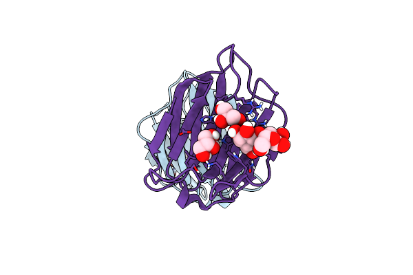
Crystal Structure Of G16C Human Galectin-7 Mutant
Organism: Homo sapiens
Method: X-RAY DIFFRACTION Resolution:2.30 Å Release Date: 2021-08-25 Classification: SUGAR BINDING PROTEIN Ligands: GOL |
Organism: Homo sapiens
Method: X-RAY DIFFRACTION
Release Date: 2021-08-25
Ligands: GOL
 |
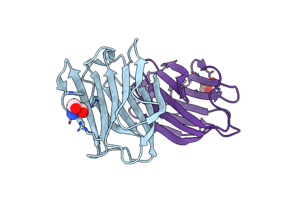
Crystal Structure Of G16C Human Galectin-7 Mutant In Complex With Lactose
Organism: Homo sapiens
Method: X-RAY DIFFRACTION Resolution:1.95 Å Release Date: 2021-08-25 Classification: SUGAR BINDING PROTEIN Ligands: GOL |
Organism: Homo sapiens
Method: X-RAY DIFFRACTION
Release Date: 2021-08-25
Ligands: GOL
 |
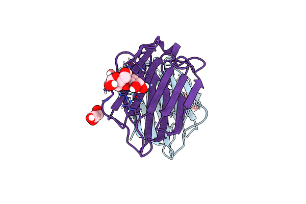
Crystal Structure Of G16S Human Galectin-7 Mutant
Organism: Homo sapiens
Method: X-RAY DIFFRACTION Resolution:2.30 Å Release Date: 2021-08-25 Classification: SUGAR BINDING PROTEIN Ligands: TRS, GOL |
Organism: Homo sapiens
Method: X-RAY DIFFRACTION
Release Date: 2021-08-25
Ligands: TRS, GOL
 |
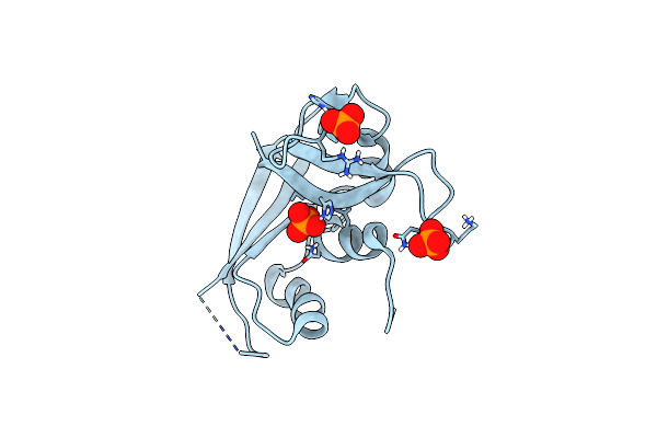
Crystal Structure Of G16S Human Galectin-7 Mutant In Complex With Lactose
Organism: Homo sapiens
Method: X-RAY DIFFRACTION Resolution:1.90 Å Release Date: 2021-08-25 Classification: SUGAR BINDING PROTEIN Ligands: GOL |
Organism: Homo sapiens
Method: X-RAY DIFFRACTION
Release Date: 2021-08-25
Ligands: GOL
 |
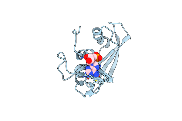
Crystal Structure Of Rnase 6
Organism: Homo sapiens
Method: X-RAY DIFFRACTION Resolution:1.50 Å Release Date: 2019-11-13 Classification: HYDROLASE Ligands: PO4 |
Organism: Homo sapiens
Method: X-RAY DIFFRACTION
Release Date: 2019-11-13
Ligands: PO4
 |
Crystal Structure Of Rnase 6
Organism: Homo sapiens
Method: X-RAY DIFFRACTION Resolution:2.59 Å Release Date: 2019-11-13 Classification: HYDROLASE Ligands: AMP |
Organism: Homo sapiens
Method: X-RAY DIFFRACTION
Release Date: 2019-11-13
Ligands: AMP
 |
Pepducin Ut-Pep2 A Biased Allosteric Agonist Of Urotensin-Ii Receptor
Organism: Homo sapiens
Method: SOLUTION NMR Release Date: 2019-10-30 Classification: PEPTIDE BINDING PROTEIN |
Organism: Homo sapiens
Method: SOLUTION NMR
Release Date: 2019-10-30