

Search Count: 145
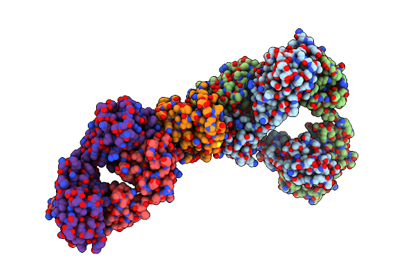 |
Cryo-Em Structure Of The Codv-Il13-Refab Triple Complex
Organism: Homo sapiens
Method: ELECTRON MICROSCOPY Release Date: 2023-06-07 Classification: IMMUNE SYSTEM |
Organism: Homo sapiens
Method: ELECTRON MICROSCOPY
Release Date: 2023-06-07
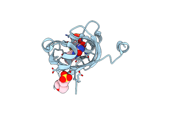 |
Room Temperature Structure Of The Ground State Of Atphot2Lov2 In Space Group P43212
Organism: Arabidopsis thaliana
Method: X-RAY DIFFRACTION Resolution:1.59 Å Release Date: 2023-03-29 Classification: PLANT PROTEIN Ligands: FMN, MES |
Organism: Arabidopsis thaliana
Method: X-RAY DIFFRACTION
Release Date: 2023-03-29
Ligands: FMN, MES
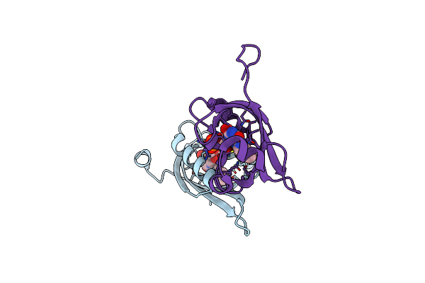 |
Room Temperature Structure Of The Ground State Of Atphot2Lov2 In Space Group P212121, As Recovered 1620 Seconds After Light Irradiation
Organism: Arabidopsis thaliana
Method: X-RAY DIFFRACTION Resolution:2.04 Å Release Date: 2023-03-29 Classification: PLANT PROTEIN Ligands: FMN |
Organism: Arabidopsis thaliana
Method: X-RAY DIFFRACTION
Release Date: 2023-03-29
Ligands: FMN
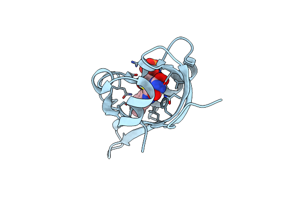 |
Room Temperature Structure Of Atphot2Lov2 In A Photostationary Equilibrium
Organism: Arabidopsis thaliana
Method: X-RAY DIFFRACTION Resolution:1.96 Å Release Date: 2023-03-29 Classification: PLANT PROTEIN Ligands: FMA, FMN |
Organism: Arabidopsis thaliana
Method: X-RAY DIFFRACTION
Release Date: 2023-03-29
Ligands: FMA, FMN
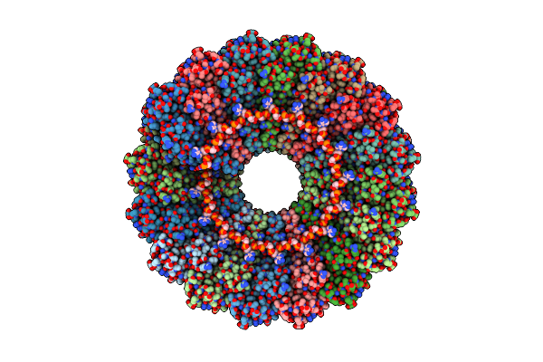 |
Cryo-Em Reconstruction Of Tmv Coat Protein
Organism: Tobacco mosaic virus
Method: ELECTRON MICROSCOPY Release Date: 2022-11-23 Classification: VIRAL PROTEIN |
Organism: Tobacco mosaic virus
Method: ELECTRON MICROSCOPY
Release Date: 2022-11-23
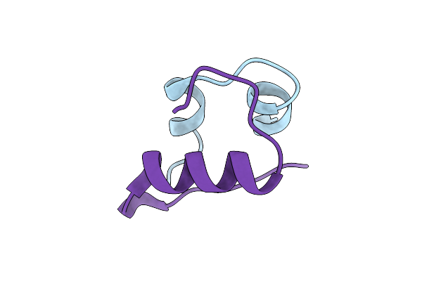 |
Cubic Insulin Sad Phasing At 14.2 Kev
Organism: Homo sapiens
Method: X-RAY DIFFRACTION Resolution:1.20 Å Release Date: 2022-06-01 Classification: HORMONE |
Organism: Homo sapiens
Method: X-RAY DIFFRACTION
Release Date: 2022-06-01
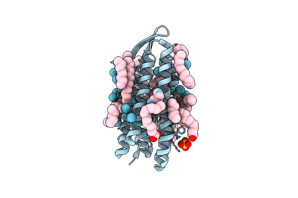 |
Crystal Structure Of The Mutant Bacteriorhodopsin Pressurized With Krypton
Organism: Halobacterium salinarum (strain atcc 700922 / jcm 11081 / nrc-1)
Method: X-RAY DIFFRACTION Resolution:2.00 Å Release Date: 2022-04-27 Classification: MEMBRANE PROTEIN Ligands: RET, LFA, OLA, HEX, SO4, KR |
Organism: Halobacterium salinarum (strain atcc 700922 / jcm 11081 / nrc-1)
Method: X-RAY DIFFRACTION
Release Date: 2022-04-27
Ligands: RET, LFA, OLA, HEX, SO4, KR
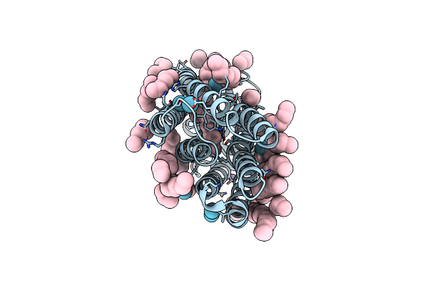 |
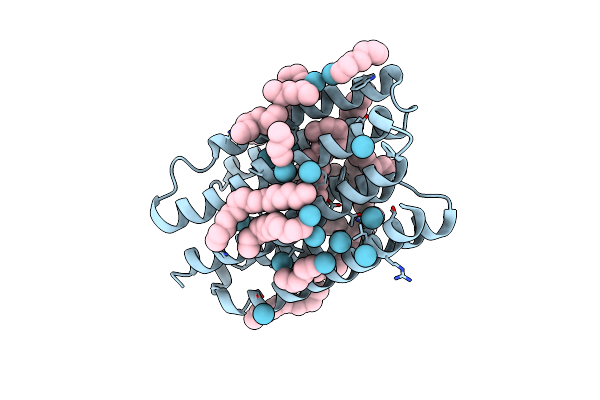
Crystal Structure Of Kr2 Sodium Pump Rhodopsin Pressurized With Krypton
Organism: Dokdonia eikasta
Method: X-RAY DIFFRACTION Resolution:2.60 Å Release Date: 2022-04-27 Classification: MEMBRANE PROTEIN Ligands: LFA, HEX, NA, KR, RET |
Organism: Dokdonia eikasta
Method: X-RAY DIFFRACTION
Release Date: 2022-04-27
Ligands: LFA, HEX, NA, KR, RET
 |
Crystal Structure Of Proton Pump Mar Rhodopsin Pressurized With Krypton
Organism: Candidatus actinomarina minuta
Method: X-RAY DIFFRACTION Resolution:2.25 Å Release Date: 2022-04-27 Classification: MEMBRANE PROTEIN Ligands: RET, LFA, HEX, KR |
Organism: Candidatus actinomarina minuta
Method: X-RAY DIFFRACTION
Release Date: 2022-04-27
Ligands: RET, LFA, HEX, KR
 |
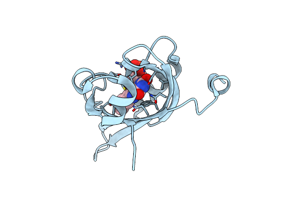
Crystal Structure Of The Mutant Bacteriorhodopsin Pressurized With Argon
Organism: Halobacterium salinarum (strain atcc 700922 / jcm 11081 / nrc-1)
Method: X-RAY DIFFRACTION Resolution:1.65 Å Release Date: 2022-04-27 Classification: MEMBRANE PROTEIN Ligands: RET, LFA, OLC, OLA, HEX, SO4, AR |
Organism: Halobacterium salinarum (strain atcc 700922 / jcm 11081 / nrc-1)
Method: X-RAY DIFFRACTION
Release Date: 2022-04-27
Ligands: RET, LFA, OLC, OLA, HEX, SO4, AR
 |
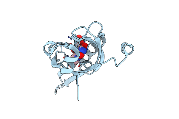
Room Temperature Structure Of The Dark State Of The Lov2 Domain Of Phototropin-2 From Arabidopsis Thaliana Determined With A Serial Crystallography Approach
Organism: Arabidopsis thaliana
Method: X-RAY DIFFRACTION Resolution:2.20 Å Release Date: 2020-07-08 Classification: PLANT PROTEIN Ligands: FMN |
Organism: Arabidopsis thaliana
Method: X-RAY DIFFRACTION
Release Date: 2020-07-08
Ligands: FMN
 |
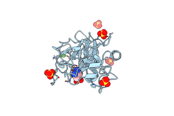
Room Temperature Structure Of The Lov2 Domain Of Phototropin-2 From Arabidopsis Thaliana 4158 Ms After Initiation Of Illumination, Determined With A Serial Crystallography Approach
Organism: Arabidopsis thaliana
Method: X-RAY DIFFRACTION Resolution:2.75 Å Release Date: 2020-07-08 Classification: PLANT PROTEIN Ligands: FMN |
Organism: Arabidopsis thaliana
Method: X-RAY DIFFRACTION
Release Date: 2020-07-08
Ligands: FMN
 |
Trypsin Fast Data Collection
Organism: Bos taurus
Method: X-RAY DIFFRACTION Resolution:1.43 Å Release Date: 2020-05-20 Classification: HYDROLASE Ligands: CA, SO4, BEN |
Organism: Bos taurus
Method: X-RAY DIFFRACTION
Release Date: 2020-05-20
Ligands: CA, SO4, BEN
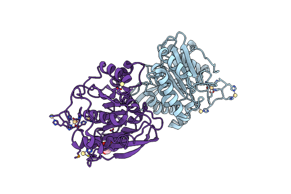 |
Selenomethionine Derivative Of Ferulic Acid Esterase (Fae)
Organism: Hungateiclostridium thermocellum
Method: X-RAY DIFFRACTION Resolution:1.80 Å Release Date: 2020-05-20 Classification: HYDROLASE Ligands: CD, GOL |
Organism: Hungateiclostridium thermocellum
Method: X-RAY DIFFRACTION
Release Date: 2020-05-20
Ligands: CD, GOL
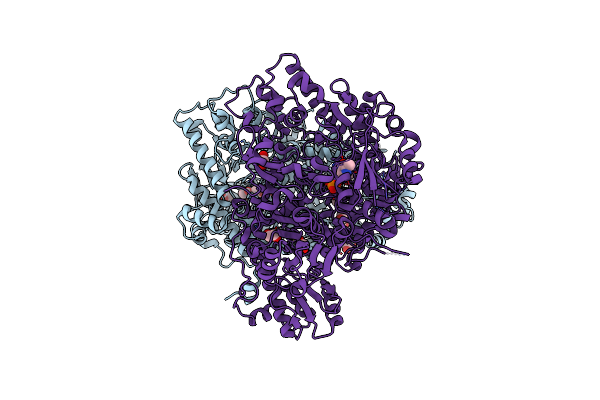 |
Crystal Structure Of Human Glycine Decarboxylase (P-Protein)
Organism: Homo sapiens
Method: X-RAY DIFFRACTION Resolution:2.30 Å Release Date: 2019-11-20 Classification: OXIDOREDUCTASE Ligands: PLP, EDO, BCT |
Organism: Homo sapiens
Method: X-RAY DIFFRACTION
Release Date: 2019-11-20
Ligands: PLP, EDO, BCT
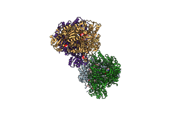 |
Crystal Structure Of Neanderthal Glycine Decarboxylase (P-Protein)
Organism: Homo sapiens neanderthalensis
Method: X-RAY DIFFRACTION Resolution:2.10 Å Release Date: 2019-11-20 Classification: OXIDOREDUCTASE Ligands: PLP, EDO, PEG, GOL |
Organism: Homo sapiens neanderthalensis
Method: X-RAY DIFFRACTION
Release Date: 2019-11-20
Ligands: PLP, EDO, PEG, GOL
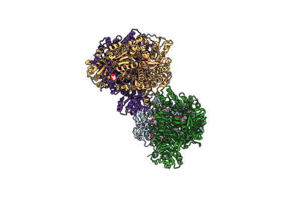 |
Crystal Structure Of Human Glycine Decarboxylase (P-Protein) Bound With Pyridoxyl-Glycine-5'-Monophosphate
Organism: Homo sapiens
Method: X-RAY DIFFRACTION Resolution:2.00 Å Release Date: 2019-11-20 Classification: OXIDOREDUCTASE Ligands: PLG, EDO, GOL, BCT, PEG |
Organism: Homo sapiens
Method: X-RAY DIFFRACTION
Release Date: 2019-11-20
Ligands: PLG, EDO, GOL, BCT, PEG
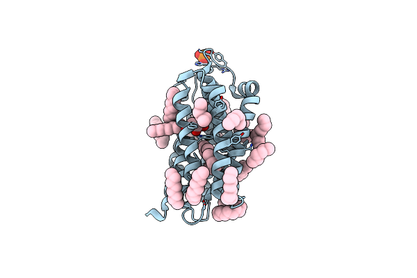 |
Crystal Structure Of The Dts-Motif Rhodopsin From Phaeocystis Globosa Virus 12T
Organism: Phaeocystis globosa virus 12t
Method: X-RAY DIFFRACTION Resolution:1.65 Å Release Date: 2019-10-02 Classification: MEMBRANE PROTEIN Ligands: RET, OCT, PO4, HEX, D10, D12, DD9, OLB |
Organism: Phaeocystis globosa virus 12t
Method: X-RAY DIFFRACTION
Release Date: 2019-10-02
Ligands: RET, OCT, PO4, HEX, D10, D12, DD9, OLB
 |
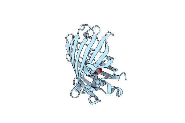
Cryogenic Temperature Structure Of The Fluorescent Protein Cerulean Recorded After An Accumulated Dose Of 290 Kgy
Organism: Aequorea victoria
Method: X-RAY DIFFRACTION Resolution:1.46 Å Release Date: 2019-06-19 Classification: FLUORESCENT PROTEIN |
Organism: Aequorea victoria
Method: X-RAY DIFFRACTION
Release Date: 2019-06-19
 |
Cryogenic Temperature Structure Of The Fluorescent Protein Cerulean Recorded After An Accumulated Dose Of 5.8 Mgy
Organism: Aequorea victoria
Method: X-RAY DIFFRACTION Resolution:1.82 Å Release Date: 2019-06-19 Classification: FLUORESCENT PROTEIN Ligands: CO2 |
Organism: Aequorea victoria
Method: X-RAY DIFFRACTION
Release Date: 2019-06-19
Ligands: CO2