
Search Count: 18,808
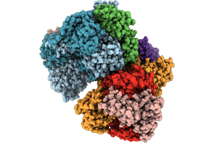 |
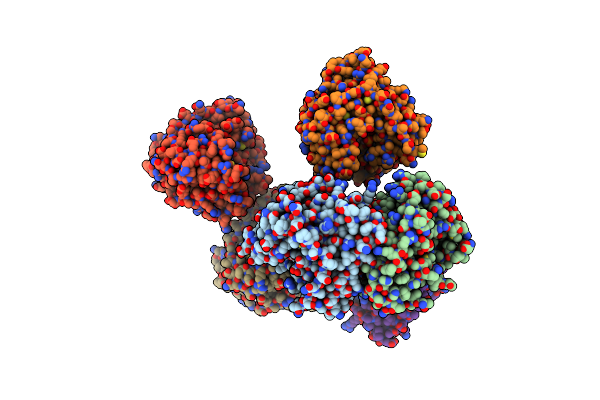
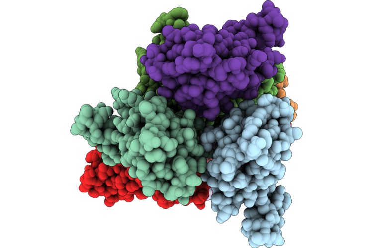
Crystal Structure Of Fused Glycerol Dehydratase A177M Variant
Organism: Klebsiella pneumoniae
Method: X-RAY DIFFRACTION Resolution:3.56 Å Release Date: 2026-06-24 Classification: LYASE Ligands: B12 |
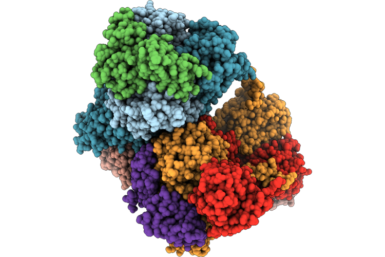 |
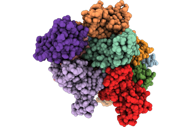
Crystal Structure Of Fused Glycerol Dehydratase M158W Variant
Organism: Klebsiella pneumoniae
Method: X-RAY DIFFRACTION Resolution:3.68 Å Release Date: 2026-06-24 Classification: LYASE Ligands: B12 |
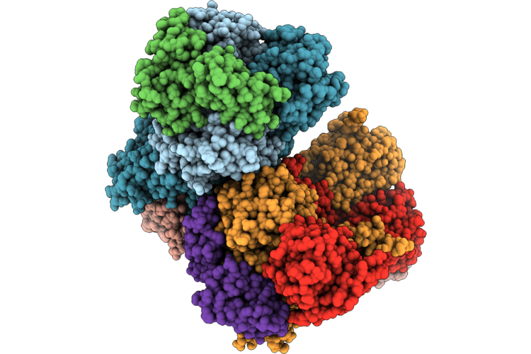 |
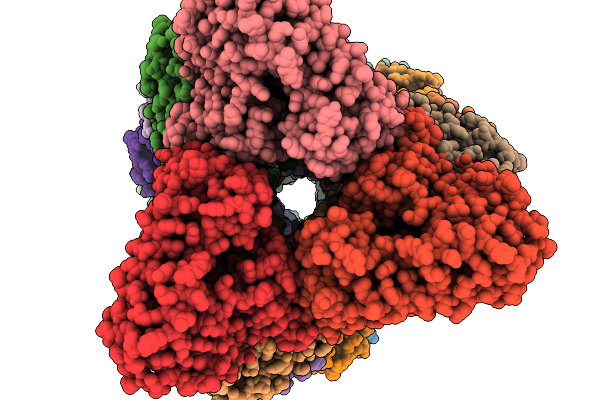
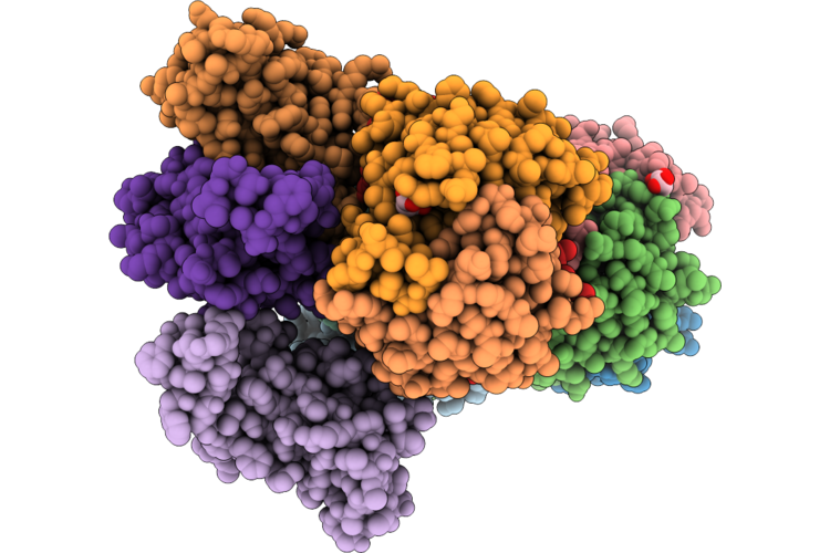
Crystal Structure Of Fused Glycerol Dehydratase
Organism: Klebsiella pneumoniae
Method: X-RAY DIFFRACTION Resolution:2.91 Å Release Date: 2026-06-24 Classification: LYASE Ligands: B12 |
 |
Crystal Structure Of A Selenomethionine-Labeled Hydropyrene Synthase (M75L Variant) In Its Closed Conformation
Organism: Streptomyces clavuligerus
Method: X-RAY DIFFRACTION Resolution:3.39 Å Release Date: 2023-10-04 Classification: LYASE Ligands: MG, AHD |
 |
Crystal Structure Of A Hydropyrene Synthase (M75L Variant) In Its Closed Conformation
Organism: Streptomyces clavuligerus
Method: X-RAY DIFFRACTION Resolution:3.04 Å Release Date: 2023-10-04 Classification: LYASE Ligands: MG, 212 |
 |
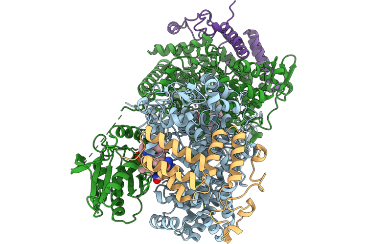
Cryo-Em Structure Of The Pt Domain Of Evss
Organism: Aspergillus stellatus
Method: ELECTRON MICROSCOPY Resolution:3.19 Å Release Date: 2026-02-11 Classification: BIOSYNTHETIC PROTEIN |
 |
Crystal Structure Of I64A Variant Of D-Dopachrome Tautomerase (D-Dt)
Organism: Homo sapiens
Method: X-RAY DIFFRACTION Resolution:1.54 Å Release Date: 2026-05-06 Classification: CYTOKINE Ligands: CA |
 |
Cryo-Em Structure Of Evss
Organism: Aspergillus stellatus
Method: ELECTRON MICROSCOPY Resolution:3.25 Å Release Date: 2026-02-11 Classification: BIOSYNTHETIC PROTEIN |
 |
Crystal Structure Of R36A Variant Of D-Dopachrome Tautomerase (D-Dt)
Organism: Homo sapiens
Method: X-RAY DIFFRACTION Resolution:1.64 Å Release Date: 2026-05-06 Classification: CYTOKINE |
 |
Crystal Structure Of S63A Variant Of D-Dopachrome Tautomerase (D-Dt)
Organism: Homo sapiens
Method: X-RAY DIFFRACTION Resolution:1.53 Å Release Date: 2026-05-06 Classification: CYTOKINE Ligands: TRS |
 |
Crystal Structure Of K109A Variant Of D-Dopachrome Tautomerase (D-Dt)
Organism: Homo sapiens
Method: X-RAY DIFFRACTION Resolution:1.49 Å Release Date: 2026-05-06 Classification: CYTOKINE Ligands: MLI |
 |
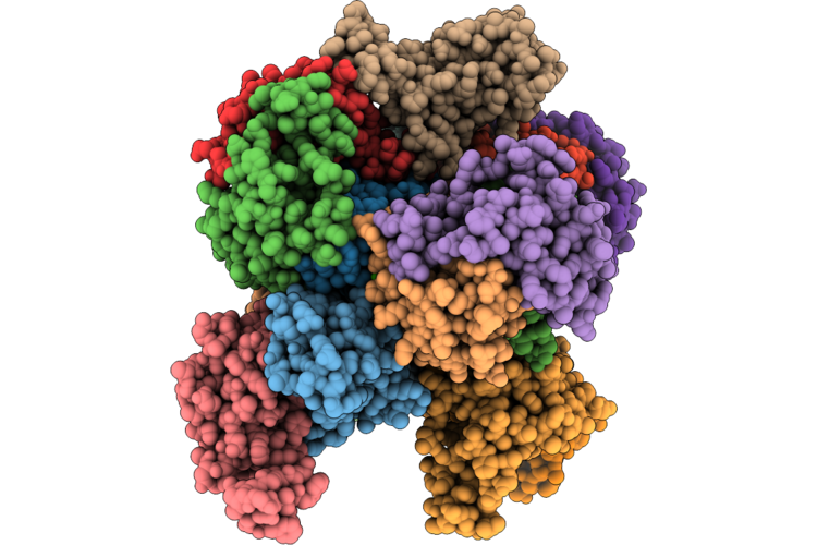
Crystal Structure Of Fused Glycerol Dehydratase L113W Variant
Organism: Klebsiella pneumoniae
Method: X-RAY DIFFRACTION Resolution:3.02 Å Release Date: 2026-06-24 Classification: LYASE Ligands: B12 |
 |
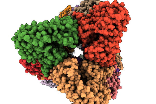
Crystal Structure Of Fused Glycerol Dehydratase A177M/M158W Variant
Organism: Klebsiella pneumoniae
Method: X-RAY DIFFRACTION Resolution:3.58 Å Release Date: 2026-06-24 Classification: LYASE Ligands: B12 |
 |
Fructose 6-Phosphate Aldolase, L107C/A129G/R134V/L163C/S166G Mutant
Organism: Escherichia coli bl21
Method: ELECTRON MICROSCOPY Release Date: 2025-03-12 Classification: LYASE |
 |
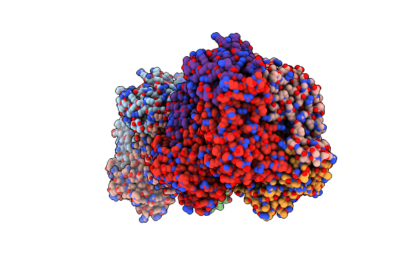
Tyrosine Ammonia-Lyase From Rhodobacter Sphaeroides
Organism: Rhodobacter sphaeroides
Method: X-RAY DIFFRACTION Resolution:1.50 Å Release Date: 2007-01-16 Classification: LYASE |
 |
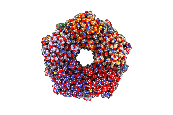
Crystal Structure Of Phenylalanine Ammonia Lyase From Rhodosporidium Toruloides
Organism: Rhodosporidium toruloides
Method: X-RAY DIFFRACTION Resolution:2.70 Å Release Date: 2004-10-12 Classification: LYASE |
 |
Tyrosine Ammonia-Lyase From Rhodobacter Sphaeroides, Complexed With Caffeate
Organism: Rhodobacter sphaeroides
Method: X-RAY DIFFRACTION Resolution:1.90 Å Release Date: 2007-01-16 Classification: LYASE Ligands: DHC |
 |
Tyrosine Ammonia-Lyase From Rhodobacter Sphaeroides, Complexed With Coumarate
Organism: Rhodobacter sphaeroides
Method: X-RAY DIFFRACTION Resolution:1.60 Å Release Date: 2007-01-16 Classification: LYASE Ligands: HC4 |
 |
Tyrosine Ammonia-Lyase From Rhodobacter Sphaeroides (His89Phe Variant) Complexed With Cinnamic Acid
Organism: Rhodobacter sphaeroides
Method: X-RAY DIFFRACTION Resolution:1.90 Å Release Date: 2007-01-16 Classification: LYASE Ligands: TCA |
 |
Tyrosine Ammonia-Lyase From Rhodobacter Sphaeroides (His89Phe Variant), Complexed With Coumaric Acid
Organism: Rhodobacter sphaeroides
Method: X-RAY DIFFRACTION Resolution:2.00 Å Release Date: 2007-01-16 Classification: LYASE Ligands: HC4 |