

Search Count: 51
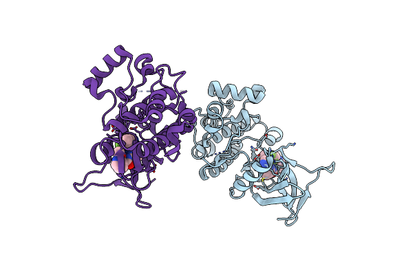 |
Ask1 Bound To Compound 28
Organism: Homo sapiens
Method: X-RAY DIFFRACTION Resolution:2.17 Å Release Date: 2024-07-17 Classification: TRANSFERASE/INHIBITOR Ligands: A1AWT |
Organism: Homo sapiens
Method: X-RAY DIFFRACTION
Release Date: 2024-07-17
Ligands: A1AWT
 |
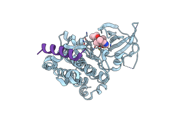
Bio-2895 (Brd0705) Bound Gsk3Alpha-Axin Complex
Organism: Homo sapiens
Method: X-RAY DIFFRACTION Resolution:1.94 Å Release Date: 2023-06-14 Classification: HYDROLASE Ligands: 6VL, CA |
Organism: Homo sapiens
Method: X-RAY DIFFRACTION
Release Date: 2023-06-14
Ligands: 6VL, CA
 |
Bio-8546 Bound Gsk3Alpha-Axin Complex
Organism: Homo sapiens
Method: X-RAY DIFFRACTION Resolution:2.40 Å Release Date: 2023-06-14 Classification: HYDROLASE Ligands: D1E |
Organism: Homo sapiens
Method: X-RAY DIFFRACTION
Release Date: 2023-06-14
Ligands: D1E
 |
Bio-8546 Bound Gsk3Beta-Axin Complex
Organism: Homo sapiens
Method: X-RAY DIFFRACTION Resolution:2.09 Å Release Date: 2023-06-14 Classification: HYDROLASE Ligands: D1E |
Organism: Homo sapiens
Method: X-RAY DIFFRACTION
Release Date: 2023-06-14
Ligands: D1E
 |
Bio-2895 (Brd0705) Bound Gsk3Beta-Axin Complex
Organism: Homo sapiens
Method: X-RAY DIFFRACTION Resolution:1.85 Å Release Date: 2023-06-14 Classification: HYDROLASE Ligands: 6VL |
Organism: Homo sapiens
Method: X-RAY DIFFRACTION
Release Date: 2023-06-14
Ligands: 6VL
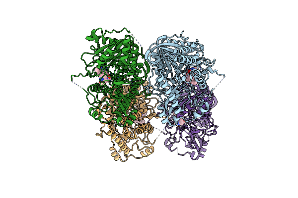 |
Structure Of Compound 34 Bound To Human Phospholipase D2 Catalytic Domain
Organism: Homo sapiens
Method: X-RAY DIFFRACTION Resolution:2.90 Å Release Date: 2023-01-25 Classification: HYDROLASE/HYDROLASE INHIBITOR Ligands: CI0 |
Organism: Homo sapiens
Method: X-RAY DIFFRACTION
Release Date: 2023-01-25
Ligands: CI0
 |
Structure Of Ttbk1 Kinase Domain In Complex With Compound 3
Organism: Homo sapiens
Method: X-RAY DIFFRACTION Resolution:1.56 Å Release Date: 2021-05-19 Classification: TRANSFERASE/INHIBITOR Ligands: VP7, NA |
Organism: Homo sapiens
Method: X-RAY DIFFRACTION
Release Date: 2021-05-19
Ligands: VP7, NA
 |
Structure Of Ttbk1 Kinase Domain In Complex With Compound 18
Organism: Homo sapiens
Method: X-RAY DIFFRACTION Resolution:2.15 Å Release Date: 2021-05-19 Classification: TRANSFERASE/INHIBITOR Ligands: VSY |
Organism: Homo sapiens
Method: X-RAY DIFFRACTION
Release Date: 2021-05-19
Ligands: VSY
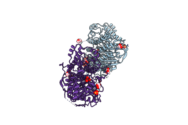 |
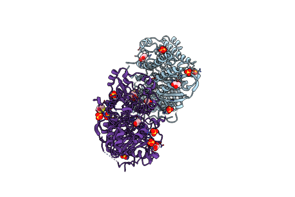
Structure Of Tungstate Bound Human Phospholipase D2 Catalytic Domain
Organism: Homo sapiens
Method: X-RAY DIFFRACTION Resolution:1.90 Å Release Date: 2020-02-19 Classification: HYDROLASE Ligands: WO4, SO4, GOL |
Organism: Homo sapiens
Method: X-RAY DIFFRACTION
Release Date: 2020-02-19
Ligands: WO4, SO4, GOL
 |
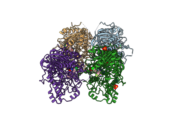
Structure Of Human Phospholipase D2 Catalytic Domain
Organism: Homo sapiens
Method: X-RAY DIFFRACTION Resolution:2.00 Å Release Date: 2020-02-19 Classification: HYDROLASE Ligands: SO4, GOL, DTT |
Organism: Homo sapiens
Method: X-RAY DIFFRACTION
Release Date: 2020-02-19
Ligands: SO4, GOL, DTT
 |
Structure Of Compound 1 (Halopemide) Bound Human Phospholipase D2 Catalytic Domain
Organism: Homo sapiens
Method: X-RAY DIFFRACTION Resolution:2.60 Å Release Date: 2020-02-19 Classification: hydrolase/hydrolase inhibitor Ligands: MJV, SO4, GOL |
Organism: Homo sapiens
Method: X-RAY DIFFRACTION
Release Date: 2020-02-19
Ligands: MJV, SO4, GOL
 |
Structure Of Compound 4 Bound Human Phospholipase D2 Catalytic Domain
Organism: Homo sapiens
Method: X-RAY DIFFRACTION Resolution:2.69 Å Release Date: 2020-02-19 Classification: hydrolase/hydrolase inhibitor Ligands: MKA, SO4 |
Organism: Homo sapiens
Method: X-RAY DIFFRACTION
Release Date: 2020-02-19
Ligands: MKA, SO4
 |
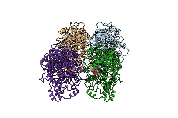
Structure Of Compound 5 Bound Human Phospholipase D1 Catalytic Domain
Organism: Homo sapiens
Method: X-RAY DIFFRACTION Resolution:3.20 Å Release Date: 2020-02-19 Classification: hydrolase/hydrolase inhibitor Ligands: MKG |
Organism: Homo sapiens
Method: X-RAY DIFFRACTION
Release Date: 2020-02-19
Ligands: MKG
 |
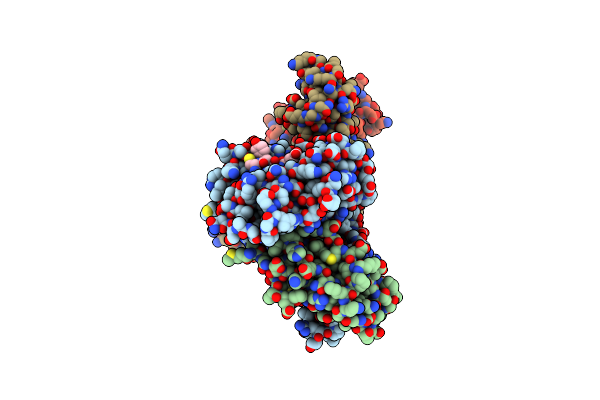
Structure Of Compound 3 (Ml299) Bound Human Phospholipase D2 Catalytic Domain
Organism: Homo sapiens
Method: X-RAY DIFFRACTION Resolution:3.20 Å Release Date: 2020-02-19 Classification: hydrolase/hydrolase inhibitor Ligands: MJY |
Organism: Homo sapiens
Method: X-RAY DIFFRACTION
Release Date: 2020-02-19
Ligands: MJY
 |
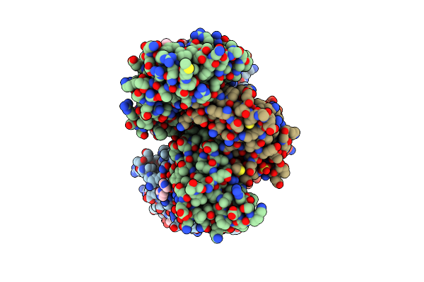
Crystal Structure Of H-2D In Complex With An Insulin Derived Peptide
Organism: Mus musculus
Method: X-RAY DIFFRACTION Resolution:2.34 Å Release Date: 2014-03-26 Classification: IMMUNE SYSTEM |
Organism: Mus musculus
Method: X-RAY DIFFRACTION
Release Date: 2014-03-26
 |
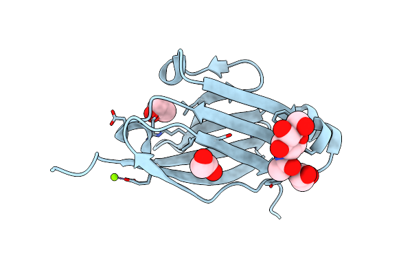
Crystal Structure Of H-2D In Complex With A Mimotopic Peptide
Organism: Mus musculus
Method: X-RAY DIFFRACTION Resolution:1.98 Å Release Date: 2014-03-26 Classification: IMMUNE SYSTEM Ligands: ZN, IMD, GOL, SO4 |
Organism: Mus musculus
Method: X-RAY DIFFRACTION
Release Date: 2014-03-26
Ligands: ZN, IMD, GOL, SO4
 |
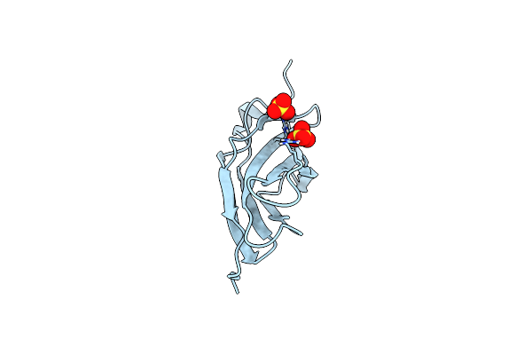
Crystal Structure Of The 1St Ig Domain From Mouse Polymeric Immunoglobulin Receptor [Psi-Nysgrc-006220]
Organism: Mus musculus
Method: X-RAY DIFFRACTION Resolution:1.51 Å Release Date: 2013-12-04 Classification: IMMUNE SYSTEM Ligands: NAG, MG, EDO |
Organism: Mus musculus
Method: X-RAY DIFFRACTION
Release Date: 2013-12-04
Ligands: NAG, MG, EDO
 |
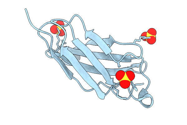
Crystal Structure Of The 4Th Fn3 Domain Of Human Protein Tyrosine Phosphatase, Receptor Type F [Psi-Nysgrc-006240]
Organism: Homo sapiens
Method: X-RAY DIFFRACTION Resolution:1.46 Å Release Date: 2013-10-30 Classification: HYDROLASE Ligands: SO4 |
Organism: Homo sapiens
Method: X-RAY DIFFRACTION
Release Date: 2013-10-30
Ligands: SO4
 |
Crystal Structure Of An Internal Fn3 Domain From Human Contactin-5 [Psi-Nysgrc-005804]
Organism: Homo sapiens
Method: X-RAY DIFFRACTION Resolution:1.80 Å Release Date: 2013-10-30 Classification: CELL ADHESION Ligands: SO4 |
Organism: Homo sapiens
Method: X-RAY DIFFRACTION
Release Date: 2013-10-30
Ligands: SO4
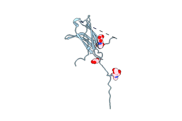 |
Crystal Structure Of A Strand Swapped Ctla-4 From Duckbill Platypus [Psi-Nysgrc-012711]
Organism: Ornithorhynchus anatinus
Method: X-RAY DIFFRACTION Resolution:2.30 Å Release Date: 2013-10-30 Classification: IMMUNE SYSTEM Ligands: GOL, NAG |
Organism: Ornithorhynchus anatinus
Method: X-RAY DIFFRACTION
Release Date: 2013-10-30
Ligands: GOL, NAG