

Search Count: 13
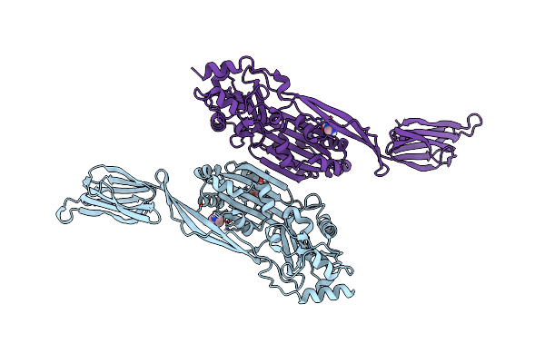 |
Crystal Structure Of Gram Negative Toxin
Organism: Desulfobulbus propionicus (strain atcc 33891 / dsm 2032 / 1pr3)
Method: X-RAY DIFFRACTION Resolution:2.30 Å Release Date: 2019-12-04 Classification: TOXIN Ligands: PG4, IMD |
Organism: Desulfobulbus propionicus (strain atcc 33891 / dsm 2032 / 1pr3)
Method: X-RAY DIFFRACTION
Release Date: 2019-12-04
Ligands: PG4, IMD
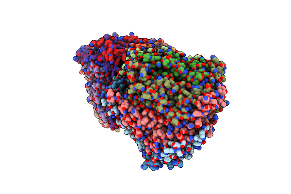 |
Crystal Structure Of The 11S Subunit Of The Plasmodium Falciparum Proteasome, Pa28
Organism: Plasmodium falciparum
Method: X-RAY DIFFRACTION Resolution:3.10 Å Release Date: 2019-08-07 Classification: PROTEIN BINDING Ligands: SO4 |
Organism: Plasmodium falciparum
Method: X-RAY DIFFRACTION
Release Date: 2019-08-07
Ligands: SO4
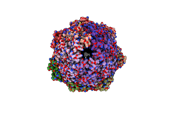 |
The Structure Of The Plasmodium Falciparum 20S Proteasome In Complex With Two Pa28 Activators
Organism: Plasmodium falciparum (isolate 3d7)
Method: ELECTRON MICROSCOPY Release Date: 2019-08-07 Classification: HYDROLASE |
Organism: Plasmodium falciparum (isolate 3d7)
Method: ELECTRON MICROSCOPY
Release Date: 2019-08-07
 |
The Structure Of The Plasmodium Falciparum 20S Proteasome.
Organism: Plasmodium falciparum (isolate 3d7)
Method: ELECTRON MICROSCOPY Release Date: 2019-08-07 Classification: HYDROLASE |
Organism: Plasmodium falciparum (isolate 3d7)
Method: ELECTRON MICROSCOPY
Release Date: 2019-08-07
 |
The Structure Of The Plasmodium Falciparum 20S Proteasome In Complex With One Pa28 Activator
Organism: Plasmodium falciparum 3d7
Method: ELECTRON MICROSCOPY Release Date: 2019-08-07 Classification: HYDROLASE |
Organism: Plasmodium falciparum 3d7
Method: ELECTRON MICROSCOPY
Release Date: 2019-08-07
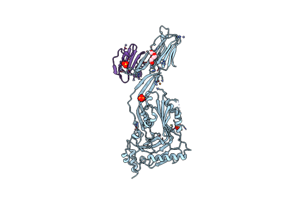 |
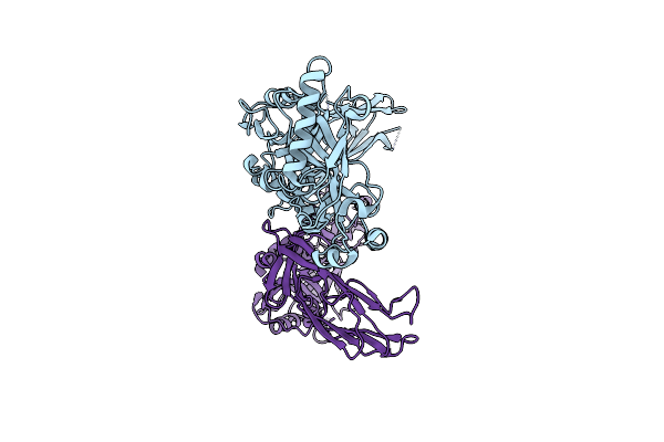
Toxin Receptor Complex
Organism: Streptococcus intermedius, Homo sapiens
Method: X-RAY DIFFRACTION Resolution:2.70 Å Release Date: 2016-08-24 Classification: TOXIN Ligands: PGE, SO4, ZN, CU |
Organism: Streptococcus intermedius, Homo sapiens
Method: X-RAY DIFFRACTION
Release Date: 2016-08-24
Ligands: PGE, SO4, ZN, CU
 |
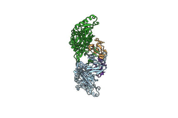
Trapped Toxin
Organism: Streptococcus intermedius
Method: X-RAY DIFFRACTION Resolution:2.89 Å Release Date: 2016-08-24 Classification: TOXIN |
Organism: Streptococcus intermedius
Method: X-RAY DIFFRACTION
Release Date: 2016-08-24
 |
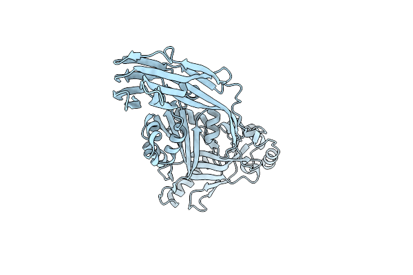
Trapped Toxin
Organism: Gardnerella vaginalis, Homo sapiens
Method: X-RAY DIFFRACTION Resolution:2.40 Å Release Date: 2016-08-24 Classification: toxin/toxin receptor |
Organism: Gardnerella vaginalis, Homo sapiens
Method: X-RAY DIFFRACTION
Release Date: 2016-08-24
 |
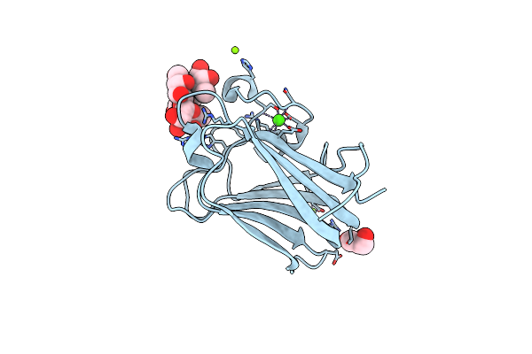
Crystal Structure Of A Cholesterol Dependent Cytolysin
Organism: Streptococcus pyogenes
Method: X-RAY DIFFRACTION Resolution:2.10 Å Release Date: 2013-10-30 Classification: TOXIN |
Organism: Streptococcus pyogenes
Method: X-RAY DIFFRACTION
Release Date: 2013-10-30
 |
His 62 Mutant Of The Lectin Binding Domain Of Lectinolysin Complexed With Lewis Y
Organism: Streptococcus mitis
Method: X-RAY DIFFRACTION Resolution:1.60 Å Release Date: 2012-11-21 Classification: SUGAR BINDING PROTEIN Ligands: MG, GOL, CA |
Organism: Streptococcus mitis
Method: X-RAY DIFFRACTION
Release Date: 2012-11-21
Ligands: MG, GOL, CA
 |
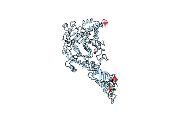
His 62 Mutant Of The Lectin Binding Domain Of Lectinolysin Complexed With Lewis B
Organism: Streptococcus mitis
Method: X-RAY DIFFRACTION Resolution:1.60 Å Release Date: 2012-11-21 Classification: SUGAR BINDING PROTEIN Ligands: MG, CA |
Organism: Streptococcus mitis
Method: X-RAY DIFFRACTION
Release Date: 2012-11-21
Ligands: MG, CA
 |
The X-Ray Crystal Structure Of Lymphocyte Perforin
Organism: Mus musculus
Method: X-RAY DIFFRACTION Resolution:2.75 Å Release Date: 2010-11-03 Classification: IMMUNE SYSTEM Ligands: GOL, CA, IOD, CL, NAG |
Organism: Mus musculus
Method: X-RAY DIFFRACTION
Release Date: 2010-11-03
Ligands: GOL, CA, IOD, CL, NAG
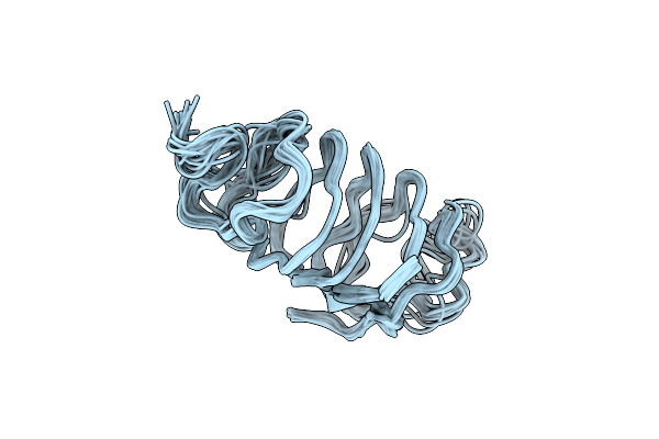 |
Solution Structure Of Spruce Budworm Antifreeze Protein At 30 Degrees Celsius
Organism: Choristoneura fumiferana
Method: SOLUTION NMR Release Date: 2000-07-27 Classification: ANTIFREEZE PROTEIN |
Organism: Choristoneura fumiferana
Method: SOLUTION NMR
Release Date: 2000-07-27