

Search Count: 28
 |
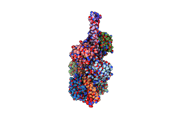
Wax Synthase 1 From Acinetobacter Baylyi (Abwsd1) Co-Crystallized With Myristic Acid
Organism: Acinetobacter baylyi adp1
Method: X-RAY DIFFRACTION Resolution:1.95 Å Release Date: 2022-03-30 Classification: BIOSYNTHETIC PROTEIN Ligands: MYR |
Organism: Acinetobacter baylyi adp1
Method: X-RAY DIFFRACTION
Release Date: 2022-03-30
Ligands: MYR
 |
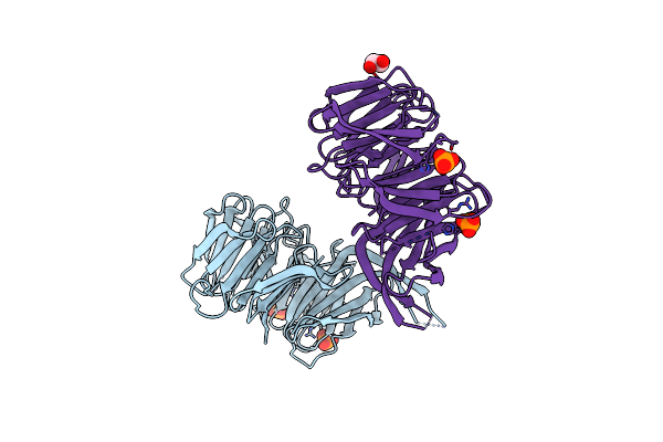
The Rab33B-Atg16L1 Crystal Structure
Organism: Mus musculus
Method: X-RAY DIFFRACTION Resolution:3.47 Å Release Date: 2020-08-05 Classification: CELL CYCLE Ligands: MG, GTP |
Organism: Mus musculus
Method: X-RAY DIFFRACTION
Release Date: 2020-08-05
Ligands: MG, GTP
 |
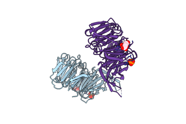
Phosphate-Bound Pichia Angusta Atg18
Organism: Pichia angusta
Method: X-RAY DIFFRACTION Resolution:2.50 Å Release Date: 2017-09-20 Classification: LIPID BINDING PROTEIN Ligands: PO4, EDO |
Organism: Pichia angusta
Method: X-RAY DIFFRACTION
Release Date: 2017-09-20
Ligands: PO4, EDO
 |
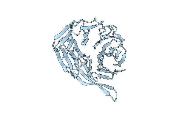
Citrate-Bound Pichia Angusta Atg18
Organism: Pichia angusta
Method: X-RAY DIFFRACTION Resolution:2.00 Å Release Date: 2017-09-20 Classification: LIPID BINDING PROTEIN Ligands: PO4, FLC |
Organism: Pichia angusta
Method: X-RAY DIFFRACTION
Release Date: 2017-09-20
Ligands: PO4, FLC
 |
Klhsv2 With Loop 6Cd Replaced By A Gly-Ser Linker
Organism: Kluyveromyces lactis
Method: X-RAY DIFFRACTION Resolution:2.80 Å Release Date: 2015-04-01 Classification: PROTEIN TRANSPORT |
Organism: Kluyveromyces lactis
Method: X-RAY DIFFRACTION
Release Date: 2015-04-01
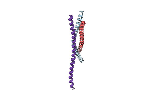 |
Human Short Coiled Coil Protein
Organism: Homo sapiens
Method: X-RAY DIFFRACTION Resolution:2.70 Å Release Date: 2013-09-11 Classification: STRUCTURAL PROTEIN |
Organism: Homo sapiens
Method: X-RAY DIFFRACTION
Release Date: 2013-09-11
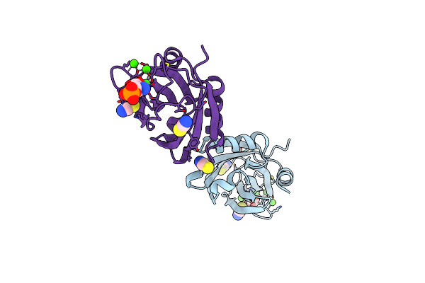 |
Synaptotagmin-1 C2B Domain With Phosphoserine
Organism: Rattus norvegicus
Method: X-RAY DIFFRACTION Resolution:1.50 Å Release Date: 2013-03-20 Classification: SIGNALING PROTEIN Ligands: CA, SEP, SCN |
Organism: Rattus norvegicus
Method: X-RAY DIFFRACTION
Release Date: 2013-03-20
Ligands: CA, SEP, SCN
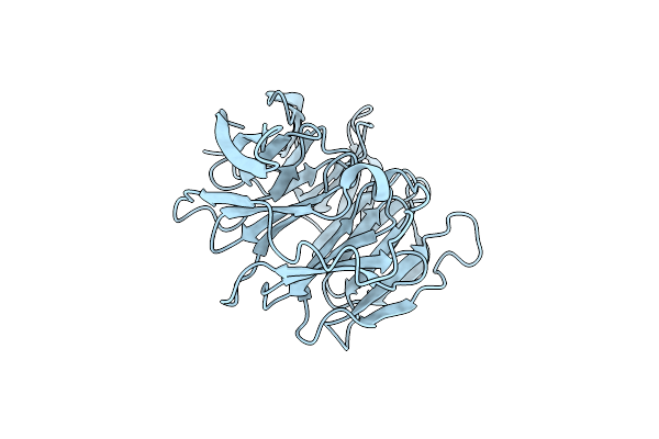 |
Kluyveromyces Lactis Hsv2 Complete Loop 6Cd
Organism: Kluyveromyces lactis
Method: X-RAY DIFFRACTION Resolution:3.35 Å Release Date: 2012-06-13 Classification: LIPID BINDING PROTEIN |
Organism: Kluyveromyces lactis
Method: X-RAY DIFFRACTION
Release Date: 2012-06-13
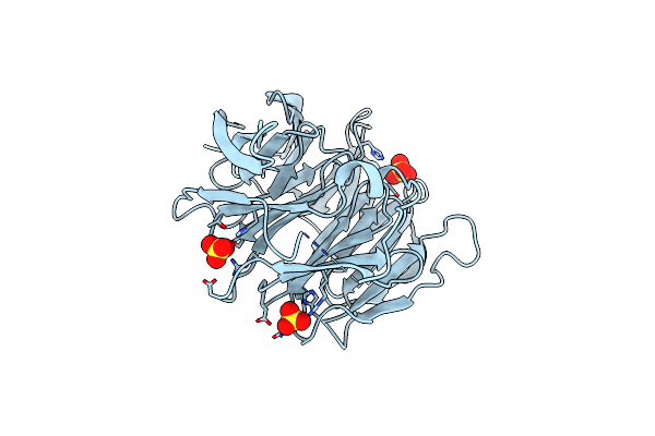 |
Kluyveromyces Lactis Hsv2
Organism: Kluyveromyces lactis
Method: X-RAY DIFFRACTION Resolution:3.00 Å Release Date: 2012-06-13 Classification: LIPID BINDING PROTEIN Ligands: SO4 |
Organism: Kluyveromyces lactis
Method: X-RAY DIFFRACTION
Release Date: 2012-06-13
Ligands: SO4
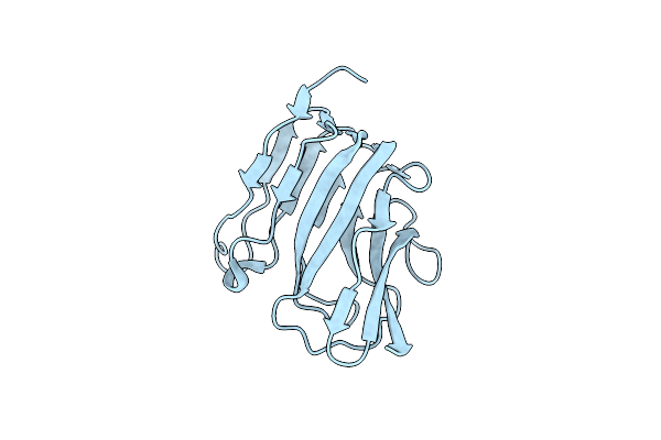 |
Crystal Structure Of The Icsa Autochaperone Region
Organism: Shigella flexneri
Method: X-RAY DIFFRACTION Resolution:2.00 Å Release Date: 2011-03-02 Classification: PROTEIN TRANSPORT |
Organism: Shigella flexneri
Method: X-RAY DIFFRACTION
Release Date: 2011-03-02
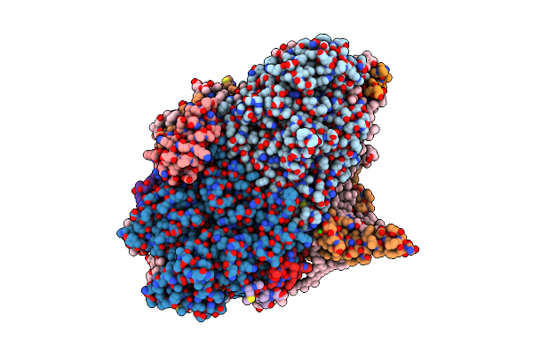 |
Femtosecond X-Ray Protein Nanocrystallography
Organism: Thermosynechococcus elongatus
Method: X-RAY DIFFRACTION Resolution:8.98 Å Release Date: 2011-02-02 Classification: PHOTOSYNTHESIS Ligands: CLA, PQN, SF4, BCR, LHG, LMG, CA |
Organism: Thermosynechococcus elongatus
Method: X-RAY DIFFRACTION
Release Date: 2011-02-02
Ligands: CLA, PQN, SF4, BCR, LHG, LMG, CA
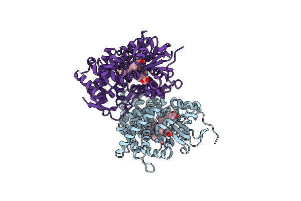 |
Retinoic Acid Bound Cyanobacterial Cyp120A1
Organism: Synechocystis sp.
Method: X-RAY DIFFRACTION Resolution:2.10 Å Release Date: 2008-04-29 Classification: OXIDOREDUCTASE Ligands: HEM, REA |
Organism: Synechocystis sp.
Method: X-RAY DIFFRACTION
Release Date: 2008-04-29
Ligands: HEM, REA
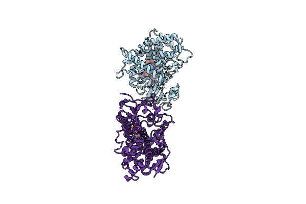 |
Substrate Free Cyanobacterial Cyp120A1
Organism: Synechocystis sp.
Method: X-RAY DIFFRACTION Resolution:2.40 Å Release Date: 2008-04-29 Classification: OXIDOREDUCTASE Ligands: HEM |
Organism: Synechocystis sp.
Method: X-RAY DIFFRACTION
Release Date: 2008-04-29
Ligands: HEM
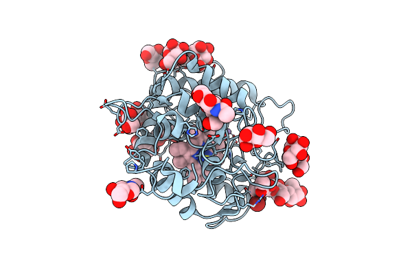 |
Chloroperoxidase Mixture Of Ferric And Ferrous States (Low Dose Data Set)
Organism: Caldariomyces fumago
Method: X-RAY DIFFRACTION Resolution:1.75 Å Release Date: 2006-12-18 Classification: OXIDOREDUCTASE Ligands: MN, HEM, NAG, MAN, BR |
Organism: Caldariomyces fumago
Method: X-RAY DIFFRACTION
Release Date: 2006-12-18
Ligands: MN, HEM, NAG, MAN, BR
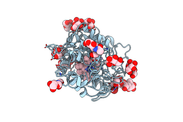 |
Ferrous Chloroperoxidase (High Dose Data Set)
Organism: Caldariomyces fumago
Method: X-RAY DIFFRACTION Resolution:1.75 Å Release Date: 2006-12-18 Classification: OXIDOREDUCTASE Ligands: MN, HEM, NAG, MAN, BR |
Organism: Caldariomyces fumago
Method: X-RAY DIFFRACTION
Release Date: 2006-12-18
Ligands: MN, HEM, NAG, MAN, BR
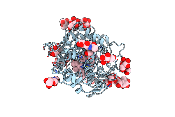 |
Structure Of Chloroperoxidase Compound 0
Organism: Leptoxyphium fumago
Method: X-RAY DIFFRACTION Resolution:1.75 Å Release Date: 2006-12-06 Classification: OXIDOREDUCTASE Ligands: MN, HEM, PEO, NAG, MAN, EDO, ACT |
Organism: Leptoxyphium fumago
Method: X-RAY DIFFRACTION
Release Date: 2006-12-06
Ligands: MN, HEM, PEO, NAG, MAN, EDO, ACT
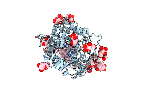 |
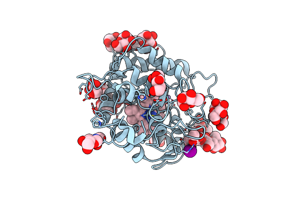
Chloroperoxidase Bromide Complex
Organism: Caldariomyces fumago
Method: X-RAY DIFFRACTION Resolution:1.80 Å Release Date: 2006-06-12 Classification: OXIDOREDUCTASE Ligands: MN, HEM, NAG, MAN, BR |
Organism: Caldariomyces fumago
Method: X-RAY DIFFRACTION
Release Date: 2006-06-12
Ligands: MN, HEM, NAG, MAN, BR
 |
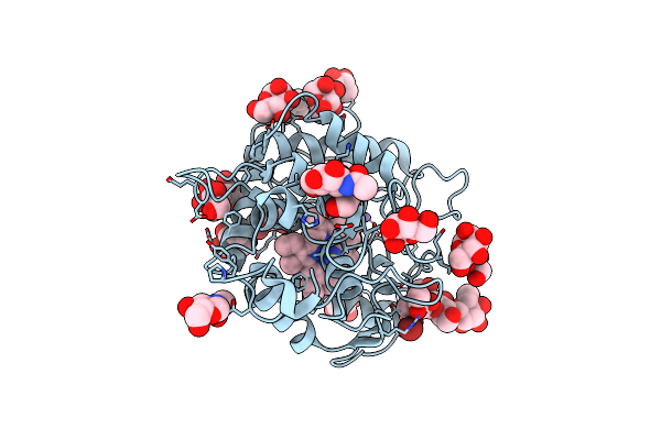
Chloroperoxidase Iodide Complex
Organism: Caldariomyces fumago
Method: X-RAY DIFFRACTION Resolution:1.15 Å Release Date: 2006-06-12 Classification: OXIDOREDUCTASE Ligands: MN, HEM, NAG, MAN, IOD |
Organism: Caldariomyces fumago
Method: X-RAY DIFFRACTION
Release Date: 2006-06-12
Ligands: MN, HEM, NAG, MAN, IOD
 |
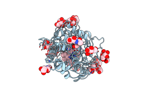
Chloroperoxidase Complexed With Cyclopentanedione
Organism: Caldariomyces fumago
Method: X-RAY DIFFRACTION Resolution:1.80 Å Release Date: 2006-06-12 Classification: OXIDOREDUCTASE Ligands: MN, HEM, NAG, MAN, EDO, CEJ, BR |
Organism: Caldariomyces fumago
Method: X-RAY DIFFRACTION
Release Date: 2006-06-12
Ligands: MN, HEM, NAG, MAN, EDO, CEJ, BR
 |
Chloroperoxidase Complexed With Cyanide And Dmso
Organism: Caldariomyces fumago
Method: X-RAY DIFFRACTION Resolution:1.70 Å Release Date: 2006-06-12 Classification: OXIDOREDUCTASE Ligands: MN, HEM, NAG, MAN, BR, EDO, CYN, DMS |
Organism: Caldariomyces fumago
Method: X-RAY DIFFRACTION
Release Date: 2006-06-12
Ligands: MN, HEM, NAG, MAN, BR, EDO, CYN, DMS