

Search Count: 16
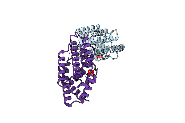 |
Crystal Structure Of Pfho From Plasmodium Falciparum At 2.78 A
Organism: Plasmodium falciparum
Method: X-RAY DIFFRACTION Resolution:2.78 Å Release Date: 2024-09-04 Classification: DNA BINDING PROTEIN Ligands: SO4 |
Organism: Plasmodium falciparum
Method: X-RAY DIFFRACTION
Release Date: 2024-09-04
Ligands: SO4
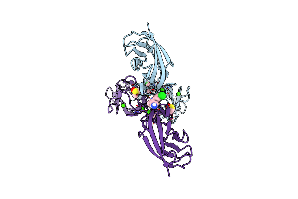 |
Crystal Structure Of Human P-Cadherin Mec12 (X Dimer) In Complex With 2-(5-Chloro-2-Methyl-1H-Indol-3-Yl)Ethan-1-Amine (Inhibitor)
Organism: Homo sapiens
Method: X-RAY DIFFRACTION Resolution:2.45 Å Release Date: 2021-09-15 Classification: CELL ADHESION Ligands: DMS, G60, CA |
Organism: Homo sapiens
Method: X-RAY DIFFRACTION
Release Date: 2021-09-15
Ligands: DMS, G60, CA
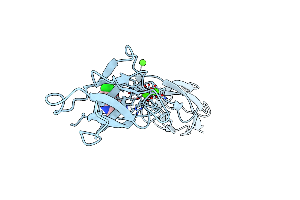 |
Crystal Structure Of Human P-Cadherin Rec12 (Monomer) In Complex With 2-(5-Chloro-2-Methyl-1H-Indol-3-Yl)Ethan-1-Amine (Inhibitor)
Organism: Homo sapiens
Method: X-RAY DIFFRACTION Resolution:2.30 Å Release Date: 2021-09-15 Classification: CELL ADHESION Ligands: CA, G60 |
Organism: Homo sapiens
Method: X-RAY DIFFRACTION
Release Date: 2021-09-15
Ligands: CA, G60
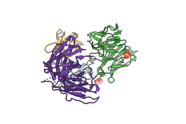 |
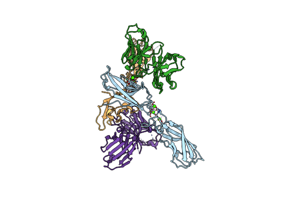
Human P-Cadherin Mec1 With Scfv Tsp7 Bound
Organism: Homo sapiens, Mus musculus
Method: X-RAY DIFFRACTION Resolution:2.55 Å Release Date: 2016-12-28 Classification: CELL ADHESION/IMMUNE SYSTEM Ligands: GOL, PO4, CL |
Organism: Homo sapiens, Mus musculus
Method: X-RAY DIFFRACTION
Release Date: 2016-12-28
Ligands: GOL, PO4, CL
 |
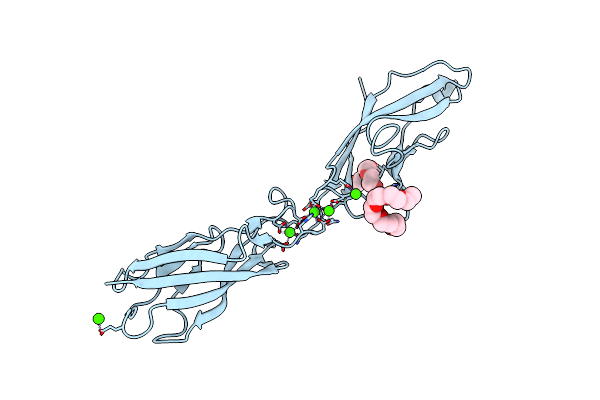
Human P-Cadherin Ec12 With Scfv Tsp11 Bound
Organism: Homo sapiens, Mus musculus
Method: X-RAY DIFFRACTION Resolution:2.45 Å Release Date: 2016-12-28 Classification: CELL ADHESION/IMMUNE SYSTEM Ligands: CA |
Organism: Homo sapiens, Mus musculus
Method: X-RAY DIFFRACTION
Release Date: 2016-12-28
Ligands: CA
 |
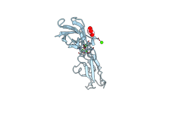
Crystal Structure Of Human P-Cadherin (Ss-Dimer)
Organism: Homo sapiens
Method: X-RAY DIFFRACTION Resolution:1.85 Å Release Date: 2016-09-07 Classification: CELL ADHESION Ligands: CA, CL, P6G, PG4 |
Organism: Homo sapiens
Method: X-RAY DIFFRACTION
Release Date: 2016-09-07
Ligands: CA, CL, P6G, PG4
 |
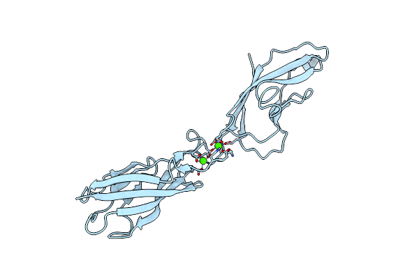
Crystal Structure Of Human P-Cadherin (Ss-Dimer Long)
Organism: Homo sapiens
Method: X-RAY DIFFRACTION Resolution:2.60 Å Release Date: 2016-09-07 Classification: CELL ADHESION Ligands: CA, GOL |
Organism: Homo sapiens
Method: X-RAY DIFFRACTION
Release Date: 2016-09-07
Ligands: CA, GOL
 |
Crystal Structure Of Human P-Cadherin (Ss-Dimer K14E)
Organism: Homo sapiens
Method: X-RAY DIFFRACTION Resolution:2.48 Å Release Date: 2016-09-07 Classification: CELL ADHESION Ligands: CA |
Organism: Homo sapiens
Method: X-RAY DIFFRACTION
Release Date: 2016-09-07
Ligands: CA
 |
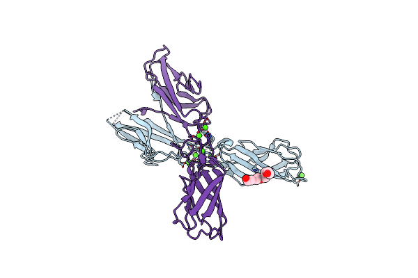
Crystal Structure Of Human P-Cadherin (Ss-Dimer Q101L)
Organism: Homo sapiens
Method: X-RAY DIFFRACTION Resolution:2.15 Å Release Date: 2016-09-07 Classification: CELL ADHESION Ligands: CA |
Organism: Homo sapiens
Method: X-RAY DIFFRACTION
Release Date: 2016-09-07
Ligands: CA
 |
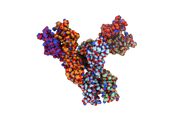
Crystal Structure Of Human P-Cadherin (Ss-X-Dimer)
Organism: Homo sapiens
Method: X-RAY DIFFRACTION Resolution:2.20 Å Release Date: 2016-09-07 Classification: CELL ADHESION Ligands: CA, 1PE |
Organism: Homo sapiens
Method: X-RAY DIFFRACTION
Release Date: 2016-09-07
Ligands: CA, 1PE
 |
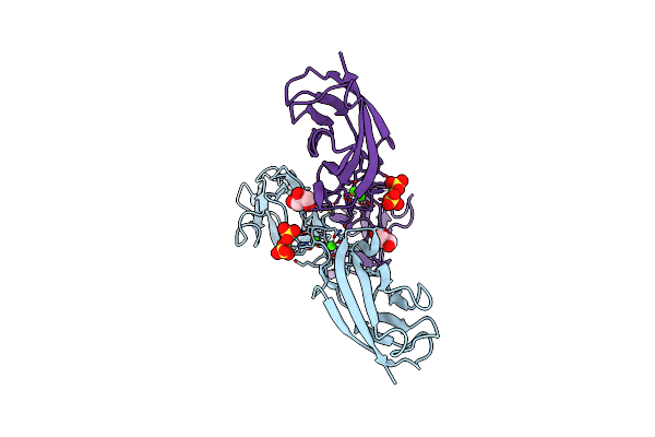
Crystal Structure Of Human P-Cadherin (Ss-X-Dimer-Long)
Organism: Homo sapiens
Method: X-RAY DIFFRACTION Resolution:2.70 Å Release Date: 2016-09-07 Classification: CELL ADHESION Ligands: CA |
Organism: Homo sapiens
Method: X-RAY DIFFRACTION
Release Date: 2016-09-07
Ligands: CA
 |
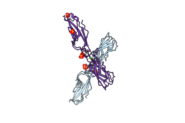
Crystal Structure Of Human P-Cadherin (Ss-X-Dimer Pocket I)
Organism: Homo sapiens
Method: X-RAY DIFFRACTION Resolution:2.40 Å Release Date: 2016-09-07 Classification: CELL ADHESION Ligands: CA, NI, SO4, GOL |
Organism: Homo sapiens
Method: X-RAY DIFFRACTION
Release Date: 2016-09-07
Ligands: CA, NI, SO4, GOL
 |
Crystal Structure Of Human P-Cadherin (Enc-X-Dimer)
Organism: Homo sapiens
Method: X-RAY DIFFRACTION Resolution:2.30 Å Release Date: 2016-09-07 Classification: CELL ADHESION Ligands: CA, SO4, NI |
Organism: Homo sapiens
Method: X-RAY DIFFRACTION
Release Date: 2016-09-07
Ligands: CA, SO4, NI
 |
Crystal Structure Of Human P-Cadherin (Int-X-Dimer)
Organism: Homo sapiens
Method: X-RAY DIFFRACTION Resolution:3.10 Å Release Date: 2016-09-07 Classification: CELL ADHESION Ligands: CA |
Organism: Homo sapiens
Method: X-RAY DIFFRACTION
Release Date: 2016-09-07
Ligands: CA
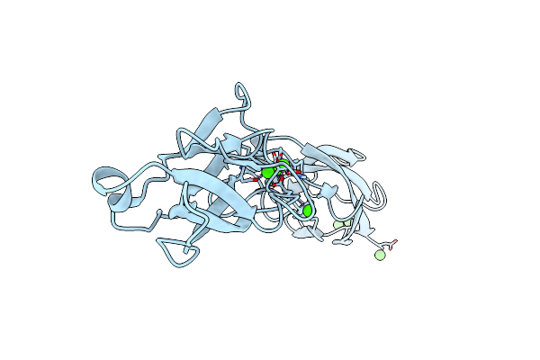 |
Crystal Structure Of Human P-Cadherin (Monomer 1)
Organism: Homo sapiens
Method: X-RAY DIFFRACTION Resolution:1.50 Å Release Date: 2016-09-07 Classification: CELL ADHESION Ligands: CA |
Organism: Homo sapiens
Method: X-RAY DIFFRACTION
Release Date: 2016-09-07
Ligands: CA
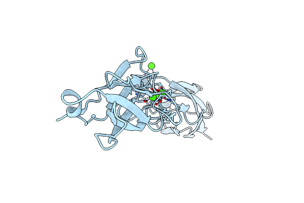 |
Crystal Structure Of Human P-Cadherin (Monomer 2)
Organism: Homo sapiens
Method: X-RAY DIFFRACTION Resolution:2.05 Å Release Date: 2016-09-07 Classification: CELL ADHESION Ligands: CA |
Organism: Homo sapiens
Method: X-RAY DIFFRACTION
Release Date: 2016-09-07
Ligands: CA