
Search Count: 241
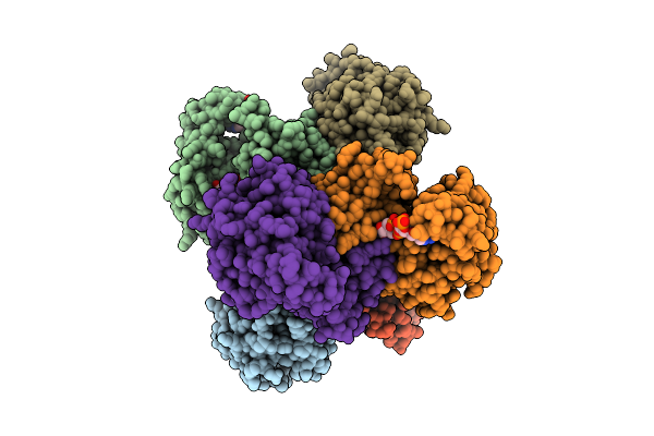 |
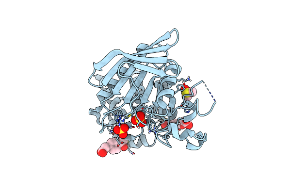
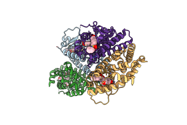
3-Hydroxybutyryl-Coa Dehydrogenase With Nad And Acetoacetyl Coa
Organism: Faecalibacterium prausnitzii l2-6
Method: X-RAY DIFFRACTION Resolution:3.29 Å Release Date: 2026-04-15 Classification: OXIDOREDUCTASE Ligands: NAD, CAA |
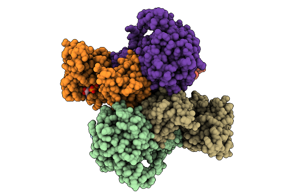 |
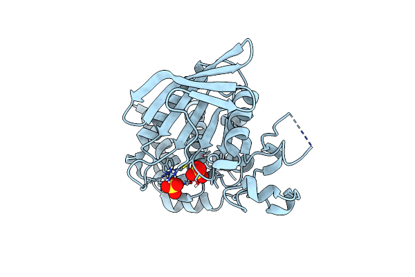
3-Hydroxybutyryl-Coa Dehydrogenase With Nad
Organism: Faecalibacterium prausnitzii l2-6
Method: X-RAY DIFFRACTION Resolution:2.71 Å Release Date: 2026-04-15 Classification: OXIDOREDUCTASE Ligands: NAD |
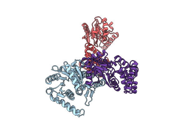 |
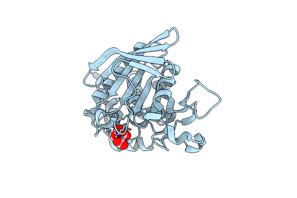
3-Hydroxybutyryl-Coa Dehydrogenase
Organism: Faecalibacterium prausnitzii l2-6
Method: X-RAY DIFFRACTION Resolution:2.29 Å Release Date: 2026-04-15 Classification: OXIDOREDUCTASE |
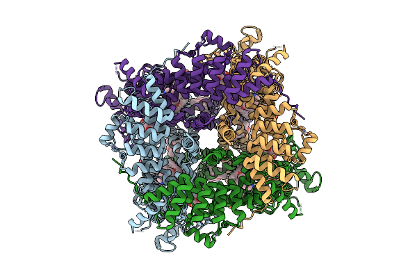 |
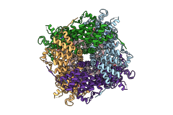
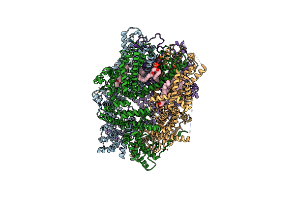
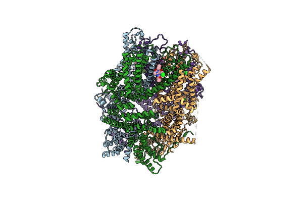
Cryo-Em Structure Of Homomeric Trpc Channel With Agonists, Class 1
Organism: Homo sapiens
Method: ELECTRON MICROSCOPY Resolution:2.43 Å Release Date: 2026-01-21 Classification: MEMBRANE PROTEIN Ligands: PTY, POV, Y01, ZN, CA, A1L55, 657 |
 |
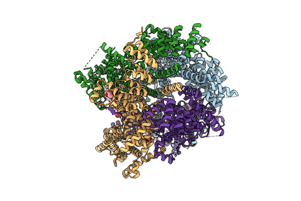
Cryo-Em Structure Of Homomeric Trpc Channel With Agonists, Class 2
Organism: Homo sapiens
Method: ELECTRON MICROSCOPY Resolution:3.03 Å Release Date: 2026-01-21 Classification: MEMBRANE PROTEIN Ligands: PTY, POV, Y01, ZN, CA, A1L55, 657 |
 |
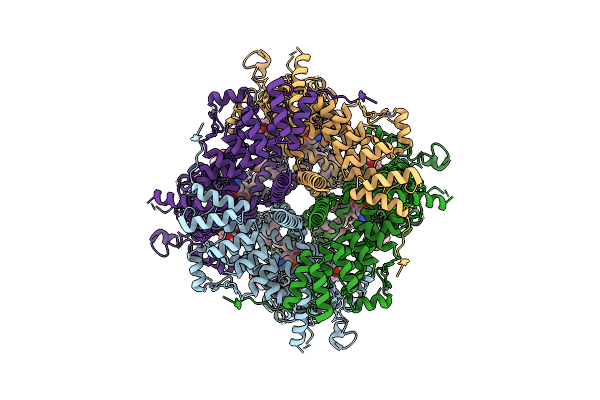
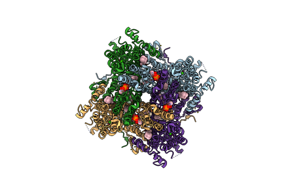
Cryo-Em Structure Of Heteromeric Trpc Channel
Organism: Homo sapiens
Method: ELECTRON MICROSCOPY Resolution:2.70 Å Release Date: 2025-11-12 Classification: MEMBRANE PROTEIN Ligands: CA, YZY, Y01, ZN |
 |
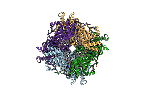
Cryo-Em Structure Of Homomeric Trpc Channel, Class 1
Organism: Homo sapiens
Method: ELECTRON MICROSCOPY Resolution:2.62 Å Release Date: 2025-11-12 Classification: MEMBRANE PROTEIN Ligands: PTY, POV, Y01, ZN, CA, YZY |
 |
Cryo-Em Structure Of Homomeric Trpc Channel, Class 2
Organism: Homo sapiens
Method: ELECTRON MICROSCOPY Resolution:2.90 Å Release Date: 2025-11-12 Classification: MEMBRANE PROTEIN Ligands: PTY, POV, Y01, ZN, CA, YZY |
 |
Zz-Domain Of The Arabidopsis Thaliana E3 Ubiquitin-Protein Ligase Prt1
Organism: Arabidopsis thaliana
Method: X-RAY DIFFRACTION Resolution:1.74 Å Release Date: 2025-05-14 Classification: LIGASE Ligands: ZN |
 |
Y-Degron Fused Zz-Domain Of The Arabidopsis Thaliana E3 Ubiquitin-Protein Ligase Prt1
Organism: Arabidopsis thaliana
Method: X-RAY DIFFRACTION Resolution:1.67 Å Release Date: 2025-05-14 Classification: LIGASE Ligands: ZN, MG |
 |
F-Degron Fused Zz-Domain Of The Arabidopsis Thaliana E3 Ubiquitin-Protein Ligase Prt1
Organism: Arabidopsis thaliana
Method: X-RAY DIFFRACTION Resolution:2.10 Å Release Date: 2025-05-14 Classification: LIGASE Ligands: ZN, MG |
 |
W-Degron Fused Zz-Domain Of The Arabidopsis Thaliana E3 Ubiquitin-Protein Ligase Prt1
Organism: Arabidopsis thaliana
Method: X-RAY DIFFRACTION Resolution:2.79 Å Release Date: 2025-05-14 Classification: LIGASE Ligands: ZN, SO4 |
 |
Step (Ptpn5) At High Resolution With Citrate Bound In Active Site
Organism: Homo sapiens
Method: X-RAY DIFFRACTION Resolution:1.27 Å Release Date: 2024-12-11 Classification: HYDROLASE Ligands: CIT |
 |
Step (Ptpn5) With Active-Site Disulfide Bond And Allosteric-Site Loop Shift
Organism: Homo sapiens
Method: X-RAY DIFFRACTION Resolution:1.75 Å Release Date: 2024-12-11 Classification: HYDROLASE Ligands: SO4 |
 |
Step (Ptpn5) With Active-Site Disulfide Bond And Covalent Ligand Bound To Distal C505 And C518
Organism: Homo sapiens
Method: X-RAY DIFFRACTION Resolution:1.60 Å Release Date: 2024-12-11 Classification: HYDROLASE Ligands: A1BHT, DMS, SO4 |
 |
Cryo-Em Structure Of The Human Trpc1/C4 Heteromer
Organism: Homo sapiens
Method: ELECTRON MICROSCOPY Release Date: 2024-10-16 Classification: METAL TRANSPORT Ligands: LPP, ZN, Y01, CA |
 |
Cryo-Em Structure Of The Human Trpc1/C4 Heteromer In Complex With Pico145
Organism: Homo sapiens
Method: ELECTRON MICROSCOPY Release Date: 2024-10-16 Classification: METAL TRANSPORT Ligands: PJQ, ZN, CA |
 |
Cryo-Em Structure Of The Human Trpc4 In Lipid Nanodiscs
Organism: Homo sapiens
Method: ELECTRON MICROSCOPY Release Date: 2024-10-16 Classification: METAL TRANSPORT Ligands: Y01, ZN, CA, LPP |
 |
Crystal Structure Of The Er-Alpha Ligand-Binding Domain (L372S, L536S) In Complex With K-411
Organism: Homo sapiens
Method: X-RAY DIFFRACTION Resolution:1.61 Å Release Date: 2024-06-12 Classification: NUCLEAR PROTEIN Ligands: A1AHV |
 |
Crystal Structure Of The Er-Alpha Ligand-Binding Domain (L372S, L536S) In Complex With K-410
Organism: Homo sapiens
Method: X-RAY DIFFRACTION Resolution:1.69 Å Release Date: 2024-06-12 Classification: NUCLEAR PROTEIN Ligands: A1AHU |