

Search Count: 89
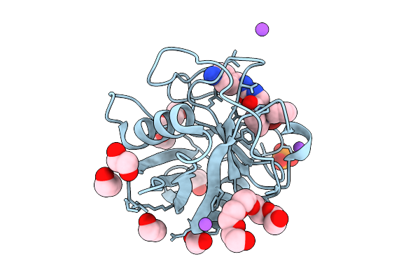 |
Scaffold-Ligand Complex
Organism: Homo sapiens
Method: X-RAY DIFFRACTION Resolution:1.45 Å Release Date: 2025-11-05 Classification: ISOMERASE Ligands: 9CK, EDO, 1PE, PGE, PO4, NA |
Organism: Homo sapiens
Method: X-RAY DIFFRACTION
Release Date: 2025-11-05
Ligands: 9CK, EDO, 1PE, PGE, PO4, NA
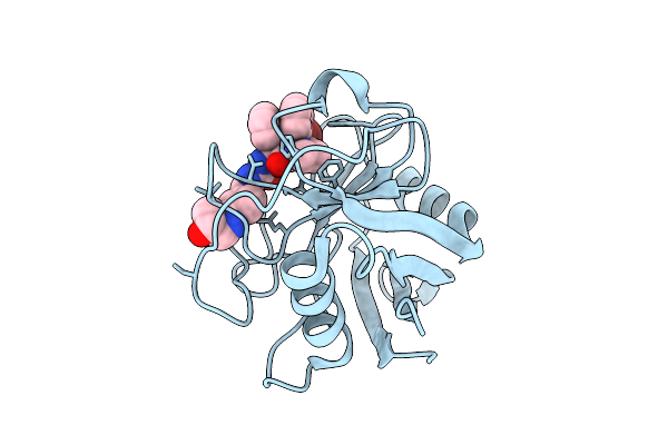 |
Structure Of Cypd In Complex With An Inhibitor.
Organism: Homo sapiens
Method: X-RAY DIFFRACTION Resolution:2.25 Å Release Date: 2025-11-05 Classification: ISOMERASE Ligands: A1IU3 |
Organism: Homo sapiens
Method: X-RAY DIFFRACTION
Release Date: 2025-11-05
Ligands: A1IU3
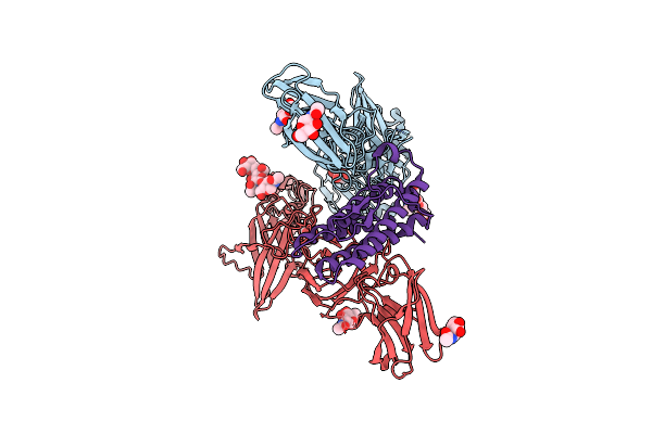 |
Cryo-Em Structure Of Murine Thrombopoietin Receptor Ectodomain In Complex With Tpo
Organism: Mus musculus, Saccharomyces cerevisiae
Method: ELECTRON MICROSCOPY Release Date: 2024-02-07 Classification: CYTOKINE/RECEPTOR Ligands: NAG, MAN |
Organism: Mus musculus, Saccharomyces cerevisiae
Method: ELECTRON MICROSCOPY
Release Date: 2024-02-07
Ligands: NAG, MAN
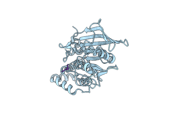 |
Crystal Structure Of Ptp1B D181A/Q262A/C215A Phosphatase Domain With Tyk2 Activation Loop Phosphopeptide
Organism: Homo sapiens
Method: X-RAY DIFFRACTION Resolution:2.99 Å Release Date: 2023-08-16 Classification: SIGNALING PROTEIN |
Organism: Homo sapiens
Method: X-RAY DIFFRACTION
Release Date: 2023-08-16
 |
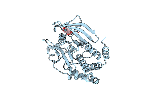
Ptp1B In Complex With 182
Organism: Homo sapiens
Method: X-RAY DIFFRACTION Resolution:1.55 Å Release Date: 2023-08-02 Classification: HYDROLASE/INHIBITOR Ligands: CL, NA, V2O, EDO |
Organism: Homo sapiens
Method: X-RAY DIFFRACTION
Release Date: 2023-08-02
Ligands: CL, NA, V2O, EDO
 |
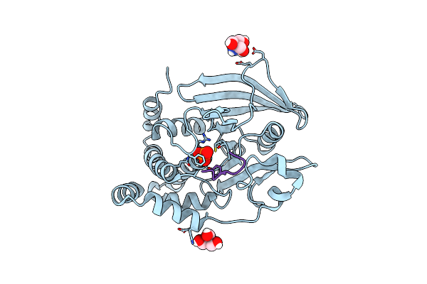
Crystal Structure Of Apo Ptp1B D181A/Q262A Phosphatase Domain
Organism: Homo sapiens
Method: X-RAY DIFFRACTION Resolution:1.60 Å Release Date: 2023-07-05 Classification: SIGNALING PROTEIN Ligands: MES |
Organism: Homo sapiens
Method: X-RAY DIFFRACTION
Release Date: 2023-07-05
Ligands: MES
 |
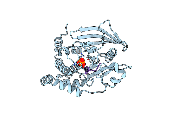
Crystal Structure Of Ptp1B D181A/Q262A Phosphatase Domain In Complex With A Jak1 Activation Loop Phosphopeptide
Organism: Homo sapiens
Method: X-RAY DIFFRACTION Resolution:2.30 Å Release Date: 2023-07-05 Classification: SIGNALING PROTEIN Ligands: PO4, TRS |
Organism: Homo sapiens
Method: X-RAY DIFFRACTION
Release Date: 2023-07-05
Ligands: PO4, TRS
 |
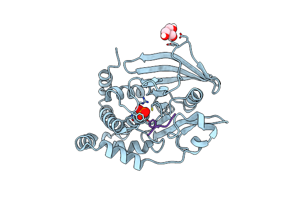
Crystal Structure Of Ptp1B D181A/Q262A Phosphatase Domain With Jak2 Activation Loop Phosphopeptide
Organism: Homo sapiens
Method: X-RAY DIFFRACTION Resolution:2.10 Å Release Date: 2023-07-05 Classification: SIGNALING PROTEIN Ligands: PO4 |
Organism: Homo sapiens
Method: X-RAY DIFFRACTION
Release Date: 2023-07-05
Ligands: PO4
 |
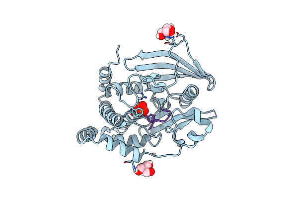
Crystal Structure Of Ptp1B D181A/Q262A Phosphatase Domain With A Jak3 Activation Loop Phosphopeptide
Organism: Homo sapiens
Method: X-RAY DIFFRACTION Resolution:2.35 Å Release Date: 2023-07-05 Classification: SIGNALING PROTEIN Ligands: PO4, TRS |
Organism: Homo sapiens
Method: X-RAY DIFFRACTION
Release Date: 2023-07-05
Ligands: PO4, TRS
 |
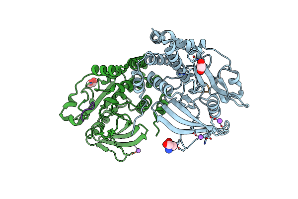
Crystal Structure Of Ptp1B D181A/Q262A Phosphatase Domain With Tyk2 Activation Loop Phosphopeptide
Organism: Homo sapiens
Method: X-RAY DIFFRACTION Resolution:2.15 Å Release Date: 2023-07-05 Classification: SIGNALING PROTEIN Ligands: PO4, TRS |
Organism: Homo sapiens
Method: X-RAY DIFFRACTION
Release Date: 2023-07-05
Ligands: PO4, TRS
 |
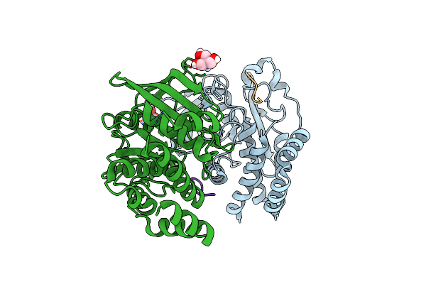
Crystal Structure Of Ptp1B D181A/Q262A/C215A Phosphatase Domain With A Jak2 Activation Loop Phosphopeptide
Organism: Homo sapiens
Method: X-RAY DIFFRACTION Resolution:2.10 Å Release Date: 2023-07-05 Classification: SIGNALING PROTEIN Ligands: TRS, EDO, CL, NA |
Organism: Homo sapiens
Method: X-RAY DIFFRACTION
Release Date: 2023-07-05
Ligands: TRS, EDO, CL, NA
 |
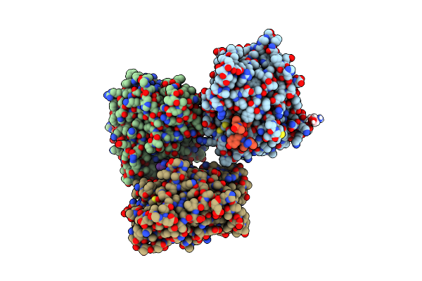
Crystal Structure Of Ptp1B D181A/Q262A/C215A Phosphatase Domain With Jak2 Activation Loop Phosphopeptide
Organism: Homo sapiens
Method: X-RAY DIFFRACTION Resolution:2.35 Å Release Date: 2023-07-05 Classification: SIGNALING PROTEIN Ligands: TRS |
Organism: Homo sapiens
Method: X-RAY DIFFRACTION
Release Date: 2023-07-05
Ligands: TRS
 |
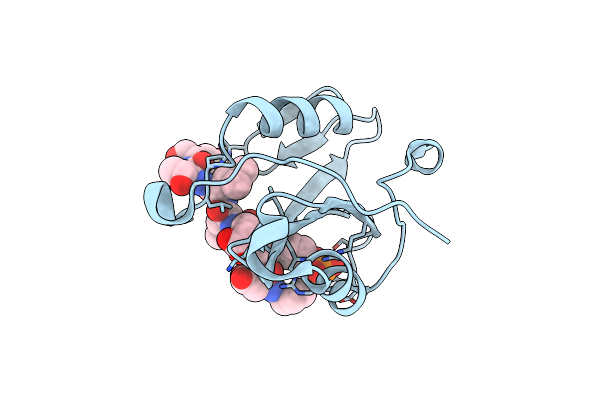
Crystal Structure Of Ptp1B D181A/Q262A/C215A Phosphatase Domain With Monophosphorylated Jak2 Activation Loop Phosphopeptide
Organism: Homo sapiens
Method: X-RAY DIFFRACTION Resolution:3.10 Å Release Date: 2023-07-05 Classification: CYTOKINE/HYDROLASE Ligands: TRS |
Organism: Homo sapiens
Method: X-RAY DIFFRACTION
Release Date: 2023-07-05
Ligands: TRS
 |
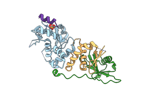
Crystal Structure Of The E372K Lnk Sh2 Domain Mutant In Complex With A Jak2 Py813 Phosphopeptide
Organism: Mus musculus, Homo sapiens
Method: X-RAY DIFFRACTION Resolution:1.65 Å Release Date: 2023-05-31 Classification: SIGNALING PROTEIN Ligands: CL |
Organism: Mus musculus, Homo sapiens
Method: X-RAY DIFFRACTION
Release Date: 2023-05-31
Ligands: CL
 |
Crystal Structure Of Socs2/Elonginb/Elonginc Bound To A Non-Canonical Peptide That Enhances Phospho-Peptide Binding
Organism: Homo sapiens, Synthetic construct
Method: X-RAY DIFFRACTION Resolution:3.19 Å Release Date: 2021-10-13 Classification: SIGNALING PROTEIN Ligands: SO4 |
Organism: Homo sapiens, Synthetic construct
Method: X-RAY DIFFRACTION
Release Date: 2021-10-13
Ligands: SO4
 |
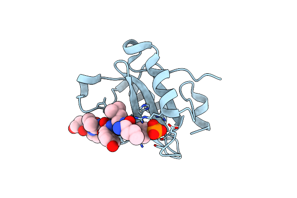
Crystal Structure Of The Lnk Sh2 Domain In Complex With A Jak2 Py813 Phosphopeptide
Organism: Mus musculus, Homo sapiens
Method: X-RAY DIFFRACTION Resolution:1.90 Å Release Date: 2021-10-06 Classification: SIGNALING PROTEIN |
Organism: Mus musculus, Homo sapiens
Method: X-RAY DIFFRACTION
Release Date: 2021-10-06
 |
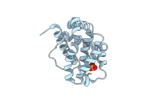
Crystal Structure Of The Lnk Sh2 Domain In Complex With An Epor Py454 Phosphopeptide
Organism: Mus musculus, Homo sapiens
Method: X-RAY DIFFRACTION Resolution:2.30 Å Release Date: 2021-10-06 Classification: SIGNALING PROTEIN |
Organism: Mus musculus, Homo sapiens
Method: X-RAY DIFFRACTION
Release Date: 2021-10-06
 |
Trichuris Suis Bcl-2
Organism: Trichuris suis
Method: X-RAY DIFFRACTION Resolution:1.60 Å Release Date: 2020-10-07 Classification: APOPTOSIS Ligands: CL, SO4 |
Organism: Trichuris suis
Method: X-RAY DIFFRACTION
Release Date: 2020-10-07
Ligands: CL, SO4
 |
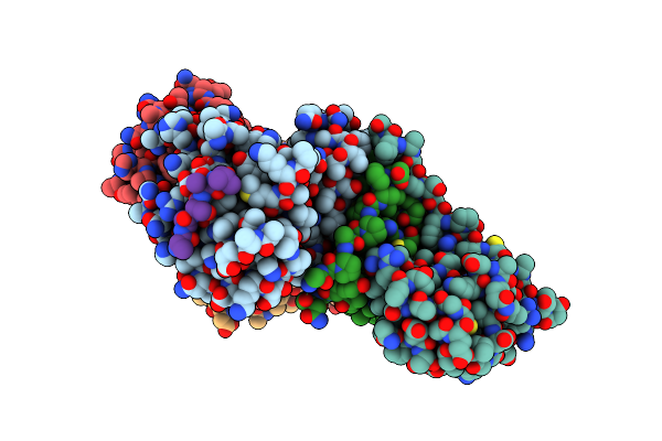
Aspartyl/Asparaginyl Beta-Hydroxylase (Asph)Oxygenase And Tpr Domains In Complex With Manganese, N-Oxalylglycine And Cyclic Peptide Substrate Mimic Of Factor X
Organism: Homo sapiens
Method: X-RAY DIFFRACTION Resolution:2.29 Å Release Date: 2019-05-08 Classification: OXIDOREDUCTASE Ligands: MN, OGA |
Organism: Homo sapiens
Method: X-RAY DIFFRACTION
Release Date: 2019-05-08
Ligands: MN, OGA
 |
Crystal Structure Of Socs1 In Complex With Elonginb And Elonginc
Organism: Homo sapiens, Xenopus laevis
Method: X-RAY DIFFRACTION Resolution:3.11 Å Release Date: 2018-05-02 Classification: SIGNALING PROTEIN Ligands: HOH |
Organism: Homo sapiens, Xenopus laevis
Method: X-RAY DIFFRACTION
Release Date: 2018-05-02
Ligands: HOH