

Search Count: 17
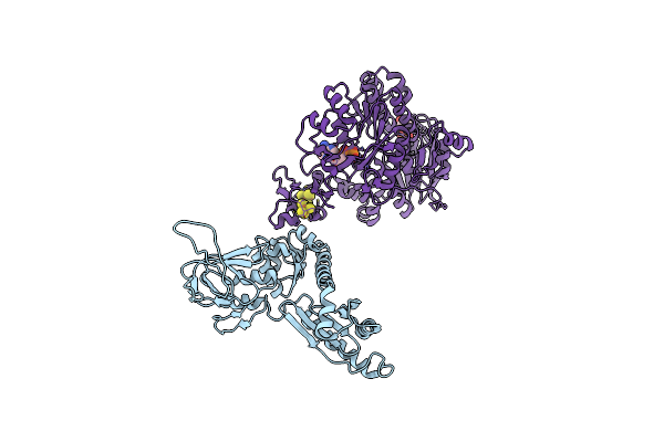 |
Model Of Ribosome-Bound Archaeal Pelota And Abce1
Organism: Thermococcus kodakarensis, Pyrococcus furiosus
Method: ELECTRON MICROSCOPY Release Date: 2012-02-22 Classification: TRANSLATION/TRANSPORT PROTEIN Ligands: ADP, SF4 |
Organism: Thermococcus kodakarensis, Pyrococcus furiosus
Method: ELECTRON MICROSCOPY
Release Date: 2012-02-22
Ligands: ADP, SF4
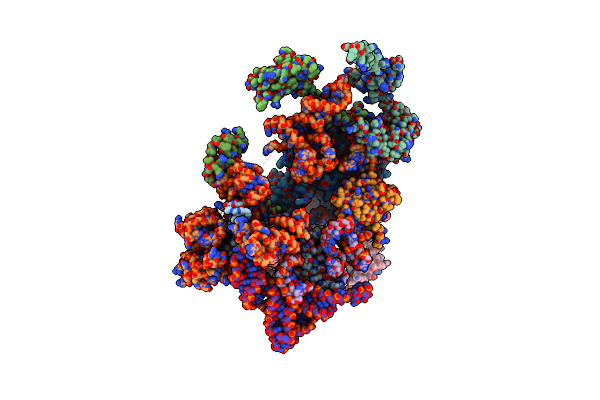 |
Models Of Ribosome-Bound Dom34P And Rli1P And Their Ribosomal Binding Partners
Organism: Saccharomyces cerevisiae
Method: ELECTRON MICROSCOPY Release Date: 2012-02-22 Classification: RIBOSOME Ligands: MG, ATP, SF4 |
Organism: Saccharomyces cerevisiae
Method: ELECTRON MICROSCOPY
Release Date: 2012-02-22
Ligands: MG, ATP, SF4
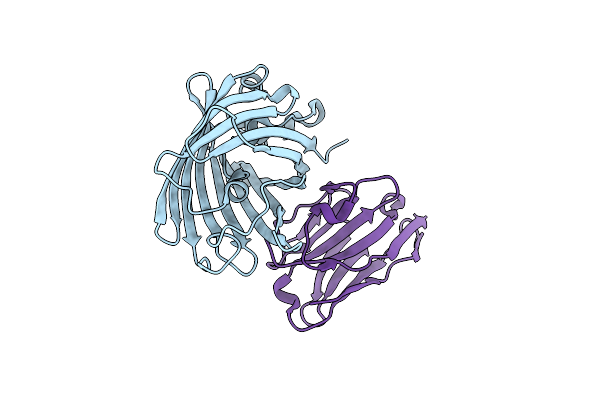 |
Green Fluorescent Protein Bound To Minimizer Nanobody
Organism: Aequorea victoria, Camelus dromedarius
Method: X-RAY DIFFRACTION Resolution:1.61 Å Release Date: 2009-12-08 Classification: FLUORESCENT PROTEIN/IMMUNE SYSTEM |
Organism: Aequorea victoria, Camelus dromedarius
Method: X-RAY DIFFRACTION
Release Date: 2009-12-08
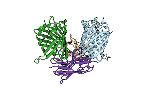 |
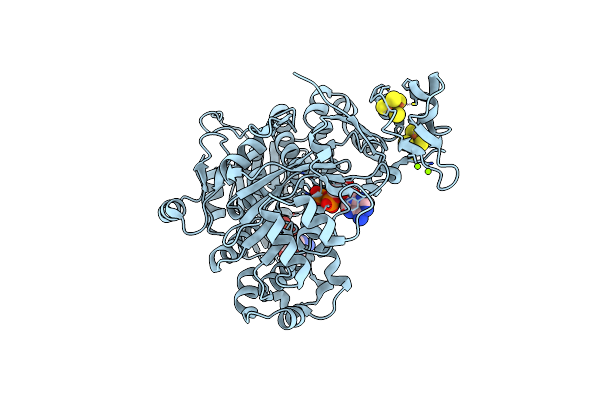
Green Fluorescent Protein Bound To Enhancer Nanobody
Organism: Aequorea victoria, Camelus dromedarius
Method: X-RAY DIFFRACTION Resolution:2.15 Å Release Date: 2009-12-08 Classification: LUMINESCENT PROTEIN/IMMUNE SYSTEM |
Organism: Aequorea victoria, Camelus dromedarius
Method: X-RAY DIFFRACTION
Release Date: 2009-12-08
 |
Structure Of The Complete Abce1/Rnaase-L Inhibitor Protein From Pyrococcus Abysii
Organism: Pyrococcus abyssi
Method: X-RAY DIFFRACTION Resolution:2.80 Å Release Date: 2007-12-25 Classification: HYDROLYASE/TRANSLATION Ligands: MG, SF4, ADP |
Organism: Pyrococcus abyssi
Method: X-RAY DIFFRACTION
Release Date: 2007-12-25
Ligands: MG, SF4, ADP
 |
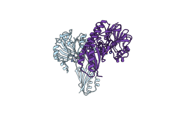
Rnase-L Inhibitor
Organism: Pyrococcus furiosus
Method: X-RAY DIFFRACTION Resolution:1.90 Å Release Date: 2005-04-19 Classification: hydrolyase/translation Ligands: MG, ADP |
Organism: Pyrococcus furiosus
Method: X-RAY DIFFRACTION
Release Date: 2005-04-19
Ligands: MG, ADP
 |
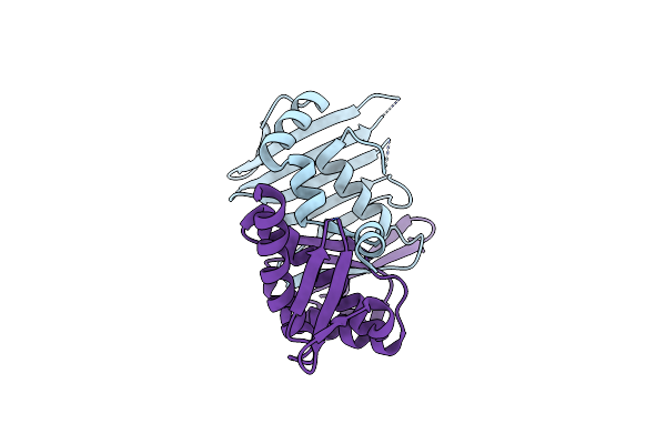
Crystal Structure Of Mre11-3
Organism: Pyrococcus furiosus
Method: X-RAY DIFFRACTION Resolution:2.30 Å Release Date: 2004-08-10 Classification: REPLICATION Ligands: MN |
Organism: Pyrococcus furiosus
Method: X-RAY DIFFRACTION
Release Date: 2004-08-10
Ligands: MN
 |
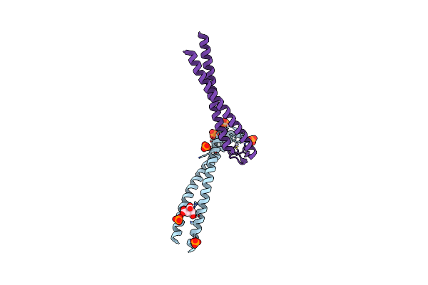
The Rad50 Signature Motif: Essential To Atp Binding And Biological Function
Organism: Pyrococcus furiosus
Method: X-RAY DIFFRACTION Resolution:2.10 Å Release Date: 2003-11-25 Classification: DNA REPAIR |
Organism: Pyrococcus furiosus
Method: X-RAY DIFFRACTION
Release Date: 2003-11-25
 |
Rad50 Coiled-Coil Zn Hook
Organism: Pyrococcus furiosus
Method: X-RAY DIFFRACTION Resolution:2.20 Å Release Date: 2002-08-28 Classification: REPLICATION Ligands: HG, PO4, CIT |
Organism: Pyrococcus furiosus
Method: X-RAY DIFFRACTION
Release Date: 2002-08-28
Ligands: HG, PO4, CIT
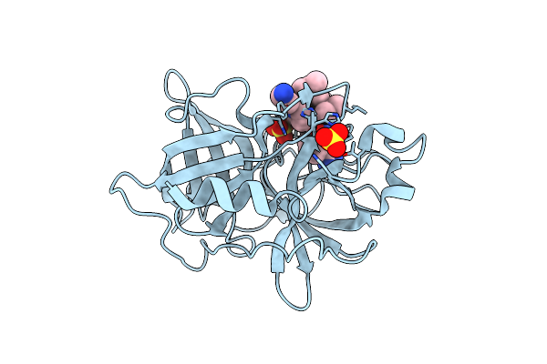 |
Urokinase Plasminogen Activator B Chain-Uki-1D Complex
Organism: Homo sapiens
Method: X-RAY DIFFRACTION Resolution:2.60 Å Release Date: 2001-07-06 Classification: HYDROLASE Ligands: SO4, UKP |
Organism: Homo sapiens
Method: X-RAY DIFFRACTION
Release Date: 2001-07-06
Ligands: SO4, UKP
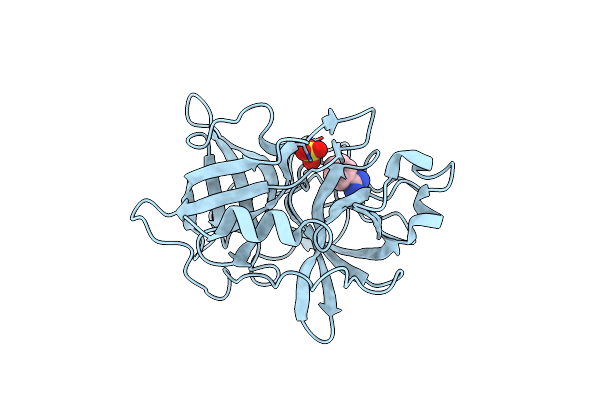 |
Urokinase Plasminogen Activator B-Chain-Benzamidine Complex
Organism: Homo sapiens
Method: X-RAY DIFFRACTION Resolution:1.80 Å Release Date: 2001-06-15 Classification: HYDROLASE Ligands: SO4, BEN |
Organism: Homo sapiens
Method: X-RAY DIFFRACTION
Release Date: 2001-06-15
Ligands: SO4, BEN
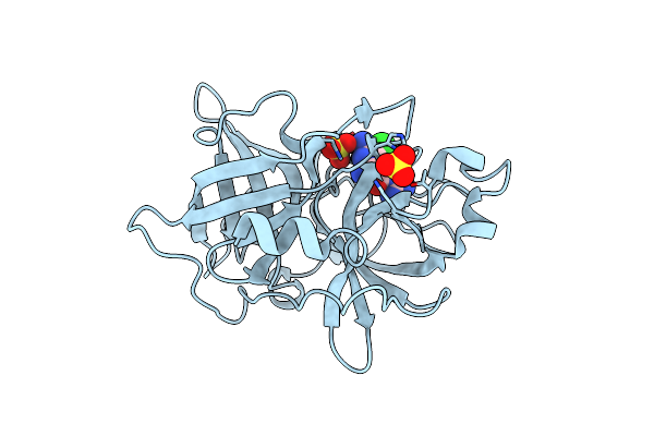 |
Urokinase Plasminogen Activator B-Chain-Amiloride Complex
Organism: Homo sapiens
Method: X-RAY DIFFRACTION Resolution:2.10 Å Release Date: 2001-06-15 Classification: HYDROLASE Ligands: SO4, AMR |
Organism: Homo sapiens
Method: X-RAY DIFFRACTION
Release Date: 2001-06-15
Ligands: SO4, AMR
 |
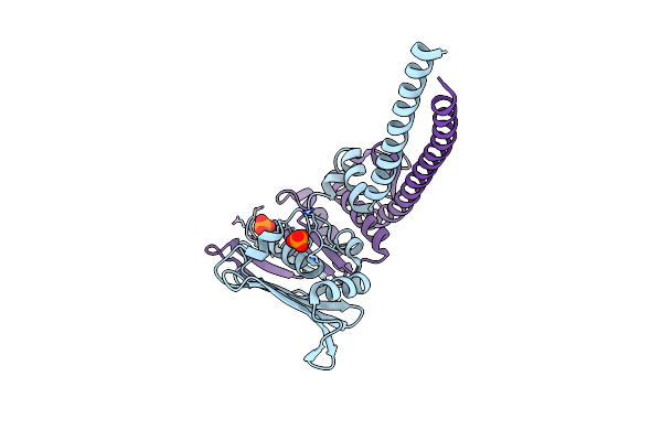
Crystal Structure Of P. Furiosus Mre11 With Manganese And Damp
Organism: Pyrococcus furiosus
Method: X-RAY DIFFRACTION Resolution:2.20 Å Release Date: 2001-05-30 Classification: REPLICATION Ligands: MN, SO4, D5M, PO4 |
Organism: Pyrococcus furiosus
Method: X-RAY DIFFRACTION
Release Date: 2001-05-30
Ligands: MN, SO4, D5M, PO4
 |
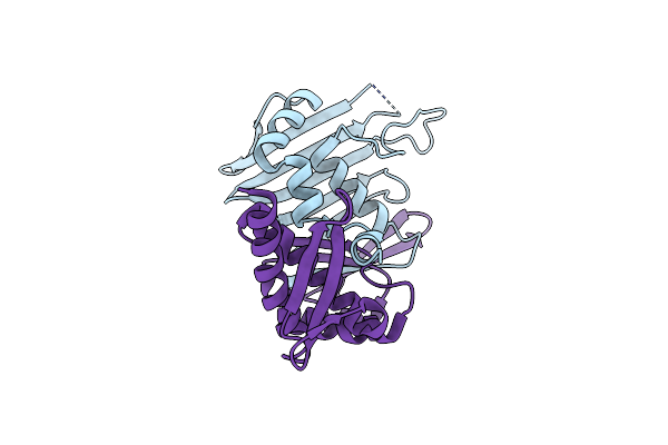
Crystal Structure Of The P. Furiosus Rad50 Atpase Domain
Organism: Pyrococcus furiosus
Method: X-RAY DIFFRACTION Resolution:3.02 Å Release Date: 2001-05-30 Classification: REPLICATION Ligands: PO4 |
Organism: Pyrococcus furiosus
Method: X-RAY DIFFRACTION
Release Date: 2001-05-30
Ligands: PO4
 |
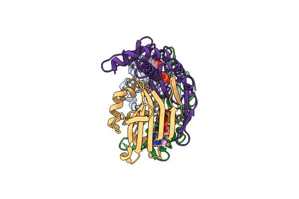
Crystal Structure Of Atp-Free Rad50 Abc-Atpase
Organism: Pyrococcus furiosus
Method: X-RAY DIFFRACTION Resolution:1.60 Å Release Date: 2000-08-02 Classification: REPLICATION |
Organism: Pyrococcus furiosus
Method: X-RAY DIFFRACTION
Release Date: 2000-08-02
 |
Crystal Structure Of Rad50 Abc-Atpase
Organism: Pyrococcus furiosus
Method: X-RAY DIFFRACTION Resolution:1.60 Å Release Date: 2000-08-02 Classification: REPLICATION Ligands: MG, ATP |
Organism: Pyrococcus furiosus
Method: X-RAY DIFFRACTION
Release Date: 2000-08-02
Ligands: MG, ATP
 |
Human Coagulation Factor Ixa In Complex With P-Amino Benzamidine
Organism: Homo sapiens
Method: X-RAY DIFFRACTION Resolution:2.80 Å Release Date: 1999-09-01 Classification: COAGULATION FACTOR Ligands: CA, PBZ, TBU |
Organism: Homo sapiens
Method: X-RAY DIFFRACTION
Release Date: 1999-09-01
Ligands: CA, PBZ, TBU