

Search Count: 24
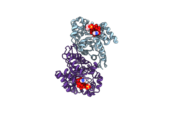 |
Fgd2 (Rv0132C) From Mycobacterium Tuberculosis Crystallised With Anderson-Evans Polyoxotungstate
Organism: Mycobacterium tuberculosis h37rv
Method: X-RAY DIFFRACTION Resolution:1.45 Å Release Date: 2025-06-25 Classification: OXIDOREDUCTASE Ligands: TEW, IMD, MES |
Organism: Mycobacterium tuberculosis h37rv
Method: X-RAY DIFFRACTION
Release Date: 2025-06-25
Ligands: TEW, IMD, MES
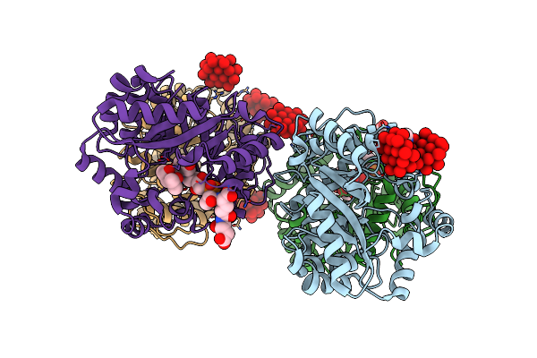 |
Fgd2 (Rv0132C) From Mycobacterium Tuberculosis With Cofactor F420 Crystallised With Anderson-Evans Polyoxotungstate
Organism: Mycobacterium tuberculosis h37rv
Method: X-RAY DIFFRACTION Resolution:2.35 Å Release Date: 2025-06-25 Classification: OXIDOREDUCTASE Ligands: TEW, IMD, MPD, F42 |
Organism: Mycobacterium tuberculosis h37rv
Method: X-RAY DIFFRACTION
Release Date: 2025-06-25
Ligands: TEW, IMD, MPD, F42
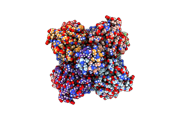 |
Cryo-Em Structure Of The C Terminal Region Of Ptx3 With A Section Of Coiled-Coil
Organism: Homo sapiens
Method: ELECTRON MICROSCOPY Release Date: 2025-01-22 Classification: IMMUNE SYSTEM Ligands: NAG |
Organism: Homo sapiens
Method: ELECTRON MICROSCOPY
Release Date: 2025-01-22
Ligands: NAG
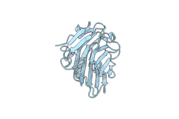 |
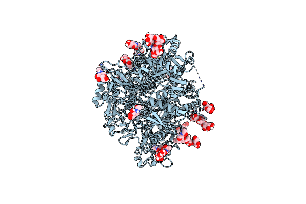
Crystal Structure Of Human Ptx3 C-Terminal Domain
Organism: Homo sapiens
Method: X-RAY DIFFRACTION Resolution:2.43 Å Release Date: 2024-07-24 Classification: IMMUNE SYSTEM |
Organism: Homo sapiens
Method: X-RAY DIFFRACTION
Release Date: 2024-07-24
 |
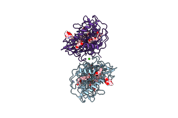
Cryoem Structure Of The Ectodomain Of Human Pla2R
Organism: Homo sapiens
Method: ELECTRON MICROSCOPY Release Date: 2022-07-20 Classification: MEMBRANE PROTEIN Ligands: NAG |
Organism: Homo sapiens
Method: ELECTRON MICROSCOPY
Release Date: 2022-07-20
Ligands: NAG
 |
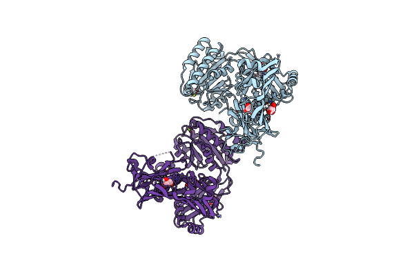
The Crystal Structure Of Trichuris Muris P43
Organism: Trichuris muris
Method: X-RAY DIFFRACTION Resolution:1.65 Å Release Date: 2019-06-12 Classification: UNKNOWN FUNCTION Ligands: NAG, ACT, P6G, CA, PEG |
Organism: Trichuris muris
Method: X-RAY DIFFRACTION
Release Date: 2019-06-12
Ligands: NAG, ACT, P6G, CA, PEG
 |
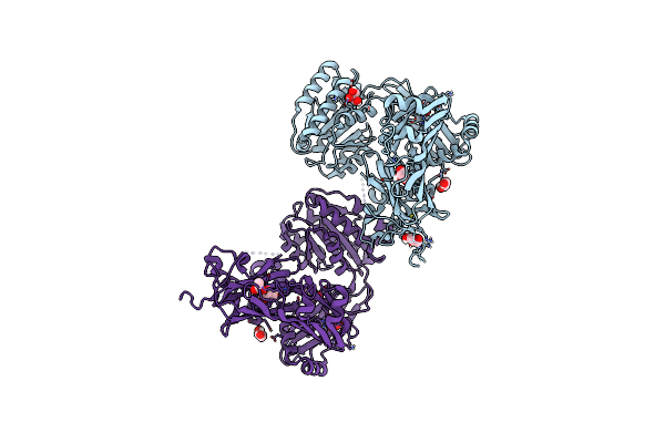
Inter-Alpha-Inhibitor Heavy Chain 1, Wild Type
Organism: Homo sapiens
Method: X-RAY DIFFRACTION Resolution:2.34 Å Release Date: 2019-02-27 Classification: STRUCTURAL PROTEIN Ligands: MG, GOL |
Organism: Homo sapiens
Method: X-RAY DIFFRACTION
Release Date: 2019-02-27
Ligands: MG, GOL
 |
Inter-Alpha-Inhibitor Heavy Chain 1, D298A
Organism: Homo sapiens
Method: X-RAY DIFFRACTION Resolution:2.20 Å Release Date: 2019-02-27 Classification: STRUCTURAL PROTEIN Ligands: GOL, ACT |
Organism: Homo sapiens
Method: X-RAY DIFFRACTION
Release Date: 2019-02-27
Ligands: GOL, ACT
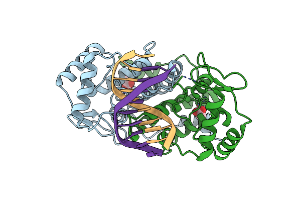 |
Icp4 Dna-Binding Domain, Lacking Intrinsically Disordered Region, In Complex With 12Mer Dna Duplex From Its Own Promoter
Organism: Human herpesvirus 1 (strain 17), Human herpesvirus 1
Method: X-RAY DIFFRACTION Resolution:2.12 Å Release Date: 2017-05-24 Classification: TRANSCRIPTION Ligands: ACY, CL |
Organism: Human herpesvirus 1 (strain 17), Human herpesvirus 1
Method: X-RAY DIFFRACTION
Release Date: 2017-05-24
Ligands: ACY, CL
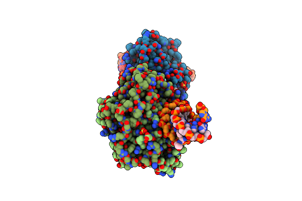 |
Icp4 Dna-Binding Domain In Complex With 19Mer Dna Duplex From Its Own Promoter
Organism: Human herpesvirus 1, Human herpesvirus 1
Method: X-RAY DIFFRACTION Resolution:2.28 Å Release Date: 2017-05-24 Classification: TRANSCRIPTION Ligands: MG, SO4, CL |
Organism: Human herpesvirus 1, Human herpesvirus 1
Method: X-RAY DIFFRACTION
Release Date: 2017-05-24
Ligands: MG, SO4, CL
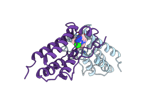 |
Bivalent Binding To Bet Bromodomains
Organism: Homo sapiens
Method: X-RAY DIFFRACTION Resolution:2.01 Å Release Date: 2016-09-28 Classification: TRANSCRIPTION Ligands: ETU |
Organism: Homo sapiens
Method: X-RAY DIFFRACTION
Release Date: 2016-09-28
Ligands: ETU
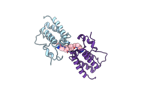 |
Bivalent Binding To Bet Bromodomains
Organism: Homo sapiens
Method: X-RAY DIFFRACTION Resolution:1.49 Å Release Date: 2016-09-28 Classification: TRANSCRIPTION Ligands: K6K |
Organism: Homo sapiens
Method: X-RAY DIFFRACTION
Release Date: 2016-09-28
Ligands: K6K
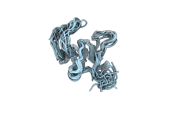 |
Solution Structure Of The Link Module Of Human Tsg-6 In Presence Of A Chondroitin 4-Sulfate Hexasaccharide
|
Organism: Homo sapiens
Method: SOLUTION NMR
Release Date: 2016-01-13
 |
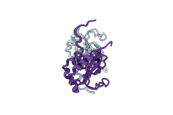
The Structure Of The Folded Domain Of The Signature Multifunctional Protein Icp27 From Herpes Simplex Virus-1 Reveals An Intertwined Dimer.
Organism: Human herpesvirus 1 (strain 17)
Method: X-RAY DIFFRACTION Resolution:1.92 Å Release Date: 2015-06-17 Classification: VIRAL PROTEIN Ligands: ZN |
Organism: Human herpesvirus 1 (strain 17)
Method: X-RAY DIFFRACTION
Release Date: 2015-06-17
Ligands: ZN
 |
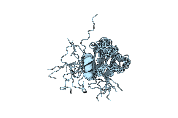
Solution Structure Of Agr2 Residues 41-175
|
Organism: Homo sapiens
Method: SOLUTION NMR
Release Date: 2013-01-09
 |
Solution Structure Of E60A Mutant Agr2
|
Organism: Homo sapiens
Method: SOLUTION NMR
Release Date: 2013-01-09
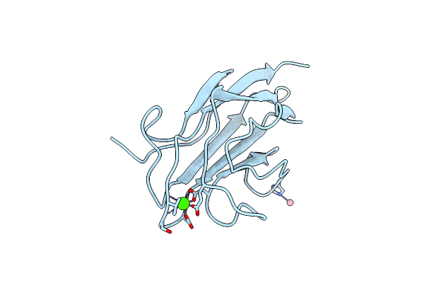 |
X-Ray Structure Of Cub_C Domain From Tsg-6
Organism: Homo sapiens
Method: X-RAY DIFFRACTION Resolution:2.30 Å Release Date: 2010-09-01 Classification: CELL ADHESION Ligands: CO, CA |
Organism: Homo sapiens
Method: X-RAY DIFFRACTION
Release Date: 2010-09-01
Ligands: CO, CA
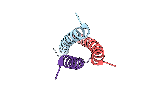 |
Crystal Structure Of Mutant Coiled Coil Gcn4 Leucine Zipper
Organism: Saccharomyces cerevisiae
Method: X-RAY DIFFRACTION Resolution:2.00 Å Release Date: 2010-03-09 Classification: TRANSCRIPTION |
Organism: Saccharomyces cerevisiae
Method: X-RAY DIFFRACTION
Release Date: 2010-03-09
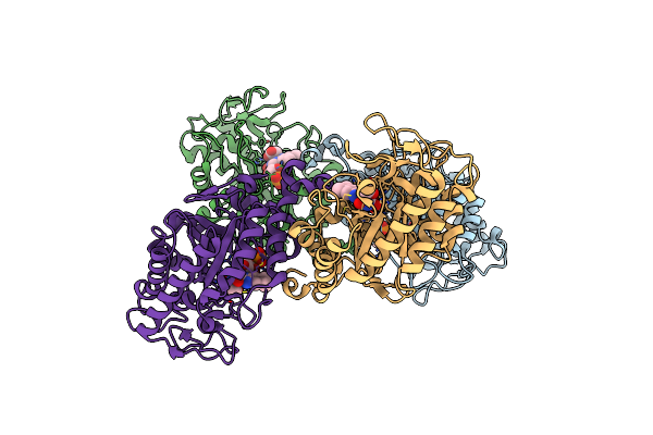 |
Crystal Structure Of The Thermostable Old Yellow Enzyme From Thermoanaerobacter Pseudethanolicus E39
Organism: Thermoanaerobacter pseudethanolicus atcc 33223
Method: X-RAY DIFFRACTION Resolution:1.60 Å Release Date: 2009-12-08 Classification: OXIDOREDUCTASE Ligands: FMN, ACT |
Organism: Thermoanaerobacter pseudethanolicus atcc 33223
Method: X-RAY DIFFRACTION
Release Date: 2009-12-08
Ligands: FMN, ACT
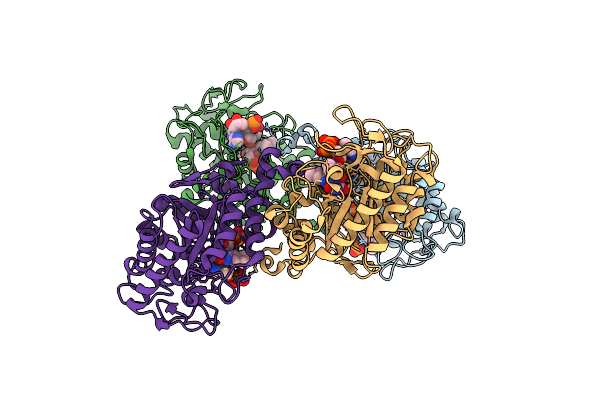 |
Crystal Structure Of The Thermostable Nadh4-Bound Old Yellow Enzyme From Thermoanaerobacter Pseudethanolicus E39
Organism: Thermoanaerobacter pseudethanolicus atcc 33223
Method: X-RAY DIFFRACTION Resolution:1.80 Å Release Date: 2009-12-08 Classification: OXIDOREDUCTASE Ligands: FMN, TXD |
Organism: Thermoanaerobacter pseudethanolicus atcc 33223
Method: X-RAY DIFFRACTION
Release Date: 2009-12-08
Ligands: FMN, TXD