

Search Count: 23
 |
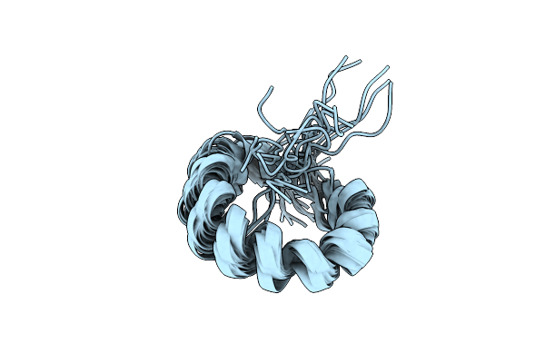
Folded Alpha Helical De Novo Proteins From Apilactobacillus Kunkeei
Organism: Apilactobacillus kunkeei
Method: SOLUTION NMR Release Date: 2024-02-21 Classification: UNKNOWN FUNCTION |
Organism: Apilactobacillus kunkeei
Method: SOLUTION NMR
Release Date: 2024-02-21
 |
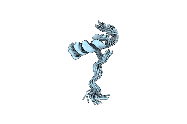
Folded Alpha Helical De Novo Proteins From Apilactobacillus Kunkeei
Organism: Apilactobacillus kunkeei
Method: SOLUTION NMR Release Date: 2024-02-21 Classification: UNKNOWN FUNCTION |
Organism: Apilactobacillus kunkeei
Method: SOLUTION NMR
Release Date: 2024-02-21
 |
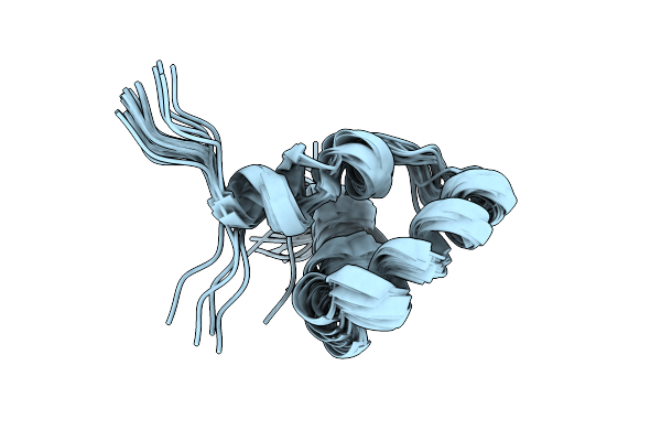
Folded Alpha Helical De Novo Proteins From Apilactobacillus Kunkeei
Organism: Apilactobacillus kunkeei
Method: SOLUTION NMR Release Date: 2024-02-21 Classification: UNKNOWN FUNCTION |
Organism: Apilactobacillus kunkeei
Method: SOLUTION NMR
Release Date: 2024-02-21
 |
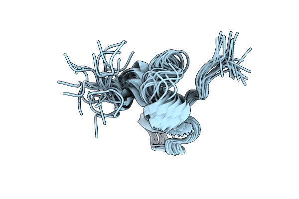
Structure And Folding Of A 600-Million-Year-Old Nuclear Coactivator Binding Domain Suggest Conservation Of Dynamic Properties
Organism: Homo sapiens
Method: SOLUTION NMR Release Date: 2022-04-20 Classification: DNA BINDING PROTEIN |
Organism: Homo sapiens
Method: SOLUTION NMR
Release Date: 2022-04-20
 |
Structure And Folding Of A 600-Million-Year-Old Nuclear Coactivator Binding Domain Suggest Conservation Of Dynamic Properties
Organism: Homo sapiens
Method: SOLUTION NMR Release Date: 2022-04-20 Classification: DNA BINDING PROTEIN |
Organism: Homo sapiens
Method: SOLUTION NMR
Release Date: 2022-04-20
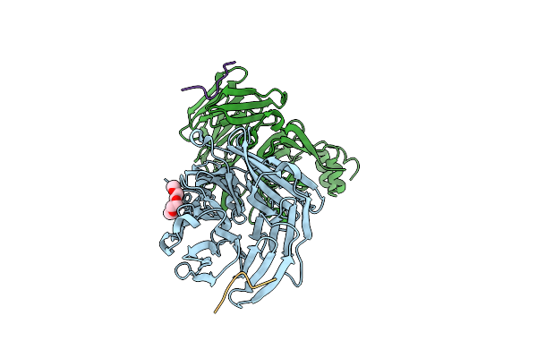 |
Clathrin Heavy Chain N-Terminal Domain Complexed With Peptide From Protein Mu-Ns Of Reovirus Type 1
Organism: Homo sapiens, Mammalian orthoreovirus 1 lang
Method: X-RAY DIFFRACTION Resolution:1.97 Å Release Date: 2022-03-02 Classification: TRANSPORT PROTEIN Ligands: PG4 |
Organism: Homo sapiens, Mammalian orthoreovirus 1 lang
Method: X-RAY DIFFRACTION
Release Date: 2022-03-02
Ligands: PG4
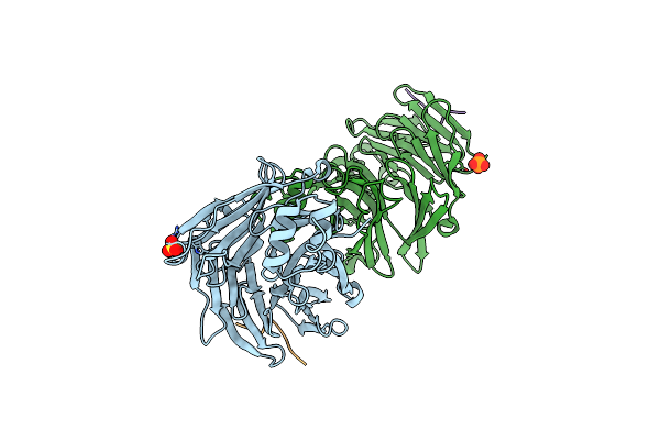 |
Clathrin Heavy Chain N-Terminal Domain Bound To Non Structured Protein 3 From Eastern Equine Encephalitis Virus
Organism: Homo sapiens, Eastern equine encephalitis virus
Method: X-RAY DIFFRACTION Resolution:1.97 Å Release Date: 2022-03-02 Classification: TRANSPORT PROTEIN Ligands: SO4, PO4 |
Organism: Homo sapiens, Eastern equine encephalitis virus
Method: X-RAY DIFFRACTION
Release Date: 2022-03-02
Ligands: SO4, PO4
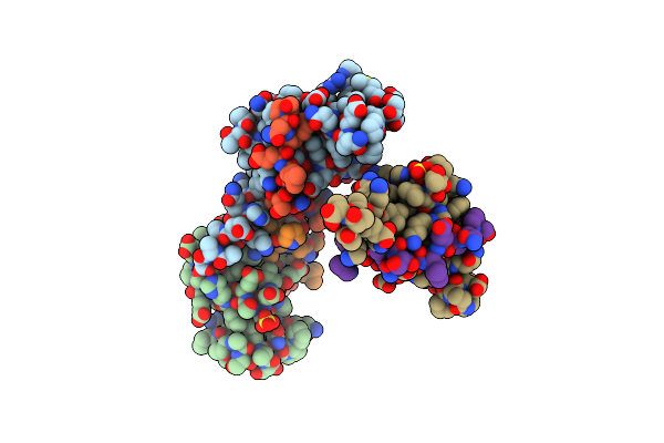 |
Crystal Structure Of C-Terminal Domain Of Pabpc1 In Complex With Nucleoprotein From Human Coronavirus 229E
Organism: Homo sapiens, Human coronavirus 229e
Method: X-RAY DIFFRACTION Resolution:1.93 Å Release Date: 2022-03-02 Classification: TRANSCRIPTION Ligands: SO4, GOL |
Organism: Homo sapiens, Human coronavirus 229e
Method: X-RAY DIFFRACTION
Release Date: 2022-03-02
Ligands: SO4, GOL
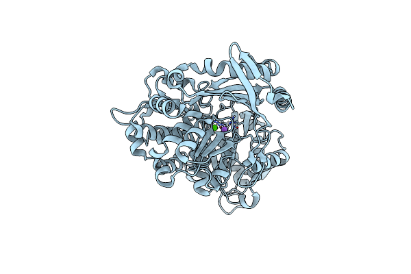 |
Structure Of Atlantic Herring (Clupea Harengus) Phosphoglucomutase 5 (Pgm5)
Organism: Clupea harengus
Method: X-RAY DIFFRACTION Resolution:2.25 Å Release Date: 2020-12-16 Classification: ISOMERASE Ligands: CA, NA |
Organism: Clupea harengus
Method: X-RAY DIFFRACTION
Release Date: 2020-12-16
Ligands: CA, NA
 |
Structure Of Baltic Herring (Clupea Harengus) Phosphoglucomutase 5 (Pgm5) With Bound Glucose-1-Phosphate
Organism: Clupea harengus
Method: X-RAY DIFFRACTION Resolution:1.95 Å Release Date: 2020-12-16 Classification: ISOMERASE Ligands: NI, CA, G1P, GOL, ACT, NA |
Organism: Clupea harengus
Method: X-RAY DIFFRACTION
Release Date: 2020-12-16
Ligands: NI, CA, G1P, GOL, ACT, NA
 |
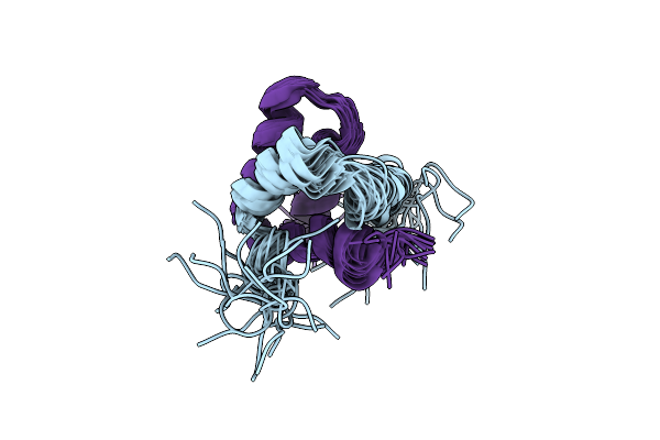
Structure Of Baltic Herring (Clupea Harengus) Phosphoglucomutase 5 (Pgm5)
Organism: Clupea harengus
Method: X-RAY DIFFRACTION Resolution:2.05 Å Release Date: 2020-12-16 Classification: ISOMERASE Ligands: NI, CA, NA, GOL, ACT |
Organism: Clupea harengus
Method: X-RAY DIFFRACTION
Release Date: 2020-12-16
Ligands: NI, CA, NA, GOL, ACT
 |
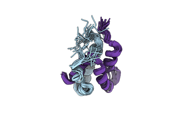
Structure And Dynamics Conspire In The Evolution Of Affinity Between Intrinsically Disordered Proteins
Organism: Homo sapiens
Method: SOLUTION NMR Release Date: 2018-10-31 Classification: DNA BINDING PROTEIN |
Organism: Homo sapiens
Method: SOLUTION NMR
Release Date: 2018-10-31
 |
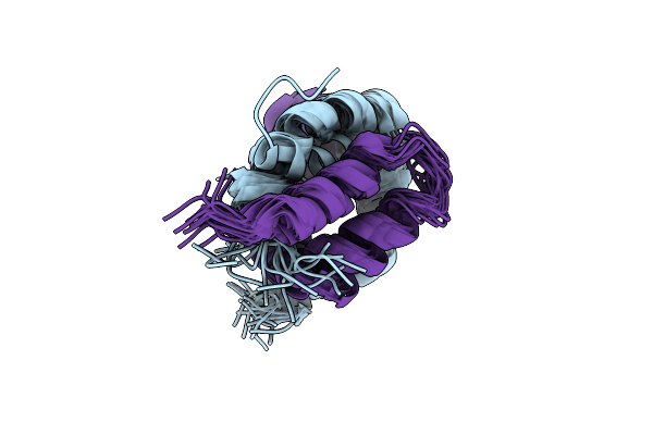
Structure And Dynamics Conspire In The Evolution Of Affinity Between Intrinsically Disordered Proteins
Organism: Homo sapiens
Method: SOLUTION NMR Release Date: 2018-10-31 Classification: DNA BINDING PROTEIN |
Organism: Homo sapiens
Method: SOLUTION NMR
Release Date: 2018-10-31
 |
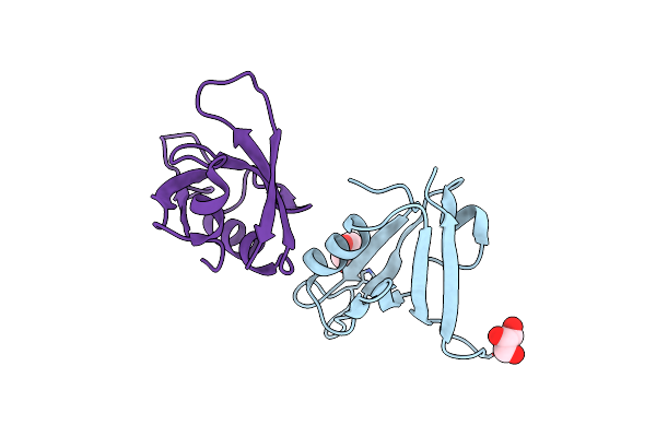
Structure And Dynamics Conspire In The Evolution Of Affinity Between Intrinsically Disordered Proteins
Organism: Homo sapiens
Method: SOLUTION NMR Release Date: 2018-10-31 Classification: DNA BINDING PROTEIN |
Organism: Homo sapiens
Method: SOLUTION NMR
Release Date: 2018-10-31
 |
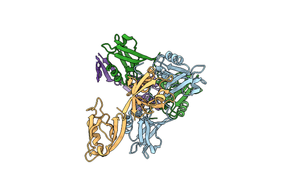
Influence Of Circular Permutation On The Folding Pathway Of A Pdz Domain
Organism: Homo sapiens
Method: X-RAY DIFFRACTION Resolution:2.30 Å Release Date: 2012-12-05 Classification: STRUCTURAL PROTEIN Ligands: TRS, GOL |
Organism: Homo sapiens
Method: X-RAY DIFFRACTION
Release Date: 2012-12-05
Ligands: TRS, GOL
 |
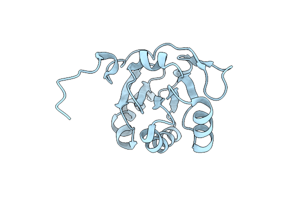
Crystal Structure Of Human Psd-95 Pdz1-2
Organism: Homo sapiens
Method: X-RAY DIFFRACTION Resolution:3.40 Å Release Date: 2012-03-21 Classification: SIGNALING PROTEIN |
Organism: Homo sapiens
Method: X-RAY DIFFRACTION
Release Date: 2012-03-21
 |
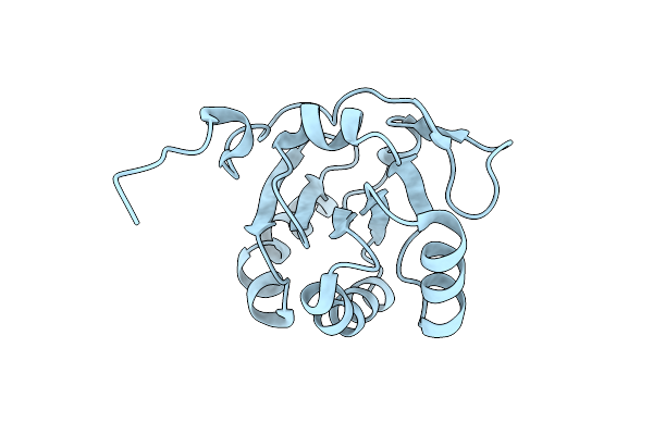
Crystal Structure Of The Periplasmic Soluble Domain Of Reduced Ccmg From Pseudomonas Aeruginosa
Organism: Pseudomonas aeruginosa
Method: X-RAY DIFFRACTION Resolution:1.75 Å Release Date: 2010-04-07 Classification: OXIDOREDUCTASE |
Organism: Pseudomonas aeruginosa
Method: X-RAY DIFFRACTION
Release Date: 2010-04-07
 |
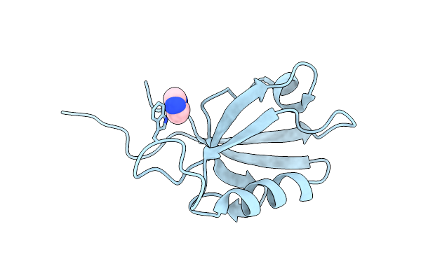
Crystal Structure Of The Periplasmic Soluble Domain Of Oxidized Ccmg From Pseudomonas Aeruginosa
Organism: Pseudomonas aeruginosa
Method: X-RAY DIFFRACTION Resolution:2.20 Å Release Date: 2010-04-07 Classification: OXIDOREDUCTASE |
Organism: Pseudomonas aeruginosa
Method: X-RAY DIFFRACTION
Release Date: 2010-04-07
 |
Crystal Structure Of The Sap97 Pdz2 I342W C378A Mutant Protein Domain
Organism: Homo sapiens
Method: X-RAY DIFFRACTION Resolution:2.00 Å Release Date: 2010-03-31 Classification: STRUCTURAL PROTEIN Ligands: NH4, IMD |
Organism: Homo sapiens
Method: X-RAY DIFFRACTION
Release Date: 2010-03-31
Ligands: NH4, IMD
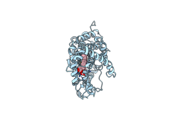 |
Structure Of The Histidine Tagged, Open Cytochrome P450 Eryk From S. Erythraea
Organism: Saccharopolyspora erythraea
Method: X-RAY DIFFRACTION Resolution:2.00 Å Release Date: 2009-07-21 Classification: OXIDOREDUCTASE Ligands: HEM |
Organism: Saccharopolyspora erythraea
Method: X-RAY DIFFRACTION
Release Date: 2009-07-21
Ligands: HEM