

Search Count: 63
 |
Crystal Structure Of Craf/Mek1 Complex With Plx4720 And Ch5126766
Organism: Homo sapiens
Method: X-RAY DIFFRACTION Resolution:2.91 Å Release Date: 2025-10-15 Classification: TRANSFERASE Ligands: 324, CHU, ANP |
Organism: Homo sapiens
Method: X-RAY DIFFRACTION
Release Date: 2025-10-15
Ligands: 324, CHU, ANP
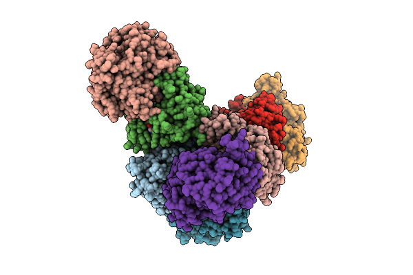 |
Crystal Structure Of Craf/Mek1 Complex With Plx4720, Ch5126766, And Amppnp
Organism: Homo sapiens
Method: X-RAY DIFFRACTION Resolution:3.50 Å Release Date: 2025-10-15 Classification: TRANSFERASE Ligands: 324, CHU, ANP |
Organism: Homo sapiens
Method: X-RAY DIFFRACTION
Release Date: 2025-10-15
Ligands: 324, CHU, ANP
 |
Cryo-Em Structure Of Craf/Mek1/14-3-3 Complex (Autoinhibited Conformation)
Organism: Homo sapiens, Spodoptera exigua
Method: ELECTRON MICROSCOPY Resolution:3.40 Å Release Date: 2025-09-10 Classification: TRANSFERASE Ligands: AGS, ZN, LCJ |
Organism: Homo sapiens, Spodoptera exigua
Method: ELECTRON MICROSCOPY
Release Date: 2025-09-10
Ligands: AGS, ZN, LCJ
 |
Cryo-Em Structure Of Craf/Mek1 Complex (Kinase Domain)
Organism: Homo sapiens
Method: ELECTRON MICROSCOPY Release Date: 2025-09-10 Classification: TRANSFERASE Ligands: AGS, MG, LCJ |
Organism: Homo sapiens
Method: ELECTRON MICROSCOPY
Release Date: 2025-09-10
Ligands: AGS, MG, LCJ
 |
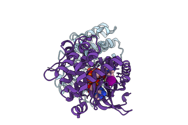
Cryo-Em Structure Of Craf/Mek1/14-3-3 Complex (Open Monomer Conformation, Craf Y340D/Y341D Mutant)
Organism: Homo sapiens, Spodoptera exigua
Method: ELECTRON MICROSCOPY Release Date: 2025-09-10 Classification: TRANSFERASE Ligands: AGS, MG, LCJ |
Organism: Homo sapiens, Spodoptera exigua
Method: ELECTRON MICROSCOPY
Release Date: 2025-09-10
Ligands: AGS, MG, LCJ
 |
Cryo-Em Structure Of Craf/Mek1 Complex (Kinase Domain, Craf Y340D/Y341D Mutant)
Organism: Homo sapiens
Method: ELECTRON MICROSCOPY Release Date: 2025-09-10 Classification: TRANSFERASE Ligands: AGS, MG, LCJ |
Organism: Homo sapiens
Method: ELECTRON MICROSCOPY
Release Date: 2025-09-10
Ligands: AGS, MG, LCJ
 |
Crystal Structure Of The Glycosyltransferase Domain Of Legionella Seta (Unbound)
Organism: Legionella pneumophila
Method: X-RAY DIFFRACTION Resolution:2.54 Å Release Date: 2025-02-19 Classification: TRANSFERASE Ligands: MG |
Organism: Legionella pneumophila
Method: X-RAY DIFFRACTION
Release Date: 2025-02-19
Ligands: MG
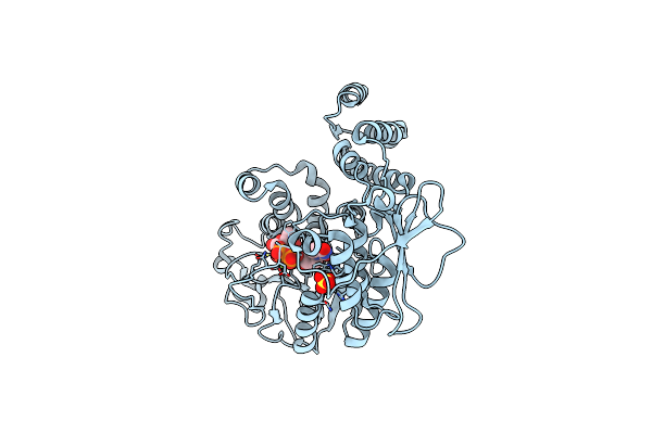 |
Crystal Structure Of The Glycosyltransferase Domain Of Legionella Seta In Complex With Updglc
Organism: Legionella pneumophila
Method: X-RAY DIFFRACTION Resolution:1.90 Å Release Date: 2025-02-19 Classification: TRANSFERASE Ligands: UDP, MG, BGC, SO4 |
Organism: Legionella pneumophila
Method: X-RAY DIFFRACTION
Release Date: 2025-02-19
Ligands: UDP, MG, BGC, SO4
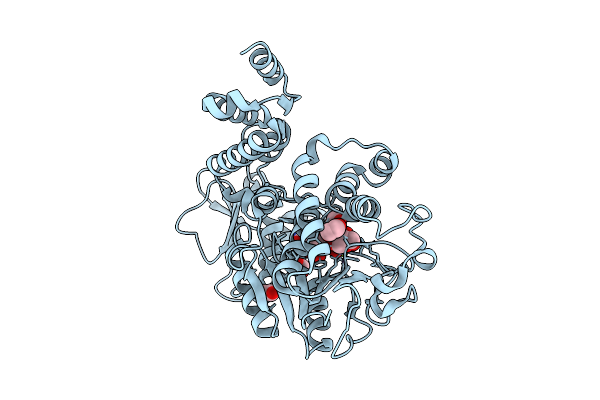 |
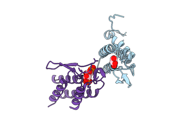
Crystal Structure Of The Glycosyltransferase Domain Of Legionella Seta In Complex With Upd
Organism: Legionella pneumophila
Method: X-RAY DIFFRACTION Resolution:1.79 Å Release Date: 2025-02-19 Classification: TRANSFERASE Ligands: UDP, MG, EDO, GOL |
Organism: Legionella pneumophila
Method: X-RAY DIFFRACTION
Release Date: 2025-02-19
Ligands: UDP, MG, EDO, GOL
 |
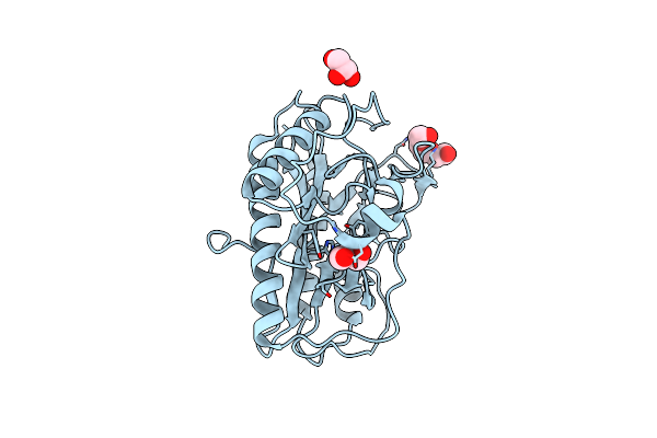
Crystal Structure Of The Pi3P-Binding Domain Of Legionella Seta In Complex With Inositol 1,3-Bisphosphate
Organism: Legionella pneumophila
Method: X-RAY DIFFRACTION Resolution:2.46 Å Release Date: 2025-02-19 Classification: TRANSFERASE Ligands: GOL, ITP |
Organism: Legionella pneumophila
Method: X-RAY DIFFRACTION
Release Date: 2025-02-19
Ligands: GOL, ITP
 |
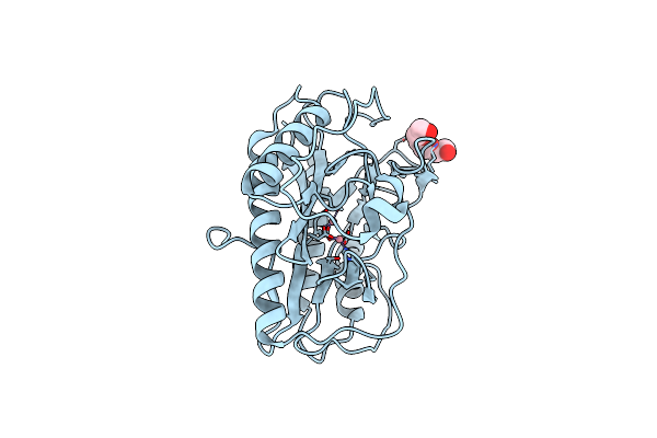
Crystal Structure Of Human Methionine Aminopeptidase 12 (Map12) In The Unbound Form
Organism: Homo sapiens
Method: X-RAY DIFFRACTION Resolution:1.39 Å Release Date: 2024-01-24 Classification: HYDROLASE Ligands: PGE, GOL |
Organism: Homo sapiens
Method: X-RAY DIFFRACTION
Release Date: 2024-01-24
Ligands: PGE, GOL
 |
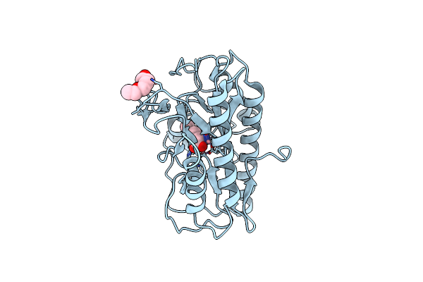
Crystal Structure Of Human Methionine Aminopeptidase 12 (Map12) In Complex With Two Cobalt Ions
Organism: Homo sapiens
Method: X-RAY DIFFRACTION Resolution:1.51 Å Release Date: 2024-01-24 Classification: HYDROLASE Ligands: CO, PGE |
Organism: Homo sapiens
Method: X-RAY DIFFRACTION
Release Date: 2024-01-24
Ligands: CO, PGE
 |
Crystal Structure Of Human Methionine Aminopeptidase 12 (Map12) In Complex With Two Cobalt Ions And Methionine
Organism: Homo sapiens
Method: X-RAY DIFFRACTION Resolution:1.45 Å Release Date: 2024-01-24 Classification: HYDROLASE Ligands: MET, CO, PGE |
Organism: Homo sapiens
Method: X-RAY DIFFRACTION
Release Date: 2024-01-24
Ligands: MET, CO, PGE
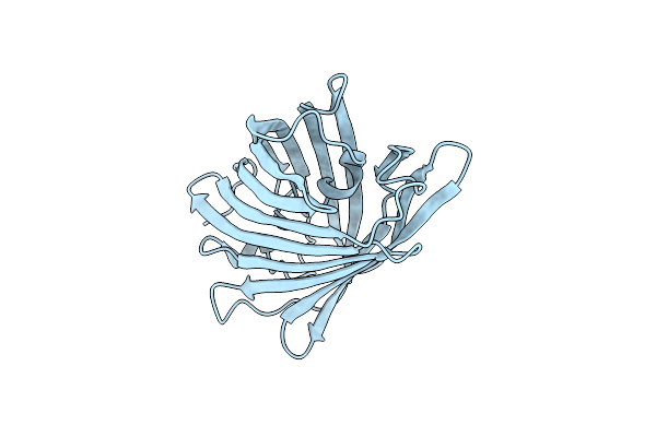 |
Rskiiro Pump Probe Structure By Tr-Sfx
Organism: Lobophyllia hemprichii
Method: X-RAY DIFFRACTION Resolution:1.50 Å Release Date: 2023-10-18 Classification: FLUORESCENT PROTEIN |
Organism: Lobophyllia hemprichii
Method: X-RAY DIFFRACTION
Release Date: 2023-10-18
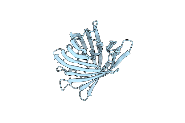 |
Rskiiro Pump Dump Probe Structure By Tr-Sfx
Organism: Lobophyllia hemprichii
Method: X-RAY DIFFRACTION Resolution:1.31 Å Release Date: 2023-10-18 Classification: FLUORESCENT PROTEIN |
Organism: Lobophyllia hemprichii
Method: X-RAY DIFFRACTION
Release Date: 2023-10-18
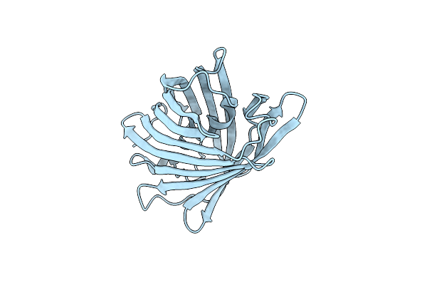 |
Rskiiro Trans Chromophore Dark Structure By Sfx
Organism: Lobophyllia hemprichii
Method: X-RAY DIFFRACTION Resolution:1.30 Å Release Date: 2023-09-27 Classification: FLUORESCENT PROTEIN |
Organism: Lobophyllia hemprichii
Method: X-RAY DIFFRACTION
Release Date: 2023-09-27
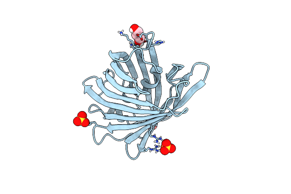 |
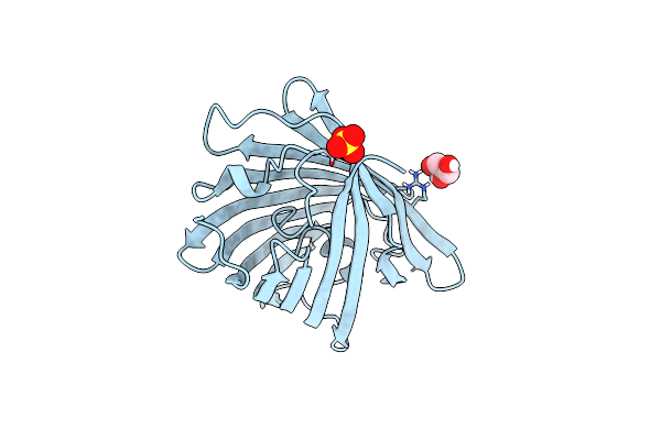
Cis Structure Of Rskiiro At 290 K
Organism: Lobophyllia hemprichii
Method: X-RAY DIFFRACTION Resolution:1.16 Å Release Date: 2023-07-05 Classification: FLUORESCENT PROTEIN Ligands: GOL, SO4 |
Organism: Lobophyllia hemprichii
Method: X-RAY DIFFRACTION
Release Date: 2023-07-05
Ligands: GOL, SO4
 |
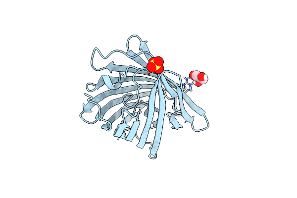
Cis Structure Intermediate Of Rskiiro Illuminated At 200 K
Organism: Lobophyllia hemprichii
Method: X-RAY DIFFRACTION Resolution:1.46 Å Release Date: 2023-07-05 Classification: FLUORESCENT PROTEIN Ligands: GOL, SO4 |
Organism: Lobophyllia hemprichii
Method: X-RAY DIFFRACTION
Release Date: 2023-07-05
Ligands: GOL, SO4
 |
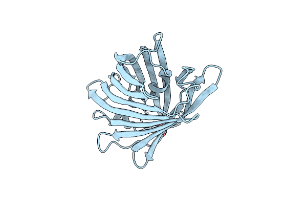
Rskiiro Thermal Annealing At 290K Of 200K Cis Intermediate
Organism: Lobophyllia hemprichii
Method: X-RAY DIFFRACTION Resolution:1.32 Å Release Date: 2023-07-05 Classification: FLUORESCENT PROTEIN Ligands: GOL, SO4 |
Organism: Lobophyllia hemprichii
Method: X-RAY DIFFRACTION
Release Date: 2023-07-05
Ligands: GOL, SO4
 |
Trans Structure Of Rskiiro Illuminated At 290 K
Organism: Lobophyllia hemprichii
Method: X-RAY DIFFRACTION Resolution:1.02 Å Release Date: 2022-11-09 Classification: FLUORESCENT PROTEIN Ligands: SO4 |
Organism: Lobophyllia hemprichii
Method: X-RAY DIFFRACTION
Release Date: 2022-11-09
Ligands: SO4