

Search Count: 20
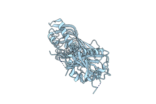 |
Arabidopsis Thaliana Bifunctional Dihydrofolate Reductase-Thymidylate Synthase
Organism: Arabidopsis thaliana
Method: X-RAY DIFFRACTION Resolution:2.00 Å Release Date: 2023-06-21 Classification: TRANSFERASE, OXIDOREDUCTASE |
Organism: Arabidopsis thaliana
Method: X-RAY DIFFRACTION
Release Date: 2023-06-21
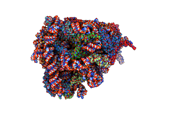 |
Human Delta-Mettl18 60S Ribosome
Organism: Homo sapiens
Method: ELECTRON MICROSCOPY Release Date: 2022-06-22 Classification: RIBOSOME Ligands: MG, ZN |
Organism: Homo sapiens
Method: ELECTRON MICROSCOPY
Release Date: 2022-06-22
Ligands: MG, ZN
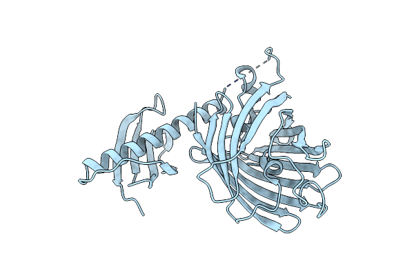 |
Crystal Structure Of Polarisact(T57S), Genetically Encoded Probe For Fluorescent Polarization
Organism: Synthetic construct
Method: X-RAY DIFFRACTION Resolution:2.50 Å Release Date: 2021-03-17 Classification: LUMINESCENT PROTEIN |
Organism: Synthetic construct
Method: X-RAY DIFFRACTION
Release Date: 2021-03-17
 |
Crystal Structure Of The Abscisic Acid Receptor Pyr1 In Complex With An Antagonist
Organism: Arabidopsis thaliana
Method: X-RAY DIFFRACTION Resolution:2.50 Å Release Date: 2018-04-18 Classification: HORMONE RECEPTOR Ligands: 8V6 |
Organism: Arabidopsis thaliana
Method: X-RAY DIFFRACTION
Release Date: 2018-04-18
Ligands: 8V6
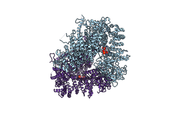 |
Crystal Structure Of Inositol 1,4,5-Trisphosphate Receptor Large Cytosolic Domain With Inositol 1,4,5-Trisphosphate
Organism: Mus musculus
Method: X-RAY DIFFRACTION Resolution:7.40 Å Release Date: 2017-04-26 Classification: METAL TRANSPORT Ligands: I3P |
Organism: Mus musculus
Method: X-RAY DIFFRACTION
Release Date: 2017-04-26
Ligands: I3P
 |
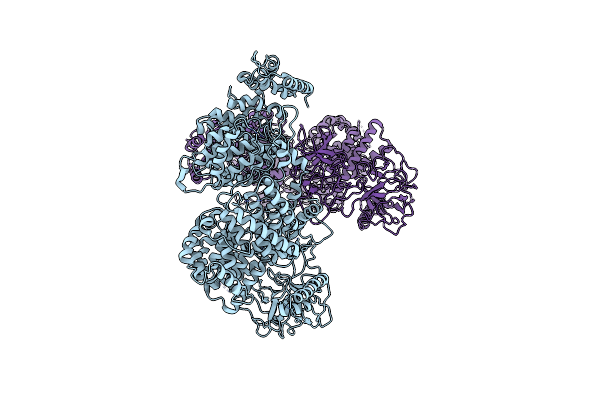
Crystal Structure Of Inositol 1,4,5-Trisphosphate Receptor Large Cytosolic Domain
Organism: Mus musculus
Method: X-RAY DIFFRACTION Resolution:7.31 Å Release Date: 2017-04-19 Classification: METAL TRANSPORT |
Organism: Mus musculus
Method: X-RAY DIFFRACTION
Release Date: 2017-04-19
 |
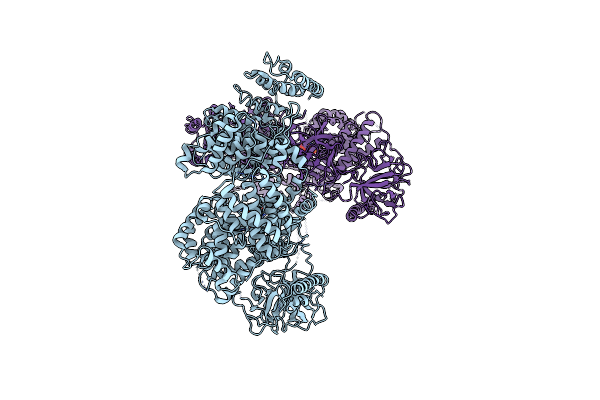
Crystal Structure Of Inositol 1,4,5-Trisphosphate Receptor Cytosolic Domain
Organism: Mus musculus
Method: X-RAY DIFFRACTION Resolution:5.81 Å Release Date: 2017-04-19 Classification: METAL TRANSPORT |
Organism: Mus musculus
Method: X-RAY DIFFRACTION
Release Date: 2017-04-19
 |
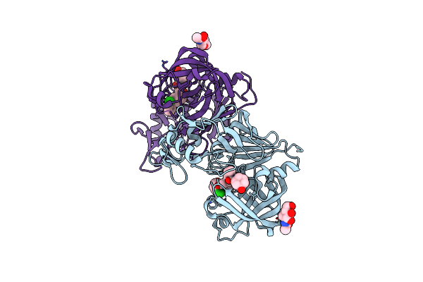
Crystal Structure Of Inositol 1,4,5-Trisphosphate Receptor Cytosolic Domain With Inositol 1,4,5-Trisphosphate
Organism: Mus musculus
Method: X-RAY DIFFRACTION Resolution:6.20 Å Release Date: 2017-04-19 Classification: METAL TRANSPORT Ligands: I3P |
Organism: Mus musculus
Method: X-RAY DIFFRACTION
Release Date: 2017-04-19
Ligands: I3P
 |
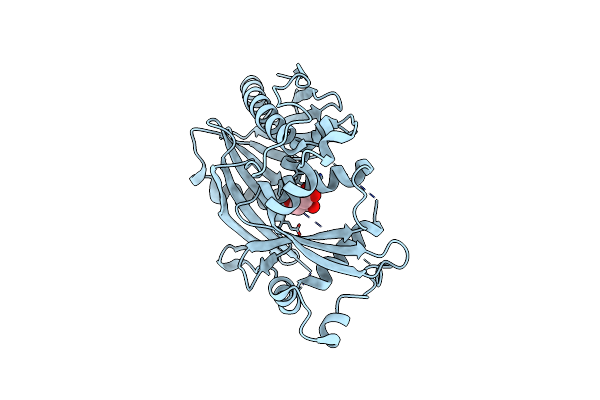
Human Renin In Complex With Compound 5
Organism: Homo sapiens
Method: X-RAY DIFFRACTION Resolution:2.60 Å Release Date: 2013-05-15 Classification: HYDROLASE/HYDROLASE INHIBITOR Ligands: NAG, HHE |
Organism: Homo sapiens
Method: X-RAY DIFFRACTION
Release Date: 2013-05-15
Ligands: NAG, HHE
 |
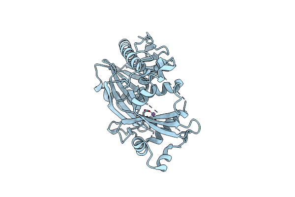
Crystal Structure Of K. Lactis Dxo1 (Ydr370C)
Organism: Kluyveromyces lactis
Method: X-RAY DIFFRACTION Resolution:2.40 Å Release Date: 2012-09-12 Classification: HYDROLASE Ligands: CIT |
Organism: Kluyveromyces lactis
Method: X-RAY DIFFRACTION
Release Date: 2012-09-12
Ligands: CIT
 |
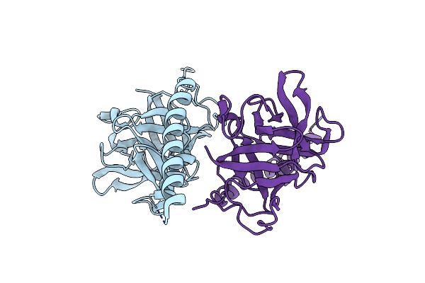
Crystal Structure Of K. Lactis Dxo1 (Ydr370C) In Complex With Manganese
Organism: Kluyveromyces lactis
Method: X-RAY DIFFRACTION Resolution:2.80 Å Release Date: 2012-09-12 Classification: HYDROLASE Ligands: MN |
Organism: Kluyveromyces lactis
Method: X-RAY DIFFRACTION
Release Date: 2012-09-12
Ligands: MN
 |
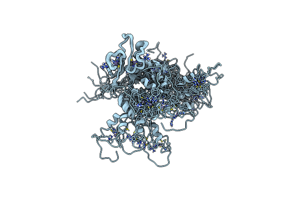
Crystal Structure Of The Ligand Binding Suppressor Domain Of Type 3 Inositol 1,4,5-Trisphosphate Receptor
Organism: Mus musculus
Method: X-RAY DIFFRACTION Resolution:1.90 Å Release Date: 2010-09-15 Classification: TRANSPORT PROTEIN |
Organism: Mus musculus
Method: X-RAY DIFFRACTION
Release Date: 2010-09-15
 |
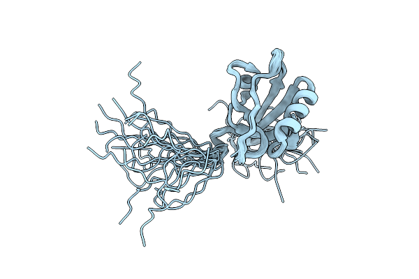
Solution Structure Of The Tandem Zf-C2H2 Domains From The Human Zinc Finger Protein Zic 3
Organism: Homo sapiens
Method: SOLUTION NMR Release Date: 2009-03-31 Classification: TRANSCRIPTION Ligands: ZN |
Organism: Homo sapiens
Method: SOLUTION NMR
Release Date: 2009-03-31
Ligands: ZN
 |
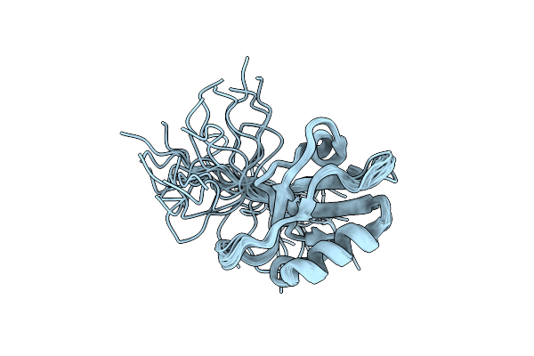
Solution Structure Of Second Pdz Domain Of Pdz Domain Containing Protein 1
Organism: Homo sapiens
Method: SOLUTION NMR Release Date: 2008-02-26 Classification: METAL BINDING PROTEIN |
Organism: Homo sapiens
Method: SOLUTION NMR
Release Date: 2008-02-26
 |
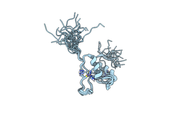
Solution Structure Of Fourth Pdz Domain Of Pdz Domain Containing Protein 1
Organism: Homo sapiens
Method: SOLUTION NMR Release Date: 2008-02-26 Classification: METAL BINDING PROTEIN |
Organism: Homo sapiens
Method: SOLUTION NMR
Release Date: 2008-02-26
 |
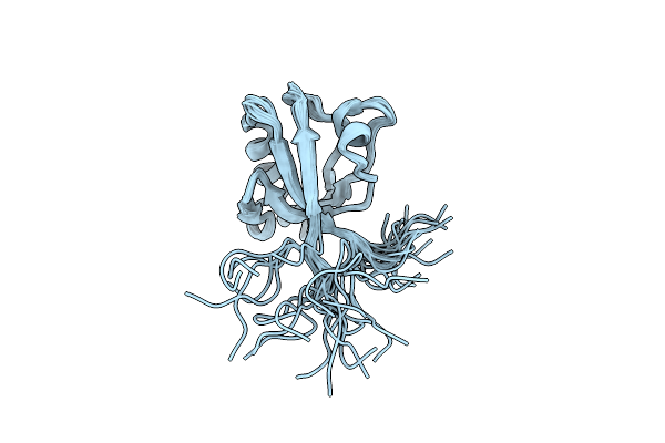
Functional And Structural Basis Of Nuclear Localization Signal In Zic3 Zinc Finger Domain: A Role Of Conserved Tryptophan Residue In The Zinc Finger Domain
Organism: Homo sapiens
Method: SOLUTION NMR Release Date: 2008-02-05 Classification: GENE REGULATION Ligands: ZN |
Organism: Homo sapiens
Method: SOLUTION NMR
Release Date: 2008-02-05
Ligands: ZN
 |
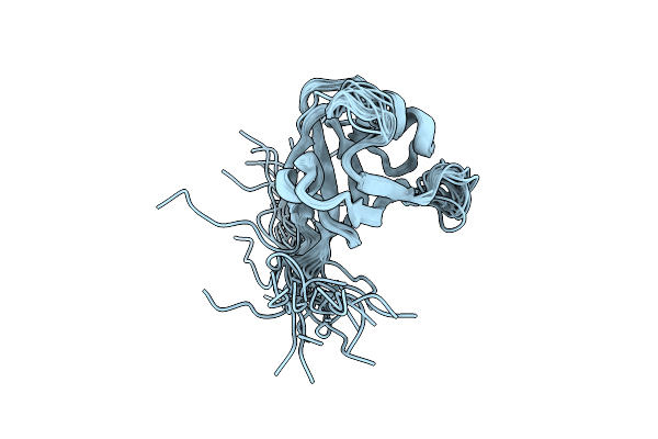
Solution Structure Of Pdz Domain Of Pdz And Lim Domain Protein
Organism: Homo sapiens
Method: SOLUTION NMR Release Date: 2007-08-21 Classification: METAL BINDING PROTEIN |
Organism: Homo sapiens
Method: SOLUTION NMR
Release Date: 2007-08-21
 |
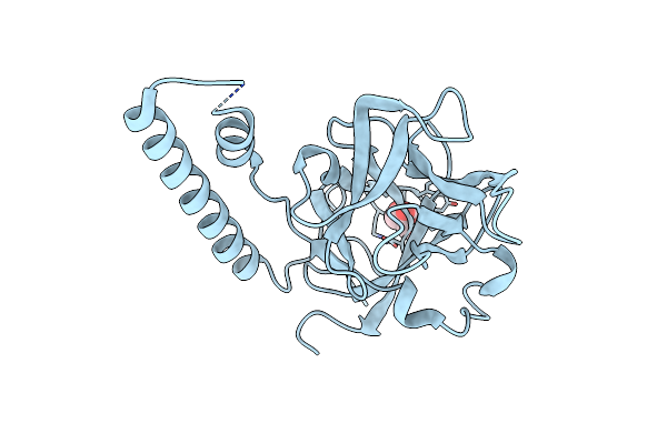
Solution Structure Of First Pdz Domain Of Pdz Domain Containing Protein 7
Organism: Homo sapiens
Method: SOLUTION NMR Release Date: 2007-08-21 Classification: METAL BINDING PROTEIN |
Organism: Homo sapiens
Method: SOLUTION NMR
Release Date: 2007-08-21
 |
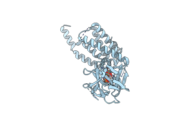
Crystal Structure Of The Ligand Binding Suppressor Domain Of Type 1 Inositol 1,4,5-Trisphosphate Receptor
Organism: Mus musculus
Method: X-RAY DIFFRACTION Resolution:1.80 Å Release Date: 2005-01-25 Classification: MEMBRANE PROTEIN Ligands: GOL |
Organism: Mus musculus
Method: X-RAY DIFFRACTION
Release Date: 2005-01-25
Ligands: GOL
 |
Crystal Structure Of The Inositol 1,4,5-Trisphosphate Receptor Binding Core In Complex With Ip3
Organism: Mus musculus
Method: X-RAY DIFFRACTION Resolution:2.20 Å Release Date: 2002-12-25 Classification: MEMBRANE PROTEIN Ligands: I3P |
Organism: Mus musculus
Method: X-RAY DIFFRACTION
Release Date: 2002-12-25
Ligands: I3P