

Search Count: 26
 |
Crystal Structure Of A Designed Coiled Coil With Two Distinct Terbium Sites
Organism: Synthetic construct
Method: X-RAY DIFFRACTION Resolution:2.76 Å Release Date: 2026-06-10 Classification: DE NOVO PROTEIN Ligands: TB |
Organism: Synthetic construct
Method: X-RAY DIFFRACTION
Release Date: 2026-06-10
Ligands: TB
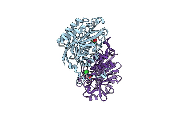 |
The C146A Variant Of An Amidase From Pyrococcus Horikoshii With Bound Acetamide
Organism: Pyrococcus horikoshii (strain atcc 700860 / dsm 12428 / jcm 9974 / nbrc 100139 / ot-3)
Method: X-RAY DIFFRACTION Resolution:1.65 Å Release Date: 2021-07-21 Classification: HYDROLASE Ligands: ACM, CL |
Organism: Pyrococcus horikoshii (strain atcc 700860 / dsm 12428 / jcm 9974 / nbrc 100139 / ot-3)
Method: X-RAY DIFFRACTION
Release Date: 2021-07-21
Ligands: ACM, CL
 |
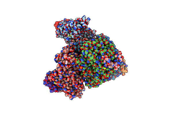
Nitn Amidase From Neterenkonia Sp. An1 After Thrombin His-Tag Removal.
Organism: Nesterenkonia sp. an1
Method: X-RAY DIFFRACTION Resolution:1.19 Å Release Date: 2016-05-18 Classification: HYDROLASE |
Organism: Nesterenkonia sp. an1
Method: X-RAY DIFFRACTION
Release Date: 2016-05-18
 |
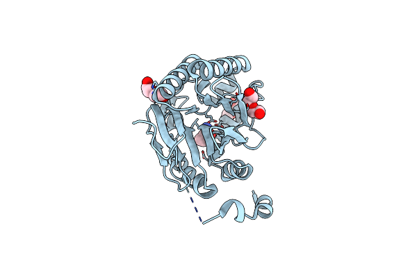
Structural Basis For The Recruitment Of Glycogen Synthase By Glycogenin
Organism: Caenorhabditis elegans
Method: X-RAY DIFFRACTION Resolution:2.60 Å Release Date: 2014-07-09 Classification: TRANSFERASE Ligands: SO4, GOL |
Organism: Caenorhabditis elegans
Method: X-RAY DIFFRACTION
Release Date: 2014-07-09
Ligands: SO4, GOL
 |
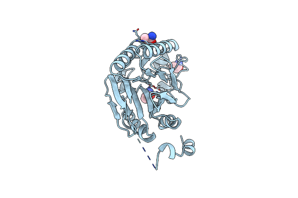
The C145A Mutant Of The Amidase From Nesterenkonia Sp. An1 In Complex With Butyramide
Organism: Nesterenkonia sp. 10004
Method: X-RAY DIFFRACTION Resolution:1.44 Å Release Date: 2014-02-12 Classification: HYDROLASE Ligands: PEG, BMD |
Organism: Nesterenkonia sp. 10004
Method: X-RAY DIFFRACTION
Release Date: 2014-02-12
Ligands: PEG, BMD
 |
The E41Q Mutant Of The Amidase From Nesterenkonia Sp. An1 Showing Covalent Addition Of The Acetamide Moiety Of Fluoroacetamide At The Active Site Cysteine
Organism: Nesterenkonia sp. 10004
Method: X-RAY DIFFRACTION Resolution:1.92 Å Release Date: 2014-02-12 Classification: hydrolase/substrate Ligands: ACM, FTM |
Organism: Nesterenkonia sp. 10004
Method: X-RAY DIFFRACTION
Release Date: 2014-02-12
Ligands: ACM, FTM
 |
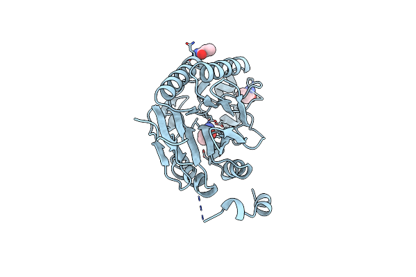
The E41Q Mutant Of The Amidase From Nesterenkonia Sp. An1 Showing The Result Of Michael Addition Of Acrylamide At The Active Site Cysteine
Organism: Nesterenkonia sp. 10004
Method: X-RAY DIFFRACTION Resolution:1.40 Å Release Date: 2014-02-12 Classification: HYDROLASE Ligands: ROP, 1HC |
Organism: Nesterenkonia sp. 10004
Method: X-RAY DIFFRACTION
Release Date: 2014-02-12
Ligands: ROP, 1HC
 |
The E41Q/C145A Double Mutant Of The Amidase From Nesterenkonia Sp. An1 In Complex With Acrylamide
Organism: Nesterenkonia sp. 10004
Method: X-RAY DIFFRACTION Resolution:1.65 Å Release Date: 2014-02-12 Classification: hydrolase/substrate Ligands: ROP, 1HC |
Organism: Nesterenkonia sp. 10004
Method: X-RAY DIFFRACTION
Release Date: 2014-02-12
Ligands: ROP, 1HC
 |
The E41L Mutant Of The Amidase From Nesterenkonia Sp. An1 Showing Covalent Addition Of The Acetamide Moiety Of Fluoroacetamide At The Active Site Cysteine
Organism: Nesterenkonia sp. 10004
Method: X-RAY DIFFRACTION Resolution:1.60 Å Release Date: 2014-02-12 Classification: HYDROLASE Ligands: ACM, FTM |
Organism: Nesterenkonia sp. 10004
Method: X-RAY DIFFRACTION
Release Date: 2014-02-12
Ligands: ACM, FTM
 |
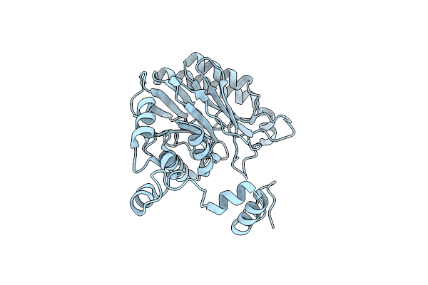
The E142L Mutant Of The Amidase From Geobacillus Pallidus Showing The Result Of Michael Addition Of Acrylamide At The Active Site Cysteine
Organism: Bacillus sp.
Method: X-RAY DIFFRACTION Resolution:1.90 Å Release Date: 2013-08-21 Classification: HYDROLASE Ligands: ROP, CL |
Organism: Bacillus sp.
Method: X-RAY DIFFRACTION
Release Date: 2013-08-21
Ligands: ROP, CL
 |
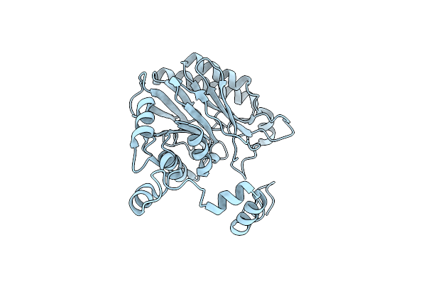
The E142L Mutant Of The Amidase From Geobacillus Pallidus
Organism: Bacillus sp.
Method: X-RAY DIFFRACTION Resolution:1.90 Å Release Date: 2013-08-21 Classification: HYDROLASE Ligands: CL |
Organism: Bacillus sp.
Method: X-RAY DIFFRACTION
Release Date: 2013-08-21
Ligands: CL
 |
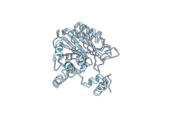
The Mechanism Of The Amidases: The Effect Of The Mutation E142L In The Amidase From Geobacillus Pallidus
Organism: Bacillus sp.
Method: X-RAY DIFFRACTION Resolution:1.85 Å Release Date: 2013-08-21 Classification: HYDROLASE Ligands: CL |
Organism: Bacillus sp.
Method: X-RAY DIFFRACTION
Release Date: 2013-08-21
Ligands: CL
 |
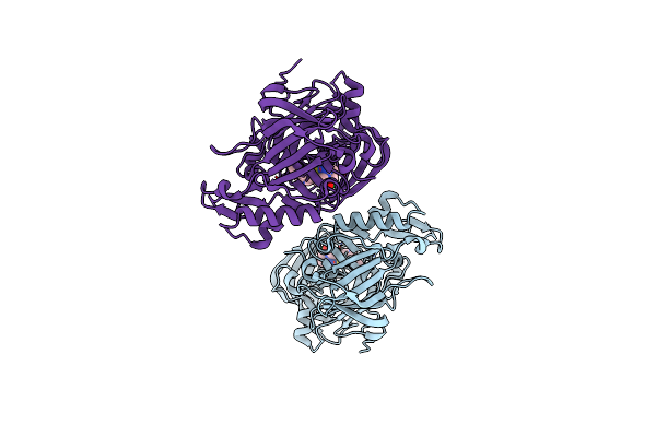
The E142D Mutant Of The Amidase From Geobacillus Pallidus
Organism: Bacillus sp.
Method: X-RAY DIFFRACTION Resolution:1.10 Å Release Date: 2013-08-21 Classification: HYDROLASE |
Organism: Bacillus sp.
Method: X-RAY DIFFRACTION
Release Date: 2013-08-21
 |
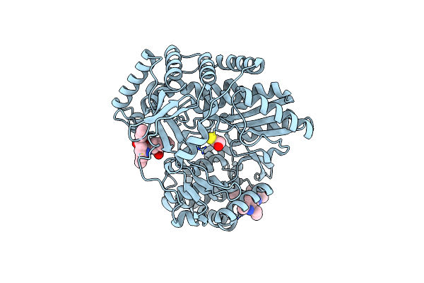
Structure Of Bace In Complex With (S)-4-(3-Chloro-5-(5-(Prop-1-Yn-1-Yl)Pyridin-3-Yl)Thiophen-2-Yl)-1,4-Dimethyl-6-Oxotetrahydropyrimidin-2(1H)-Iminium
Organism: Homo sapiens
Method: X-RAY DIFFRACTION Resolution:1.70 Å Release Date: 2012-08-08 Classification: Hydrolase/Hydrolase inhibitor Ligands: 0V6 |
Organism: Homo sapiens
Method: X-RAY DIFFRACTION
Release Date: 2012-08-08
Ligands: 0V6
 |
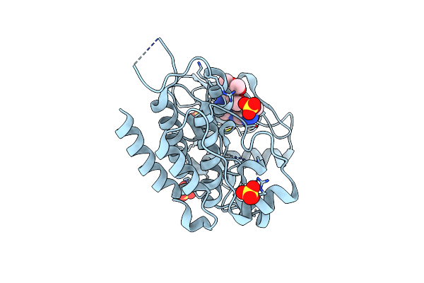
Crystal Structure Of Hcv Ns5B Rna Polymerase In Complex With Pf868554
Organism: Hepatitis c virus
Method: X-RAY DIFFRACTION Resolution:1.86 Å Release Date: 2009-03-10 Classification: TRANSFERASE Ligands: AG0, AG6, BME |
Organism: Hepatitis c virus
Method: X-RAY DIFFRACTION
Release Date: 2009-03-10
Ligands: AG0, AG6, BME
 |
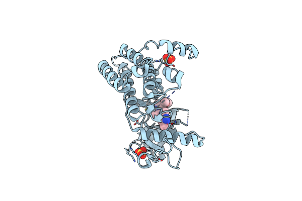
Crystal Structure Of Vegfr2 In Complex With A 3,4,5-Trimethoxy Aniline Containing Pyrimidine
Organism: Homo sapiens
Method: X-RAY DIFFRACTION Resolution:2.15 Å Release Date: 2008-10-07 Classification: TRANSFERASE Ligands: SO4, SAV |
Organism: Homo sapiens
Method: X-RAY DIFFRACTION
Release Date: 2008-10-07
Ligands: SO4, SAV
 |
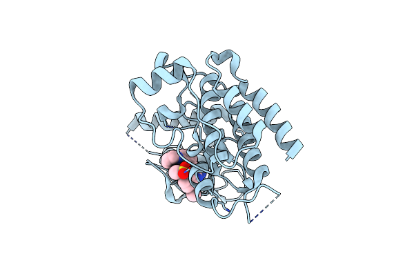
Crystal Structure Of Vegfr2 In Complex With A 3,4,5-Trimethoxy Aniline Containing Pyrimidine
Organism: Homo sapiens
Method: X-RAY DIFFRACTION Resolution:2.25 Å Release Date: 2008-10-07 Classification: TRANSFERASE Ligands: SO4, KIM |
Organism: Homo sapiens
Method: X-RAY DIFFRACTION
Release Date: 2008-10-07
Ligands: SO4, KIM
 |
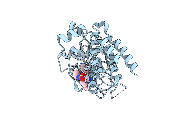
Crystal Structure Of Vegfr2 In Complex With A 2-Anilino-5-Aryl-Oxazole Inhibitor
Organism: Homo sapiens
Method: X-RAY DIFFRACTION Resolution:2.10 Å Release Date: 2005-06-07 Classification: TRANSFERASE Ligands: AAZ |
Organism: Homo sapiens
Method: X-RAY DIFFRACTION
Release Date: 2005-06-07
Ligands: AAZ
 |
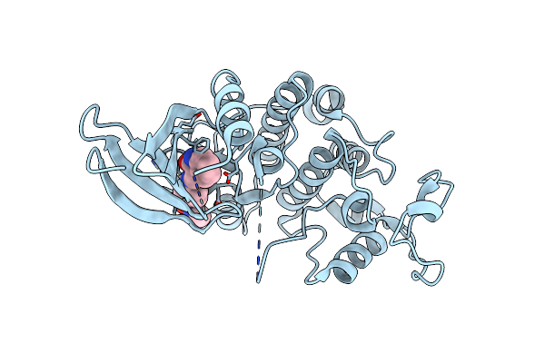
Crystal Structure Of Vegfr2 In Complex With A 2-Anilino-5-Aryl-Oxazole Inhibitor
Organism: Homo sapiens
Method: X-RAY DIFFRACTION Resolution:2.10 Å Release Date: 2005-06-07 Classification: TRANSFERASE Ligands: AAX |
Organism: Homo sapiens
Method: X-RAY DIFFRACTION
Release Date: 2005-06-07
Ligands: AAX
 |
Cdk2 Complexed With N-Methyl-4-{[(2-Oxo-1,2-Dihydro-3H-Indol-3-Ylidene)Methyl]Amino}Benzenesulfonamide
Organism: Homo sapiens
Method: X-RAY DIFFRACTION Resolution:2.20 Å Release Date: 2002-05-14 Classification: TRANSFERASE Ligands: LS1 |
Organism: Homo sapiens
Method: X-RAY DIFFRACTION
Release Date: 2002-05-14
Ligands: LS1