

Search Count: 28
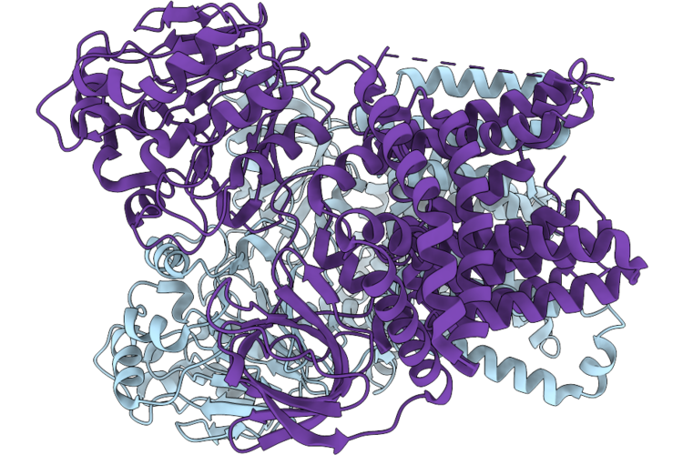 |
Cryo-Em Structure Of Full-Length Comec From Neomoorella Carbonis
Organism: Neomoorella carbonis
Method: ELECTRON MICROSCOPY Resolution:4.10 Å Release Date: 2026-08-05 Classification: MEMBRANE PROTEIN |
Organism: Neomoorella carbonis
Method: ELECTRON MICROSCOPY
Release Date: 2026-08-05
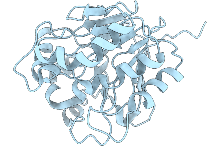 |
Cryo-Em Structure Of Beta-Lactamase-Like Domain From Neomoorella Carbonis
Organism: Neomoorella carbonis
Method: ELECTRON MICROSCOPY Resolution:4.20 Å Release Date: 2026-05-20 Classification: MEMBRANE PROTEIN |
Organism: Neomoorella carbonis
Method: ELECTRON MICROSCOPY
Release Date: 2026-05-20
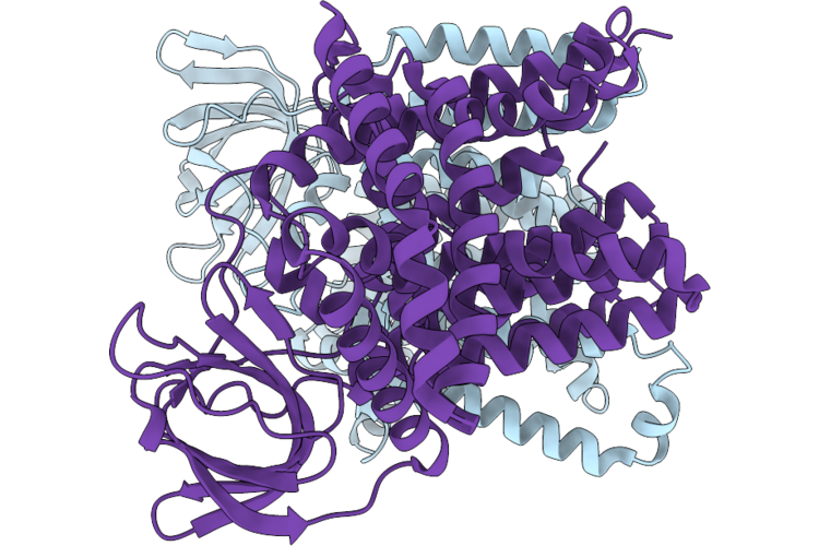 |
Cryo-Em Structure Of Comec From Neomoorella Carbonis
Organism: Neomoorella carbonis
Method: ELECTRON MICROSCOPY Resolution:4.10 Å Release Date: 2026-05-20 Classification: MEMBRANE PROTEIN |
Organism: Neomoorella carbonis
Method: ELECTRON MICROSCOPY
Release Date: 2026-05-20
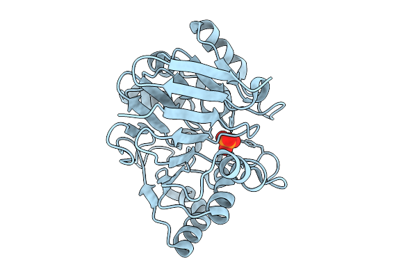 |
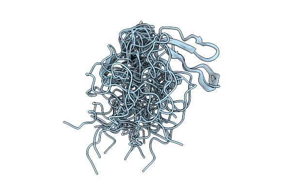
Crystal Structure Of Beta-Lactamase-Like Domain Of Comec From Moorella Glycerini
Organism: Neomoorella glycerini
Method: X-RAY DIFFRACTION Resolution:1.86 Å Release Date: 2025-10-01 Classification: METAL BINDING PROTEIN Ligands: PO4 |
Organism: Neomoorella glycerini
Method: X-RAY DIFFRACTION
Release Date: 2025-10-01
Ligands: PO4
 |
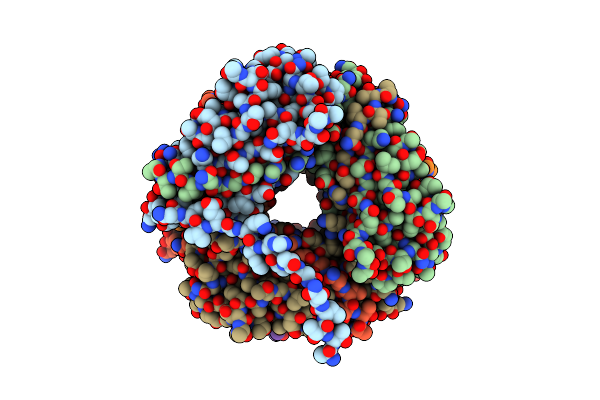
The Solution Nmr Structure Of Ob Domain Of Comec From Moorella Glycerini
Organism: Moorella glycerini
Method: SOLUTION NMR Release Date: 2025-10-01 Classification: DNA BINDING PROTEIN |
Organism: Moorella glycerini
Method: SOLUTION NMR
Release Date: 2025-10-01
 |
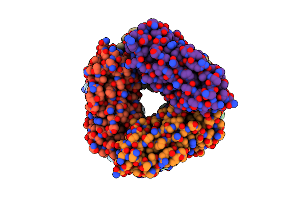
2.7 A Cryo-Em Structure Of In Vitro Assembled Type 1 Pilus Rod
Organism: Escherichia coli
Method: ELECTRON MICROSCOPY Release Date: 2024-04-10 Classification: STRUCTURAL PROTEIN |
Organism: Escherichia coli
Method: ELECTRON MICROSCOPY
Release Date: 2024-04-10
 |
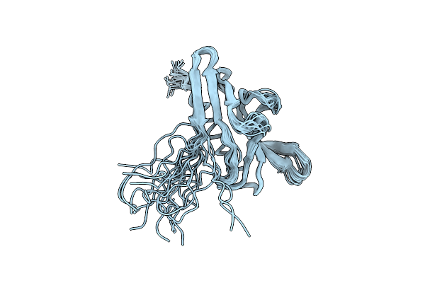
2.5 A Cryo-Em Structure Of The In Vitro Fimd-Catalyzed Assembly Of Type 1 Pilus Rod
Organism: Escherichia coli
Method: ELECTRON MICROSCOPY Release Date: 2024-04-10 Classification: STRUCTURAL PROTEIN |
Organism: Escherichia coli
Method: ELECTRON MICROSCOPY
Release Date: 2024-04-10
 |
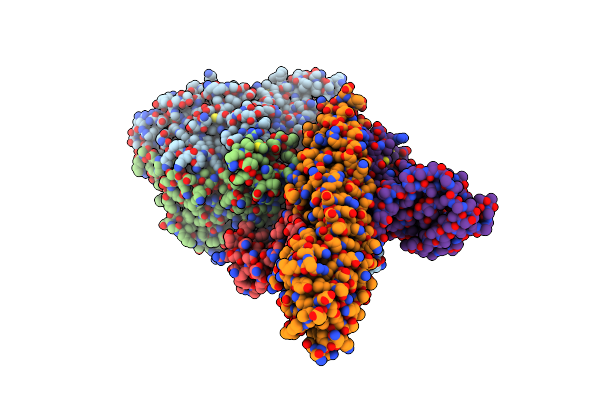
Solution Structure Of The Dna-Binding Minor Pilin Fimt From Legionella Pneumophila
Organism: Legionella pneumophila
Method: SOLUTION NMR Release Date: 2022-03-02 Classification: DNA BINDING PROTEIN |
Organism: Legionella pneumophila
Method: SOLUTION NMR
Release Date: 2022-03-02
 |
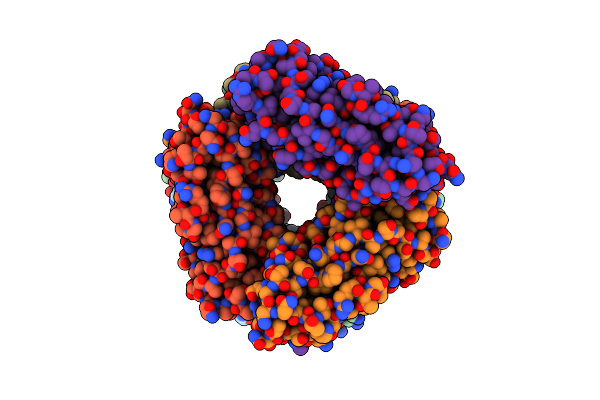
L. Pneumophila Type Iv Coupling Complex (T4Cc) With Density For Doty N-Terminal And Middle Domains
Organism: legionella pneumophila subsp. pneumophila str. philadelphia 1
Method: ELECTRON MICROSCOPY Release Date: 2021-12-01 Classification: MEMBRANE PROTEIN Ligands: ZN |
Organism: legionella pneumophila subsp. pneumophila str. philadelphia 1
Method: ELECTRON MICROSCOPY
Release Date: 2021-12-01
Ligands: ZN
 |
2.85 A Cryo-Em Structure Of The In Vivo Assembled Type 1 Pilus Rod
Organism: Escherichia coli
Method: ELECTRON MICROSCOPY Resolution:2.85 Å Release Date: 2021-03-31 Classification: STRUCTURAL PROTEIN |
Organism: Escherichia coli
Method: ELECTRON MICROSCOPY
Release Date: 2021-03-31
 |
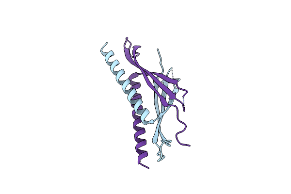
Type Iv Coupling Complex (T4Cc) From L. Pneumophila.
Organism: Legionella pneumophila
Method: ELECTRON MICROSCOPY Release Date: 2020-05-13 Classification: MEMBRANE PROTEIN Ligands: ZN |
Organism: Legionella pneumophila
Method: ELECTRON MICROSCOPY
Release Date: 2020-05-13
Ligands: ZN
 |
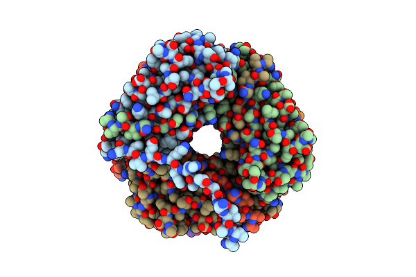
Crystal Structure Of Commd9 Comm Domain
Organism: Homo sapiens
Method: X-RAY DIFFRACTION Resolution:2.17 Å Release Date: 2018-08-15 Classification: ENDOCYTOSIS |
Organism: Homo sapiens
Method: X-RAY DIFFRACTION
Release Date: 2018-08-15
 |
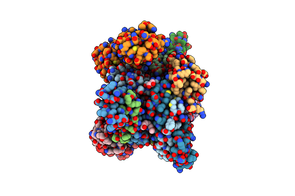
The Cryo-Electron Microscopy Structure Of The Type 1 Chaperone-Usher Pilus Rod
Organism: Escherichia coli j96
Method: ELECTRON MICROSCOPY Release Date: 2017-11-22 Classification: PROTEIN FIBRIL |
Organism: Escherichia coli j96
Method: ELECTRON MICROSCOPY
Release Date: 2017-11-22
 |
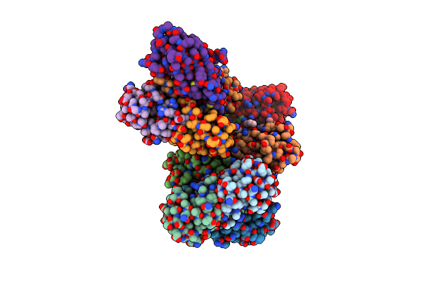
K6-Specific Affimer Bound To K6 Diub
Organism: Synthetic construct, Homo sapiens
Method: X-RAY DIFFRACTION Resolution:2.50 Å Release Date: 2017-10-04 Classification: SIGNALING PROTEIN Ligands: GOL, 15P |
Organism: Synthetic construct, Homo sapiens
Method: X-RAY DIFFRACTION
Release Date: 2017-10-04
Ligands: GOL, 15P
 |
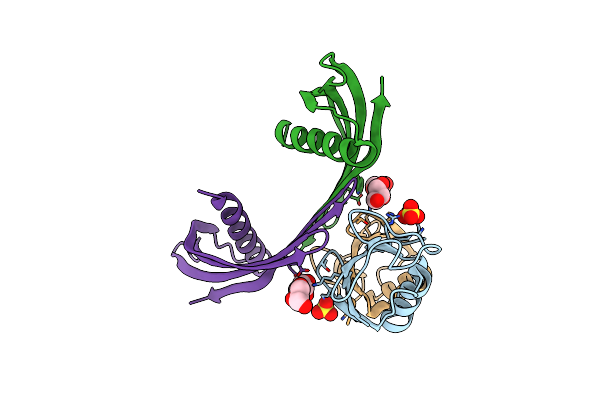
K33-Specific Affimer Bound To K33 Diub
Organism: Homo sapiens, Synthetic construct
Method: X-RAY DIFFRACTION Resolution:3.80 Å Release Date: 2017-10-04 Classification: SIGNALING PROTEIN Ligands: 15P |
Organism: Homo sapiens, Synthetic construct
Method: X-RAY DIFFRACTION
Release Date: 2017-10-04
Ligands: 15P
 |
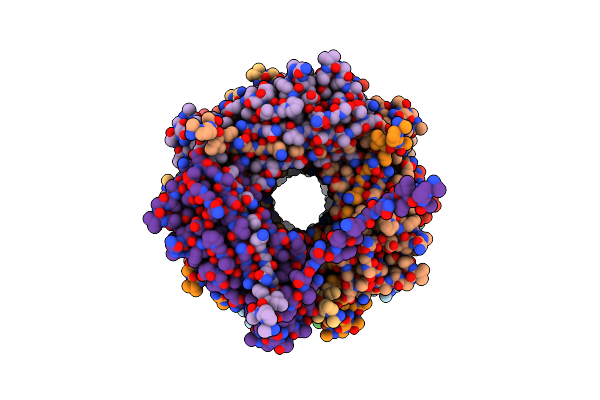
K33-Specific Affimer Bound To K33 Diub
Organism: Homo sapiens, Synthetic construct
Method: X-RAY DIFFRACTION Resolution:2.80 Å Release Date: 2017-10-04 Classification: SIGNALING PROTEIN Ligands: SO4, GOL |
Organism: Homo sapiens, Synthetic construct
Method: X-RAY DIFFRACTION
Release Date: 2017-10-04
Ligands: SO4, GOL
 |
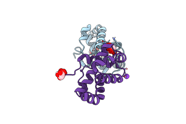
Structure Of A Chaperone-Usher Pilus Reveals The Molecular Basis Of Rod Uncoilin
Organism: Escherichia coli
Method: ELECTRON MICROSCOPY Resolution:3.80 Å Release Date: 2016-01-13 Classification: STRUCTURAL PROTEIN |
Organism: Escherichia coli
Method: ELECTRON MICROSCOPY
Release Date: 2016-01-13
 |
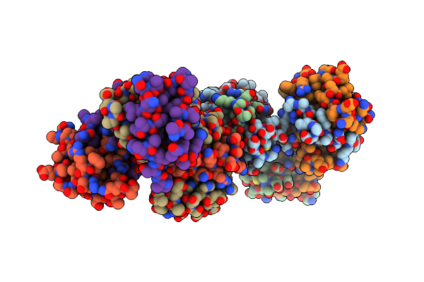
The Crystal Structure Of The N-Terminal Domain Of Commd9
Organism: Homo sapiens
Method: X-RAY DIFFRACTION Resolution:1.55 Å Release Date: 2015-03-04 Classification: PROTEIN BINDING Ligands: CIT, K |
Organism: Homo sapiens
Method: X-RAY DIFFRACTION
Release Date: 2015-03-04
Ligands: CIT, K
 |
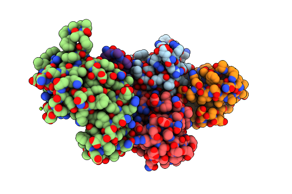
The Crystal Structure Of The N-Terminal Domain Of Commd9
Organism: Homo sapiens
Method: X-RAY DIFFRACTION Resolution:2.79 Å Release Date: 2014-11-26 Classification: PROTEIN BINDING |
Organism: Homo sapiens
Method: X-RAY DIFFRACTION
Release Date: 2014-11-26
 |
Structure Of Otud2 Otu Domain In Complex With Ubiquitin K11-Linked Peptide
Organism: Homo sapiens
Method: X-RAY DIFFRACTION Resolution:2.35 Å Release Date: 2013-07-24 Classification: HYDROLASE Ligands: NO3, MG |
Organism: Homo sapiens
Method: X-RAY DIFFRACTION
Release Date: 2013-07-24
Ligands: NO3, MG