

Search Count: 39
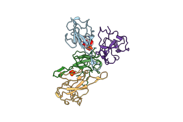 |
Crystal Structure Of Sars-Cov-2 Nucleocapsid Phosphoprotein N-Terminal Domain(N-Ntd) In Complex With Ump
Organism: Severe acute respiratory syndrome coronavirus 2
Method: X-RAY DIFFRACTION Resolution:2.12 Å Release Date: 2025-04-23 Classification: VIRAL PROTEIN Ligands: U, PO4 |
Organism: Severe acute respiratory syndrome coronavirus 2
Method: X-RAY DIFFRACTION
Release Date: 2025-04-23
Ligands: U, PO4
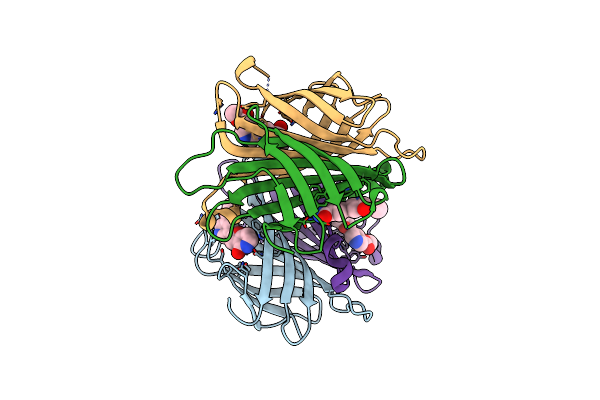 |
X-Ray Crystal Structure Of Alis4-Streptavidine Complex Without Cryo-Protectant Using A High-Pressure Cryocooling Method
Organism: Streptomyces avidinii
Method: X-RAY DIFFRACTION Resolution:1.48 Å Release Date: 2025-01-29 Classification: CYTOSOLIC PROTEIN Ligands: 6FX |
Organism: Streptomyces avidinii
Method: X-RAY DIFFRACTION
Release Date: 2025-01-29
Ligands: 6FX
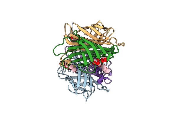 |
X-Ray Crystal Structure Of Alis4-Streptavidine Complex With 10% Glycerol Using A High-Pressure Cryocooling Method
Organism: Streptomyces avidinii
Method: X-RAY DIFFRACTION Resolution:1.45 Å Release Date: 2025-01-29 Classification: CYTOSOLIC PROTEIN Ligands: DMS, GOL, PEG |
Organism: Streptomyces avidinii
Method: X-RAY DIFFRACTION
Release Date: 2025-01-29
Ligands: DMS, GOL, PEG
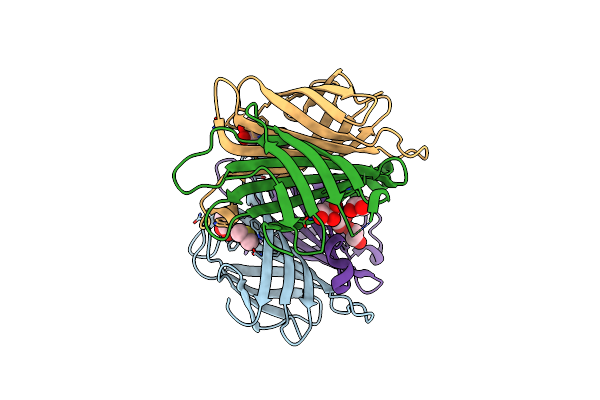 |
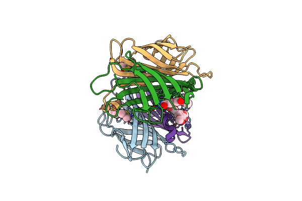
X-Ray Crystal Structure Of Alis4-Streptavidine Complex With 20% Glycerol Using A High-Pressure Cryocooling Method
Organism: Streptomyces avidinii
Method: X-RAY DIFFRACTION Resolution:1.45 Å Release Date: 2025-01-29 Classification: CYTOSOLIC PROTEIN Ligands: GOL, DMS, PEG |
Organism: Streptomyces avidinii
Method: X-RAY DIFFRACTION
Release Date: 2025-01-29
Ligands: GOL, DMS, PEG
 |
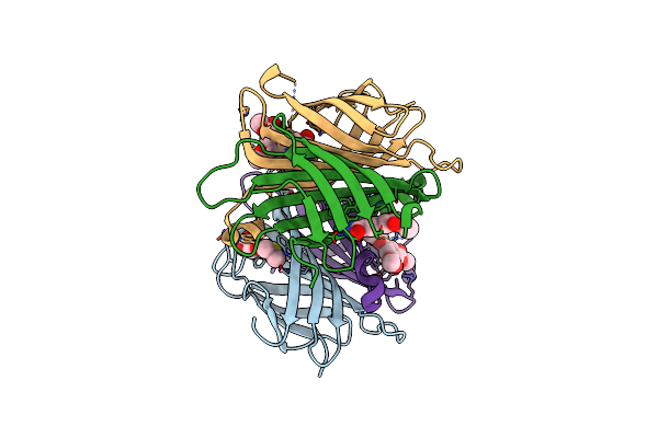
X-Ray Crystal Structure Of Alis5-Streptavidine Complex Without Cryo-Protectant Using A High-Pressure Cryocooling Method
Organism: Streptomyces avidinii
Method: X-RAY DIFFRACTION Resolution:1.23 Å Release Date: 2025-01-29 Classification: CYTOSOLIC PROTEIN Ligands: DMS, A1LXQ |
Organism: Streptomyces avidinii
Method: X-RAY DIFFRACTION
Release Date: 2025-01-29
Ligands: DMS, A1LXQ
 |
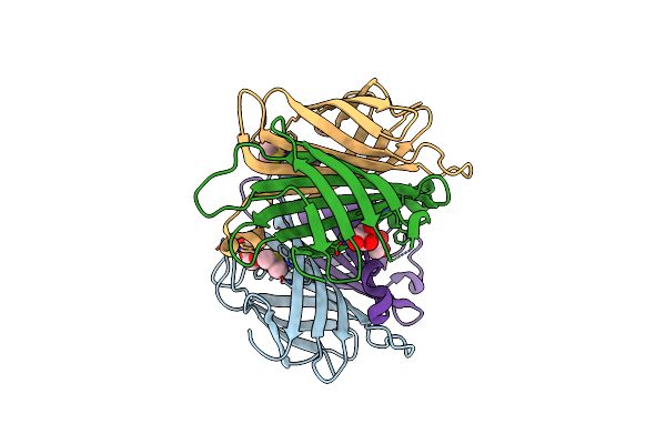
X-Ray Crystal Structure Of Alis5-Streptavidine Complex With 10% Glycerol Using A High-Pressure Cryocooling Method
Organism: Streptomyces avidinii
Method: X-RAY DIFFRACTION Resolution:1.45 Å Release Date: 2025-01-29 Classification: CYTOSOLIC PROTEIN Ligands: DMS, GOL, A1LXQ |
Organism: Streptomyces avidinii
Method: X-RAY DIFFRACTION
Release Date: 2025-01-29
Ligands: DMS, GOL, A1LXQ
 |
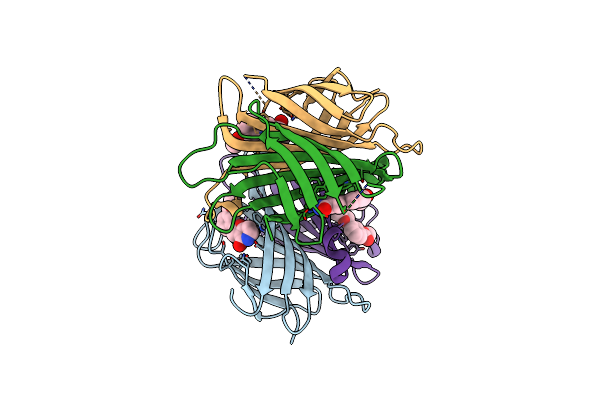
X-Ray Crystal Structure Of Alis5-Streptavidine Complex With 20% Glycerol Using A High-Pressure Cryocooling Method
Organism: Streptomyces avidinii
Method: X-RAY DIFFRACTION Resolution:1.33 Å Release Date: 2025-01-29 Classification: CYTOSOLIC PROTEIN Ligands: GOL, DMS |
Organism: Streptomyces avidinii
Method: X-RAY DIFFRACTION
Release Date: 2025-01-29
Ligands: GOL, DMS
 |
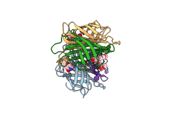
X-Ray Crystal Structure Of Alis2-Streptavidine Complex Without Cryo-Protectant Using A High-Pressure Cryocooling Method
Organism: Streptomyces avidinii
Method: X-RAY DIFFRACTION Resolution:1.42 Å Release Date: 2025-01-29 Classification: CYTOSOLIC PROTEIN Ligands: MT6 |
Organism: Streptomyces avidinii
Method: X-RAY DIFFRACTION
Release Date: 2025-01-29
Ligands: MT6
 |
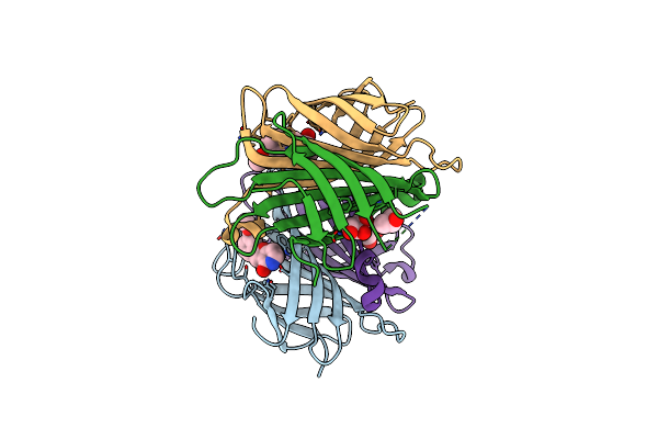
X-Ray Crystal Structure Of Alis2-Streptavidine Complex With 10% Glycerol Using A High-Pressure Cryocooling Method
Organism: Streptomyces avidinii
Method: X-RAY DIFFRACTION Resolution:1.34 Å Release Date: 2025-01-29 Classification: CYTOSOLIC PROTEIN Ligands: MT6 |
Organism: Streptomyces avidinii
Method: X-RAY DIFFRACTION
Release Date: 2025-01-29
Ligands: MT6
 |
X-Ray Crystal Structure Of Alis2-Streptavidine Complex With 20% Glycerol Using A High-Pressure Cryocooling Method
Organism: Streptomyces avidinii
Method: X-RAY DIFFRACTION Resolution:1.50 Å Release Date: 2025-01-29 Classification: CYTOSOLIC PROTEIN Ligands: MT6, GOL |
Organism: Streptomyces avidinii
Method: X-RAY DIFFRACTION
Release Date: 2025-01-29
Ligands: MT6, GOL
 |
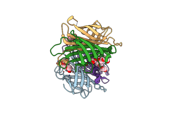
X-Ray Crystal Structure Of Streptavidin Flash-Cooled In 30% Glycerol At Ambient Pressure
Organism: Streptomyces avidinii
Method: X-RAY DIFFRACTION Resolution:1.38 Å Release Date: 2024-12-18 Classification: CYTOSOLIC PROTEIN Ligands: PG6, PG4, GOL, PEG |
Organism: Streptomyces avidinii
Method: X-RAY DIFFRACTION
Release Date: 2024-12-18
Ligands: PG6, PG4, GOL, PEG
 |
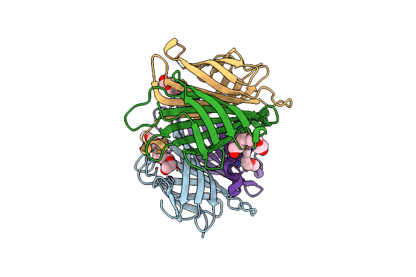
X-Ray Crystal Structure Of Streptavidin Flash-Cooled In 30% Peg1000 At Ambient Pressure
Organism: Streptomyces avidinii
Method: X-RAY DIFFRACTION Resolution:1.40 Å Release Date: 2024-12-18 Classification: CYTOSOLIC PROTEIN Ligands: PG4, PEG |
Organism: Streptomyces avidinii
Method: X-RAY DIFFRACTION
Release Date: 2024-12-18
Ligands: PG4, PEG
 |
X-Ray Crystal Structure Of Streptavidine Without Cryo-Protectant Using A High-Pressure Cryocooling Method
Organism: Streptomyces avidinii
Method: X-RAY DIFFRACTION Resolution:1.31 Å Release Date: 2024-12-18 Classification: CYTOSOLIC PROTEIN Ligands: PG6, PG4 |
Organism: Streptomyces avidinii
Method: X-RAY DIFFRACTION
Release Date: 2024-12-18
Ligands: PG6, PG4
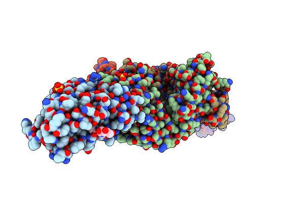 |
Crystal Structure Of Sars-Cov-2 Spike Receptor-Binding Domain (Rbd) In Complex With Ncv2Sg53 Fab
Organism: Severe acute respiratory syndrome coronavirus 2, Homo sapiens
Method: X-RAY DIFFRACTION Resolution:2.35 Å Release Date: 2023-04-19 Classification: VIRAL PROTEIN/IMMUNE SYSTEM Ligands: SO4, NAG |
Organism: Severe acute respiratory syndrome coronavirus 2, Homo sapiens
Method: X-RAY DIFFRACTION
Release Date: 2023-04-19
Ligands: SO4, NAG
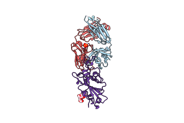 |
Crystal Structure Of Sars-Cov-2 Spike Receptor-Binding Domain (Rbd) In Complex With Ncv2Sg48 Fab
Organism: Severe acute respiratory syndrome coronavirus 2, Homo sapiens
Method: X-RAY DIFFRACTION Resolution:2.18 Å Release Date: 2023-04-19 Classification: VIRAL PROTEIN/IMMUNE SYSTEM Ligands: SO4 |
Organism: Severe acute respiratory syndrome coronavirus 2, Homo sapiens
Method: X-RAY DIFFRACTION
Release Date: 2023-04-19
Ligands: SO4
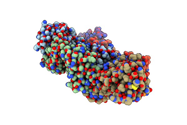 |
Crystal Structure Of Sars-Cov-2 Omicron Variant Spike Receptor-Binding Domain (Rbd) In Complex With Ncv2Sg48 Fab
Organism: Homo sapiens, Severe acute respiratory syndrome coronavirus 2
Method: X-RAY DIFFRACTION Resolution:3.30 Å Release Date: 2023-04-19 Classification: IMMUNE SYSTEM/VIRAL PROTEIN Ligands: SO4 |
Organism: Homo sapiens, Severe acute respiratory syndrome coronavirus 2
Method: X-RAY DIFFRACTION
Release Date: 2023-04-19
Ligands: SO4
 |
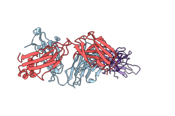
Crystal Structure Of Sars-Cov-2 Delta Variant Spike Receptor-Binding Domain (Rbd) In Complex With Ncv2Sg48 Fab
Organism: Severe acute respiratory syndrome coronavirus 2, Homo sapiens
Method: X-RAY DIFFRACTION Resolution:2.38 Å Release Date: 2023-04-19 Classification: VIRAL PROTEIN/IMMUNE SYSTEM Ligands: NAG |
Organism: Severe acute respiratory syndrome coronavirus 2, Homo sapiens
Method: X-RAY DIFFRACTION
Release Date: 2023-04-19
Ligands: NAG
 |
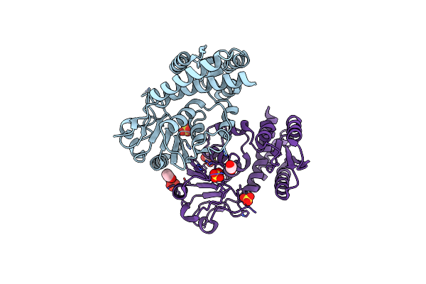
Crystal Structure Of Sars-Cov-2 Delta Variant Spike Receptor-Binding Domain (Rbd) In Complex With Ncv2Sg53 Fab
Organism: Severe acute respiratory syndrome coronavirus 2, Homo sapiens
Method: X-RAY DIFFRACTION Resolution:3.06 Å Release Date: 2023-04-19 Classification: VIRAL PROTEIN/IMMUNE SYSTEM |
Organism: Severe acute respiratory syndrome coronavirus 2, Homo sapiens
Method: X-RAY DIFFRACTION
Release Date: 2023-04-19
 |
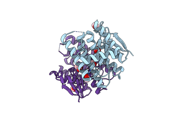
Molecular Structure Of A Novel 5,10-Methylenetetrahydrofolate Dehydrogenase From The Silkworm, Bombyx Mori
Organism: Bombyx mori
Method: X-RAY DIFFRACTION Resolution:1.75 Å Release Date: 2019-01-23 Classification: OXIDOREDUCTASE Ligands: SO4, EDO |
Organism: Bombyx mori
Method: X-RAY DIFFRACTION
Release Date: 2019-01-23
Ligands: SO4, EDO
 |
Crystal Structure Of A Diazinon-Metabolizing Glutathione S-Transferase In The Silkworm, Bombyx Mori
Organism: Bombyx mori
Method: X-RAY DIFFRACTION Resolution:1.70 Å Release Date: 2018-09-12 Classification: TRANSFERASE Ligands: EDO, ACT |
Organism: Bombyx mori
Method: X-RAY DIFFRACTION
Release Date: 2018-09-12
Ligands: EDO, ACT