

Search Count: 67
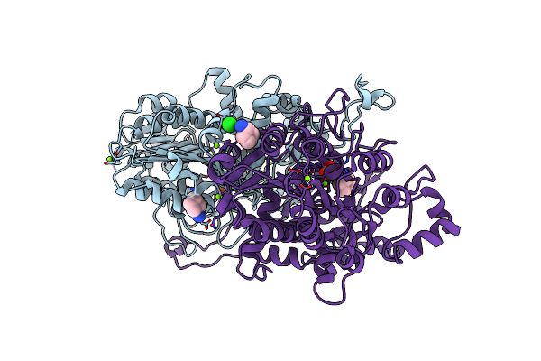 |
X-Ray Crystal Structure Of Vibrio Alkaline Phosphatase With The Non-Competitive Inhibitor Cyclohexylamine
Organism: Vibrio sp. g15-21
Method: X-RAY DIFFRACTION Resolution:2.27 Å Release Date: 2020-10-21 Classification: HYDROLASE Ligands: ZN, MG, CL, PO4, HAI |
Organism: Vibrio sp. g15-21
Method: X-RAY DIFFRACTION
Release Date: 2020-10-21
Ligands: ZN, MG, CL, PO4, HAI
 |
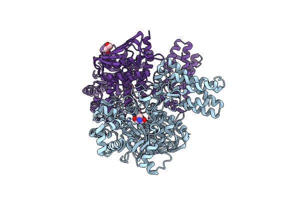
Crystal Structure Of The Gh18 Chitinase Chib From The Chitin Utilization Locus Of Flavobacterium Johnsoniae
Organism: Flavobacterium johnsoniae (strain atcc 17061 / dsm 2064 / uw101)
Method: X-RAY DIFFRACTION Resolution:1.63 Å Release Date: 2020-09-02 Classification: HYDROLASE Ligands: EDO, FMT |
Organism: Flavobacterium johnsoniae (strain atcc 17061 / dsm 2064 / uw101)
Method: X-RAY DIFFRACTION
Release Date: 2020-09-02
Ligands: EDO, FMT
 |
X-Ray Structure Of Flavobacterium Johnsoniae Chitobiase (Fjgh20)
Organism: Flavobacterium johnsoniae (strain atcc 17061 / dsm 2064 / uw101)
Method: X-RAY DIFFRACTION Resolution:1.70 Å Release Date: 2020-09-02 Classification: HYDROLASE Ligands: TRS, GOL |
Organism: Flavobacterium johnsoniae (strain atcc 17061 / dsm 2064 / uw101)
Method: X-RAY DIFFRACTION
Release Date: 2020-09-02
Ligands: TRS, GOL
 |
X-Ray Crystal Structure Of The R336L Vibrio Alkaline Phosphatase Variant.
Organism: Vibrio splendidus
Method: X-RAY DIFFRACTION Resolution:2.00 Å Release Date: 2020-01-29 Classification: HYDROLASE Ligands: DTB, ZN, MG, SO4 |
Organism: Vibrio splendidus
Method: X-RAY DIFFRACTION
Release Date: 2020-01-29
Ligands: DTB, ZN, MG, SO4
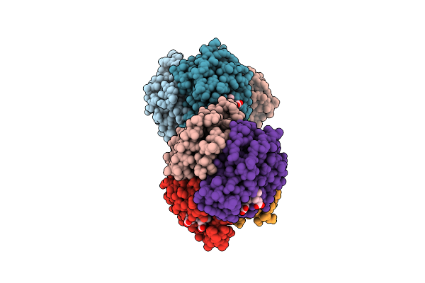 |
The Crystal Structure Of Haemoglobin From Atlantic Cod
Organism: Gadus morhua
Method: X-RAY DIFFRACTION Resolution:2.50 Å Release Date: 2019-08-21 Classification: OXYGEN TRANSPORT Ligands: HEM |
Organism: Gadus morhua
Method: X-RAY DIFFRACTION
Release Date: 2019-08-21
Ligands: HEM
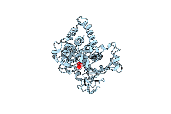 |
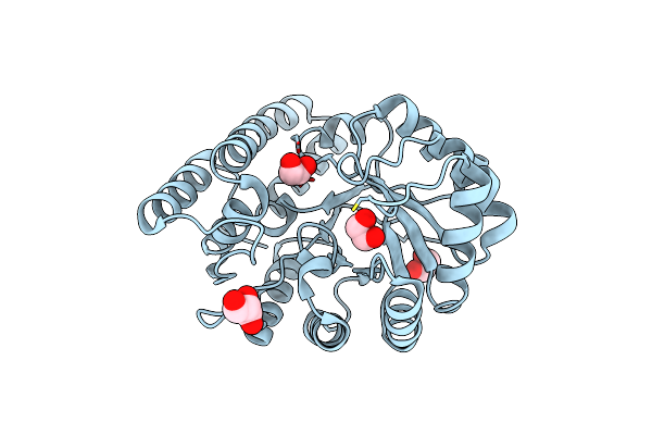
Structural Insight Into A Promiscuous Ce15 Esterase From The Marine Bacterial Metagenome
Organism: Unknown prokaryotic organism
Method: X-RAY DIFFRACTION Resolution:1.90 Å Release Date: 2018-03-21 Classification: HYDROLASE Ligands: GOL |
Organism: Unknown prokaryotic organism
Method: X-RAY DIFFRACTION
Release Date: 2018-03-21
Ligands: GOL
 |
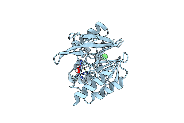
Native Structure Of N-Acetylneuramininate Lyase (Sialic Acid Aldolase) From Aliivibrio Salmonicida
Organism: Aliivibrio salmonicida
Method: X-RAY DIFFRACTION Resolution:1.65 Å Release Date: 2016-03-02 Classification: LYASE Ligands: GOL, EDO |
Organism: Aliivibrio salmonicida
Method: X-RAY DIFFRACTION
Release Date: 2016-03-02
Ligands: GOL, EDO
 |
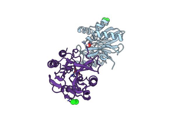
Vim-2-Nat, Discovery Of Novel Inhibitor Scaffolds Against The Metallo- Beta-Lactamase Vim-2 By Spr Based Fragment Screening
Organism: Pseudomonas aeruginosa
Method: X-RAY DIFFRACTION Resolution:2.10 Å Release Date: 2015-11-04 Classification: HYDROLASE Ligands: OH, ZN, CL |
Organism: Pseudomonas aeruginosa
Method: X-RAY DIFFRACTION
Release Date: 2015-11-04
Ligands: OH, ZN, CL
 |
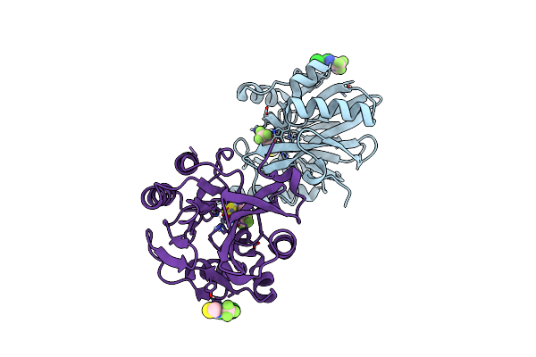
Vim-2-Ox, Discovery Of Novel Inhibitor Scaffolds Against The Metallo- Beta-Lactamase Vim-2 By Spr Based Fragment Screening
Organism: Pseudomonas aeruginosa
Method: X-RAY DIFFRACTION Resolution:1.96 Å Release Date: 2015-11-04 Classification: HYDROLASE Ligands: OH, ZN, CL |
Organism: Pseudomonas aeruginosa
Method: X-RAY DIFFRACTION
Release Date: 2015-11-04
Ligands: OH, ZN, CL
 |
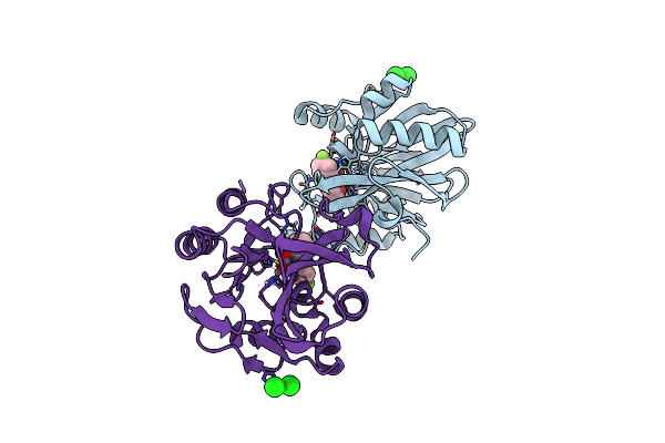
Vim-2-1, Discovery Of Novel Inhibitor Scaffolds Against The Metallo- Beta-Lactamase Vim-2 By Spr Based Fragment Screening
Organism: Pseudomonas aeruginosa
Method: X-RAY DIFFRACTION Resolution:1.80 Å Release Date: 2015-11-04 Classification: HYDROLASE Ligands: ZN, RHU, CL, OH |
Organism: Pseudomonas aeruginosa
Method: X-RAY DIFFRACTION
Release Date: 2015-11-04
Ligands: ZN, RHU, CL, OH
 |
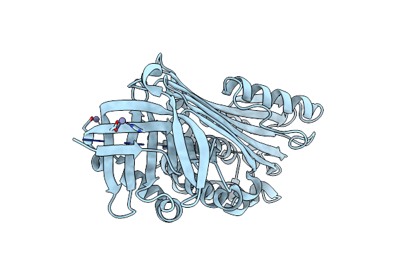
Vim-2-2, Discovery Of Novel Inhibitor Scaffolds Against The Metallo- Beta-Lactamase Vim-2 By Spr Based Fragment Screening
Organism: Pseudomonas aeruginosa
Method: X-RAY DIFFRACTION Resolution:1.80 Å Release Date: 2015-11-04 Classification: HYDROLASE Ligands: ZN, WL3, OH, CL |
Organism: Pseudomonas aeruginosa
Method: X-RAY DIFFRACTION
Release Date: 2015-11-04
Ligands: ZN, WL3, OH, CL
 |
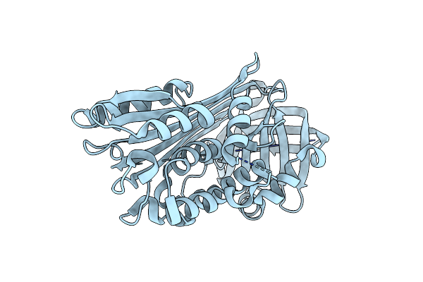
Psoriasis Pathogenesis - Pso P27 Constitute A Compact Structure Forming Large Aggregates. High Ph Structure
Organism: Homo sapiens
Method: X-RAY DIFFRACTION Resolution:2.15 Å Release Date: 2015-07-01 Classification: IMMUNE SYSTEM Ligands: ZN |
Organism: Homo sapiens
Method: X-RAY DIFFRACTION
Release Date: 2015-07-01
Ligands: ZN
 |
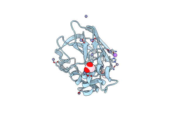
Psoriasis Pathogenesis - Pso P27 Constitute A Compact Structure Forming Large Aggregates. Low Ph Structure
Organism: Homo sapiens
Method: X-RAY DIFFRACTION Resolution:2.00 Å Release Date: 2015-07-01 Classification: IMMUNE SYSTEM |
Organism: Homo sapiens
Method: X-RAY DIFFRACTION
Release Date: 2015-07-01
 |
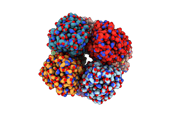
Structure Of Sclpmo10B In Complex With Copper.
Organism: Streptomyces coelicolor
Method: X-RAY DIFFRACTION Resolution:1.29 Å Release Date: 2014-05-28 Classification: OXIDOREDUCTASE Ligands: CU, ZN, NA, ACT |
Organism: Streptomyces coelicolor
Method: X-RAY DIFFRACTION
Release Date: 2014-05-28
Ligands: CU, ZN, NA, ACT
 |
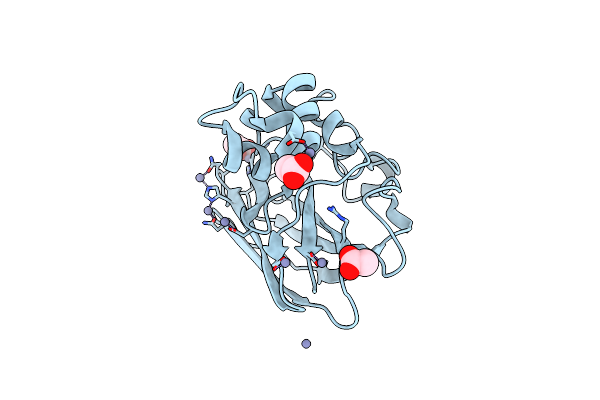
Structure Of Cellulose Active Lpmo Cels2 (Sclpmo10C) In Complex With Copper.
Organism: Streptomyces coelicolor
Method: X-RAY DIFFRACTION Resolution:1.50 Å Release Date: 2014-05-28 Classification: OXIDOREDUCTASE Ligands: CU, CA |
Organism: Streptomyces coelicolor
Method: X-RAY DIFFRACTION
Release Date: 2014-05-28
Ligands: CU, CA
 |
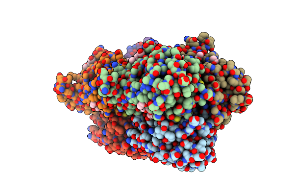
Structure Of Sclpmo10B In Complex With Zinc.
Organism: Streptomyces coelicolor
Method: X-RAY DIFFRACTION Resolution:1.40 Å Release Date: 2014-05-28 Classification: OXIDOREDUCTASE Ligands: ZN, ACT |
Organism: Streptomyces coelicolor
Method: X-RAY DIFFRACTION
Release Date: 2014-05-28
Ligands: ZN, ACT
 |
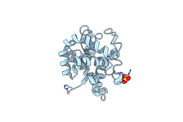
Cora Is A Surface-Associated Copper-Binding Protein Important In Methylomicrobium Album Bg8 Copper Acquisition
Organism: Methylomicrobium album bg8
Method: X-RAY DIFFRACTION Resolution:1.60 Å Release Date: 2014-02-19 Classification: COPPER-BINDING PROTEIN Ligands: CU1, CA, PG4, P6G |
Organism: Methylomicrobium album bg8
Method: X-RAY DIFFRACTION
Release Date: 2014-02-19
Ligands: CU1, CA, PG4, P6G
 |
Thermodynamics And Structure Of A Salmon Cold-Active Goose-Type Lysozyme
Organism: Salmo salar
Method: X-RAY DIFFRACTION Resolution:1.75 Å Release Date: 2010-05-05 Classification: HYDROLASE Ligands: CO, SO4 |
Organism: Salmo salar
Method: X-RAY DIFFRACTION
Release Date: 2010-05-05
Ligands: CO, SO4
 |
The Crystal Structure Of G-Type Lysozyme From Atlantic Cod (Gadus Morhua L.) In Complex With Nag Oligomers Sheds New Light On Substrate Binding And The Catalytic Mechanism. Native Structure To 1.9
Organism: Gadus morhua
Method: X-RAY DIFFRACTION Resolution:1.90 Å Release Date: 2009-10-20 Classification: HYDROLASE Ligands: CO |
Organism: Gadus morhua
Method: X-RAY DIFFRACTION
Release Date: 2009-10-20
Ligands: CO
 |
The Crystal Structure Of G-Type Lysozyme From Atlantic Cod (Gadus Morhua L.) In Complex With Nag Oligomers Sheds New Light On Substrate Binding And The Catalytic Mechanism. Structure With Nag To 1.7
Organism: Gadus morhua
Method: X-RAY DIFFRACTION Resolution:1.70 Å Release Date: 2009-10-20 Classification: HYDROLASE |
Organism: Gadus morhua
Method: X-RAY DIFFRACTION
Release Date: 2009-10-20