

Search Count: 25
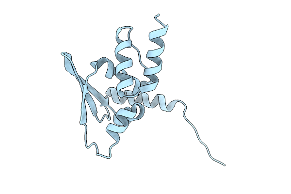 |
Crystal Structure Of The Btb Domain Mouse Keap1
Organism: Mus musculus
Method: X-RAY DIFFRACTION Resolution:2.80 Å Release Date: 2025-10-22 Classification: TRANSCRIPTION |
Organism: Mus musculus
Method: X-RAY DIFFRACTION
Release Date: 2025-10-22
 |
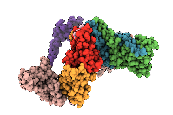
Crystal Structure Of The Btb Domain Mouse Keap1 In Complex With Cddo-Im
Organism: Mus musculus
Method: X-RAY DIFFRACTION Resolution:2.44 Å Release Date: 2025-10-22 Classification: TRANSCRIPTION Ligands: SXJ, CA |
Organism: Mus musculus
Method: X-RAY DIFFRACTION
Release Date: 2025-10-22
Ligands: SXJ, CA
 |
Crystal Structure Of The Btb Domain Mouse Keap1 In Complex With Cddo-Me
Organism: Mus musculus
Method: X-RAY DIFFRACTION Resolution:3.43 Å Release Date: 2025-10-22 Classification: TRANSCRIPTION Ligands: A1L9B |
Organism: Mus musculus
Method: X-RAY DIFFRACTION
Release Date: 2025-10-22
Ligands: A1L9B
 |
Crystal Structure Of Cgl1 From Crassostrea Gigas, Mannobiose-Bound Form (Cgl1/Man(Alpha)1-2Man)
Organism: Crassostrea gigas
Method: X-RAY DIFFRACTION Resolution:1.00 Å Release Date: 2023-10-25 Classification: SUGAR BINDING PROTEIN Ligands: ACY, MAN, CAC |
Organism: Crassostrea gigas
Method: X-RAY DIFFRACTION
Release Date: 2023-10-25
Ligands: ACY, MAN, CAC
 |
Crystal Structure Of Cgl1 From Crassostrea Gigas, Mannotriose-Bound Form (Cgl1/Man(Alpha)1-2Man(Alpha)1-2Man)
Organism: Crassostrea gigas
Method: X-RAY DIFFRACTION Resolution:0.97 Å Release Date: 2023-10-25 Classification: SUGAR BINDING PROTEIN Ligands: ACY, MG, CAC, MAN |
Organism: Crassostrea gigas
Method: X-RAY DIFFRACTION
Release Date: 2023-10-25
Ligands: ACY, MG, CAC, MAN
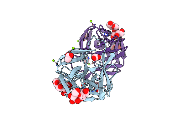 |
Crystal Structure Of Cgl1 From Crassostrea Gigas, Mannotetraose-Bound Form (Cgl1/Man(Alpha)1-2Man(Alpha)1-2Man(Alpha)1-6Man)
Organism: Crassostrea gigas
Method: X-RAY DIFFRACTION Resolution:1.30 Å Release Date: 2023-10-25 Classification: SUGAR BINDING PROTEIN Ligands: ACY, MG |
Organism: Crassostrea gigas
Method: X-RAY DIFFRACTION
Release Date: 2023-10-25
Ligands: ACY, MG
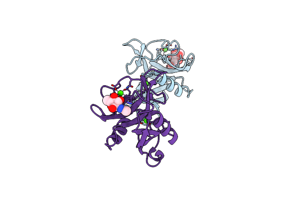 |
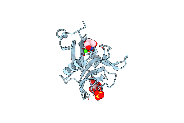
Spl-1 - Glcnac Complex
Organism: Saxidomus purpuratus
Method: X-RAY DIFFRACTION Resolution:1.70 Å Release Date: 2020-06-10 Classification: SUGAR BINDING PROTEIN Ligands: CA, NAG |
Organism: Saxidomus purpuratus
Method: X-RAY DIFFRACTION
Release Date: 2020-06-10
Ligands: CA, NAG
 |
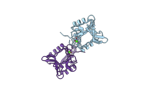
Ca2+-Independent C-Type Lectin Spl-2 From Saxidomus Purpuratus
Organism: Saxidomus purpuratus
Method: X-RAY DIFFRACTION Resolution:2.00 Å Release Date: 2019-03-20 Classification: SUGAR BINDING PROTEIN Ligands: NGA, TRS, CA, SO4 |
Organism: Saxidomus purpuratus
Method: X-RAY DIFFRACTION
Release Date: 2019-03-20
Ligands: NGA, TRS, CA, SO4
 |
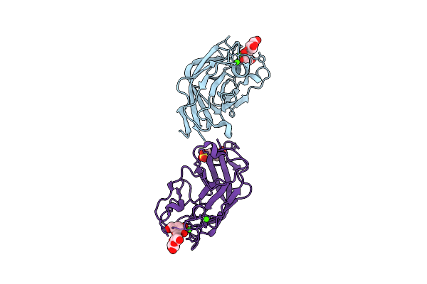
Ca2+-Independent C-Type Lectin Spl-1 From Saxidomus Purpuratus
Organism: Saxidomus purpuratus
Method: X-RAY DIFFRACTION Resolution:1.60 Å Release Date: 2019-03-20 Classification: SUGAR BINDING PROTEIN Ligands: CA |
Organism: Saxidomus purpuratus
Method: X-RAY DIFFRACTION
Release Date: 2019-03-20
Ligands: CA
 |
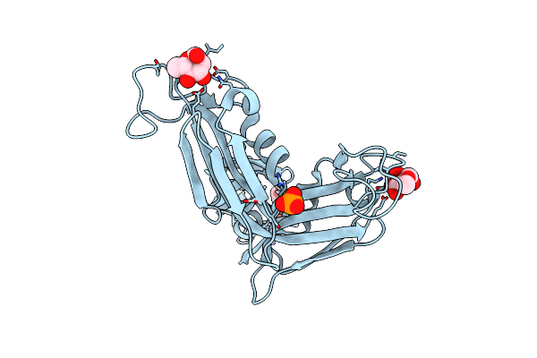
Ajlec From The Sea Anemone Anthopleura Japonica
Organism: Anthopleura japonica
Method: X-RAY DIFFRACTION Resolution:1.20 Å Release Date: 2018-07-25 Classification: SUGAR BINDING PROTEIN Ligands: CA, EPE |
Organism: Anthopleura japonica
Method: X-RAY DIFFRACTION
Release Date: 2018-07-25
Ligands: CA, EPE
 |
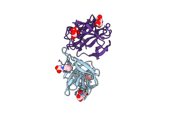
Crystal Structure Of A Rhamnose-Binding Lectin Sul-I From The Toxopneustid Sea Urchin Toxopneustes Pileolus
Organism: Toxopneustes pileolus
Method: X-RAY DIFFRACTION Resolution:1.80 Å Release Date: 2017-05-17 Classification: SUGAR BINDING PROTEIN Ligands: RAM, PO4 |
Organism: Toxopneustes pileolus
Method: X-RAY DIFFRACTION
Release Date: 2017-05-17
Ligands: RAM, PO4
 |
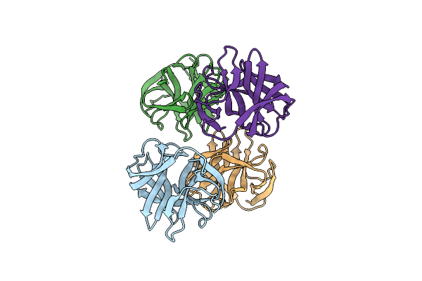
Crystal Structure Of Cgl1 From Crassostrea Gigas, Mannose-Bound Form (Cgl1/Man2)
Organism: Crassostrea gigas
Method: X-RAY DIFFRACTION Resolution:1.00 Å Release Date: 2016-07-06 Classification: SUGAR BINDING PROTEIN Ligands: BMA, MAN, NHE |
Organism: Crassostrea gigas
Method: X-RAY DIFFRACTION
Release Date: 2016-07-06
Ligands: BMA, MAN, NHE
 |
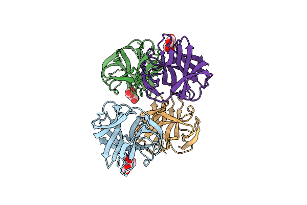
Crystal Structure Of Cgl1 From Crassostrea Gigas, Ligand Free Form (Cgl1/Free)
Organism: Crassostrea gigas
Method: X-RAY DIFFRACTION Resolution:1.10 Å Release Date: 2016-06-29 Classification: SUGAR BINDING PROTEIN |
Organism: Crassostrea gigas
Method: X-RAY DIFFRACTION
Release Date: 2016-06-29
 |
Crystal Structure Of Cgl1 From Crassostrea Gigas, Mannose-Bound Form (Cgl1/Man)
Organism: Crassostrea gigas
Method: X-RAY DIFFRACTION Resolution:1.10 Å Release Date: 2016-06-29 Classification: SUGAR BINDING PROTEIN Ligands: BMA |
Organism: Crassostrea gigas
Method: X-RAY DIFFRACTION
Release Date: 2016-06-29
Ligands: BMA
 |
Structure Of Epnh Mutant Of Cel-I
Organism: Cucumaria echinata
Method: X-RAY DIFFRACTION Resolution:1.70 Å Release Date: 2015-04-29 Classification: SUGAR BINDING PROTEIN Ligands: CA, MAN |
Organism: Cucumaria echinata
Method: X-RAY DIFFRACTION
Release Date: 2015-04-29
Ligands: CA, MAN
 |
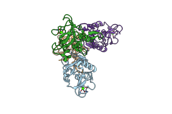
Pore-Forming Cel-Iii
Organism: Cucumaria echinata
Method: X-RAY DIFFRACTION Resolution:2.90 Å Release Date: 2014-03-19 Classification: TOXIN Ligands: CA, MG |
Organism: Cucumaria echinata
Method: X-RAY DIFFRACTION
Release Date: 2014-03-19
Ligands: CA, MG
 |
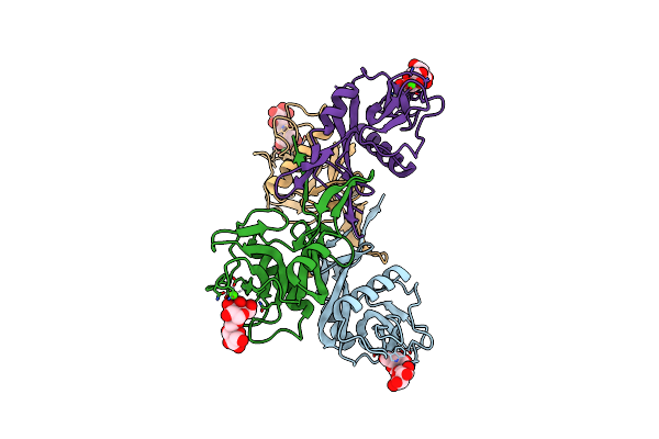
Crystal Structure Of Cel-Iv
Organism: Cucumaria echinata
Method: X-RAY DIFFRACTION Resolution:3.00 Å Release Date: 2011-01-19 Classification: SUGAR BINDING PROTEIN Ligands: CA |
Organism: Cucumaria echinata
Method: X-RAY DIFFRACTION
Release Date: 2011-01-19
Ligands: CA
 |
Crystal Structure Of Cel-Iv Complexed With Melibiose
Organism: Cucumaria echinata
Method: X-RAY DIFFRACTION Resolution:2.50 Å Release Date: 2011-01-19 Classification: SUGAR BINDING PROTEIN Ligands: CA |
Organism: Cucumaria echinata
Method: X-RAY DIFFRACTION
Release Date: 2011-01-19
Ligands: CA
 |
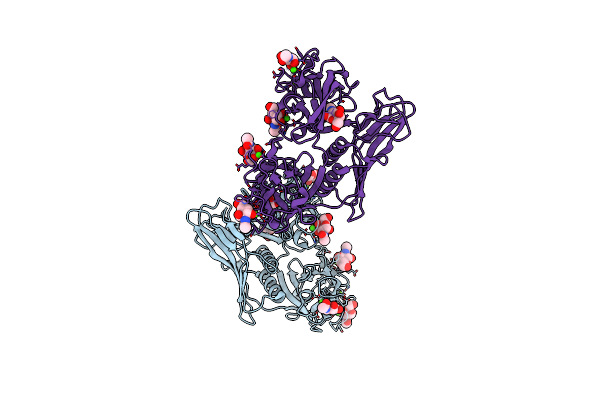
Crystal Structure Of Cel-Iv Complexed With Raffinose
Organism: Cucumaria echinata
Method: X-RAY DIFFRACTION Resolution:1.65 Å Release Date: 2011-01-19 Classification: SUGAR BINDING PROTEIN Ligands: EDO, CA |
Organism: Cucumaria echinata
Method: X-RAY DIFFRACTION
Release Date: 2011-01-19
Ligands: EDO, CA
 |
Crystal Structure Of Hemolytic Lectin Cel-Iii Complexed With Galnac
Organism: Cucumaria echinata
Method: X-RAY DIFFRACTION Resolution:1.70 Å Release Date: 2007-10-30 Classification: TOXIN Ligands: A2G, NGA, CA, MG |
Organism: Cucumaria echinata
Method: X-RAY DIFFRACTION
Release Date: 2007-10-30
Ligands: A2G, NGA, CA, MG