

Search Count: 32
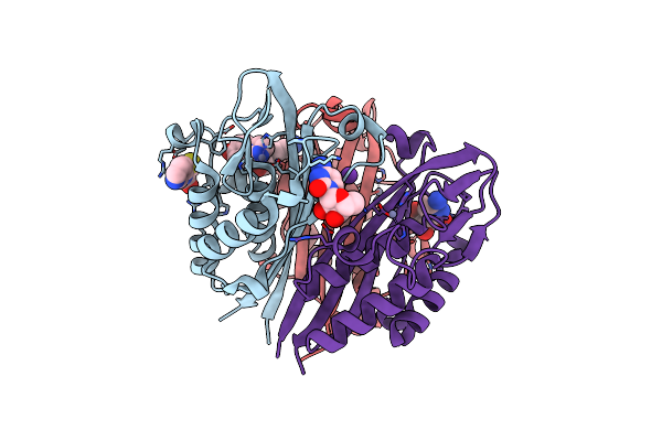 |
Crystal Structure Of 2-C-Methyl-D-Erythritol 2,4-Cyclodiphosphate Synthase From Burkholderia Pseudomallei Bound To Cytidine, Fol795 And Fol955
Organism: Burkholderia pseudomallei
Method: X-RAY DIFFRACTION Resolution:1.70 Å Release Date: 2011-02-09 Classification: LYASE Ligands: ZN, CL, CTN, MSR, 795, K |
Organism: Burkholderia pseudomallei
Method: X-RAY DIFFRACTION
Release Date: 2011-02-09
Ligands: ZN, CL, CTN, MSR, 795, K
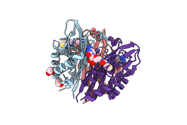 |
Crystal Structure Of 2C-Methyl-D-Erythritol 2,4-Cyclodiphosphate Synthase From Burkholderia Pseudomallei In Complex With Cytidine Derivative Ebsi01028
Organism: Burkholderia pseudomallei
Method: X-RAY DIFFRACTION Resolution:1.75 Å Release Date: 2011-01-19 Classification: LYASE Ligands: ZN, AO9, K, EDO |
Organism: Burkholderia pseudomallei
Method: X-RAY DIFFRACTION
Release Date: 2011-01-19
Ligands: ZN, AO9, K, EDO
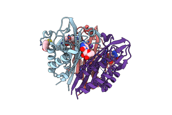 |
Crystal Structure Of 2-C-Methyl-D-Erythritol 2,4-Cyclodiphosphate Synthase From Burkholderia Pseudomallei With Cytidine And Fol694, 2-(Thiophen-2-Yl)Phenyl Methanol
Organism: Burkholderia pseudomallei
Method: X-RAY DIFFRACTION Resolution:1.70 Å Release Date: 2010-11-10 Classification: LYASE Ligands: CL, ZN, CTN, F69, K |
Organism: Burkholderia pseudomallei
Method: X-RAY DIFFRACTION
Release Date: 2010-11-10
Ligands: CL, ZN, CTN, F69, K
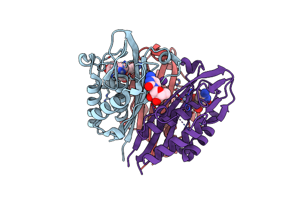 |
Crystal Structure Of 2-C-Methyl-D-Erythritol 2,4-Cyclodiphosphate Synthase From Burkholderia Pseudomallei With Cytidine And Fol955, 4-(1H-Imidazol)-1-Yl)Phenol
Organism: Burkholderia pseudomallei
Method: X-RAY DIFFRACTION Resolution:1.95 Å Release Date: 2010-10-20 Classification: LYASE Ligands: CL, ZN, MSR, CTN, K |
Organism: Burkholderia pseudomallei
Method: X-RAY DIFFRACTION
Release Date: 2010-10-20
Ligands: CL, ZN, MSR, CTN, K
 |
Co-Crystal Structure Of Dihydrofolate Reductase-Thymidylate Synthase From Babesia Bovis With Dump, Raltitrexed And Nadp
Organism: Babesia bovis
Method: X-RAY DIFFRACTION Resolution:1.80 Å Release Date: 2010-07-14 Classification: TRANSFERASE Ligands: CL, NAP, D16, UMP, EDO, GOL, PO4 |
Organism: Babesia bovis
Method: X-RAY DIFFRACTION
Release Date: 2010-07-14
Ligands: CL, NAP, D16, UMP, EDO, GOL, PO4
 |
Crystal Structure Of 2C-Methyl-D-Erythritol 2,4-Cyclodiphosphate Synthase From Burkholderia Pseudomallei With Cytosine And Fol Fragment 717, Imidazo[2,1-B][1,3]Thiazol-6-Ylmethanol
Organism: Burkholderia pseudomallei
Method: X-RAY DIFFRACTION Resolution:1.80 Å Release Date: 2010-04-07 Classification: LYASE Ligands: ZN, CYT, 717, TRS |
Organism: Burkholderia pseudomallei
Method: X-RAY DIFFRACTION
Release Date: 2010-04-07
Ligands: ZN, CYT, 717, TRS
 |
Crystal Structure Of Phosphoglyceromutase From Burkholderia Pseudomallei 1710B With Bound Malonic Acid
Organism: Burkholderia pseudomallei
Method: X-RAY DIFFRACTION Resolution:2.10 Å Release Date: 2010-02-09 Classification: ISOMERASE |
Organism: Burkholderia pseudomallei
Method: X-RAY DIFFRACTION
Release Date: 2010-02-09
 |
Crystal Structure Of 2C-Methyl-D-Erythritol 2,4-Cyclodiphosphate Synthase From Burkholderia Pseudomallei In Complex With A Fragment-Nucleoside Fusion D000161829
Organism: Burkholderia pseudomallei
Method: X-RAY DIFFRACTION Resolution:2.05 Å Release Date: 2009-11-10 Classification: LYASE Ligands: ZN, 829, K |
Organism: Burkholderia pseudomallei
Method: X-RAY DIFFRACTION
Release Date: 2009-11-10
Ligands: ZN, 829, K
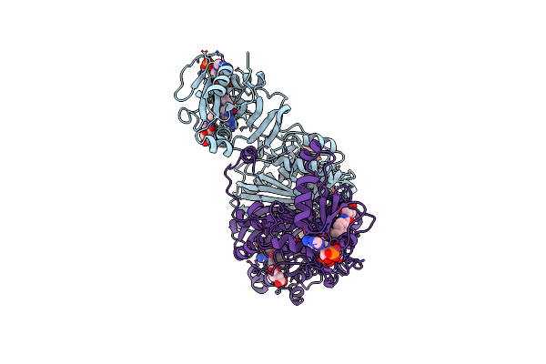 |
Co-Crystal Structure Of Dihydrofolate Reductase/Thymidylate Synthase From Babesia Bovis With Dump, Pemetrexed And Nadp
Organism: Babesia bovis
Method: X-RAY DIFFRACTION Resolution:2.20 Å Release Date: 2009-10-13 Classification: TRANSFERASE Ligands: UMP, LYA, NAP, EDO |
Organism: Babesia bovis
Method: X-RAY DIFFRACTION
Release Date: 2009-10-13
Ligands: UMP, LYA, NAP, EDO
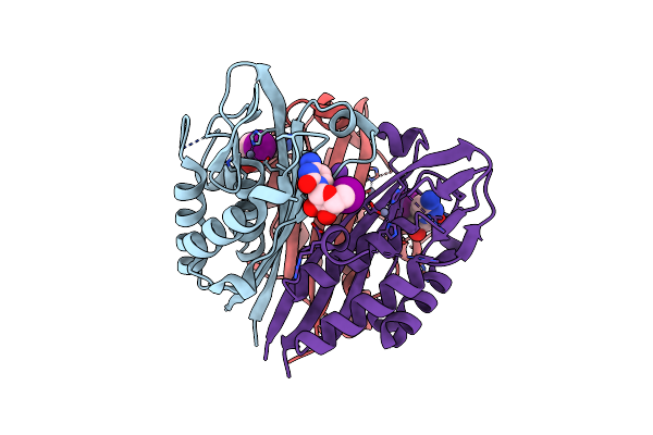 |
Crystal Structure Of 2C-Methyl-D-Erythritol 2,4-Cyclodiphosphate Synthase From Burkholderia Pseudomallei In Complex With 5'-Iodo-Cytosine
Organism: Burkholderia pseudomallei
Method: X-RAY DIFFRACTION Resolution:1.85 Å Release Date: 2009-10-13 Classification: LYASE Ligands: ZN, I5A, CL, K |
Organism: Burkholderia pseudomallei
Method: X-RAY DIFFRACTION
Release Date: 2009-10-13
Ligands: ZN, I5A, CL, K
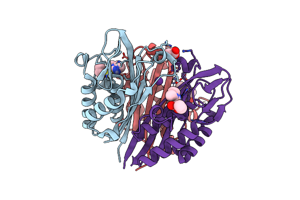 |
Co-Crystal Structure Of 2C-Methyl-D-Erythritol 2,4-Cyclodiphosphate Synthase From Burkholderia Pseudomallei With Fol Fragment 535, Ethyl 3-Methyl-5,6-Dihydroimidazo[2,1-B][1,3]Thiazole-2-Carboxylate
Organism: Burkholderia pseudomallei
Method: X-RAY DIFFRACTION Resolution:1.70 Å Release Date: 2009-10-06 Classification: LYASE Ligands: ZN, 535, CL, K, ACT |
Organism: Burkholderia pseudomallei
Method: X-RAY DIFFRACTION
Release Date: 2009-10-06
Ligands: ZN, 535, CL, K, ACT
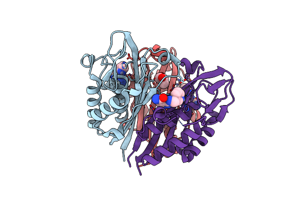 |
Crystal Structure Of 2C-Methyl-D-Erythritol-2,4-Cyclodiphosphate Synthase From Burkholderia Pseudomallei With Fol Fragment 8395
Organism: Burkholderia pseudomallei
Method: X-RAY DIFFRACTION Resolution:1.69 Å Release Date: 2009-09-29 Classification: LYASE Ligands: ZN, HHV, GOL |
Organism: Burkholderia pseudomallei
Method: X-RAY DIFFRACTION
Release Date: 2009-09-29
Ligands: ZN, HHV, GOL
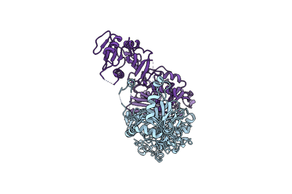 |
X-Ray Structure Dihydrofolate Reductase/Thymidylate Synthase From Babesia Bovis At 2.35A Resolution
Organism: Babesia bovis
Method: X-RAY DIFFRACTION Resolution:2.35 Å Release Date: 2009-08-18 Classification: TRANSFERASE Ligands: CL |
Organism: Babesia bovis
Method: X-RAY DIFFRACTION
Release Date: 2009-08-18
Ligands: CL
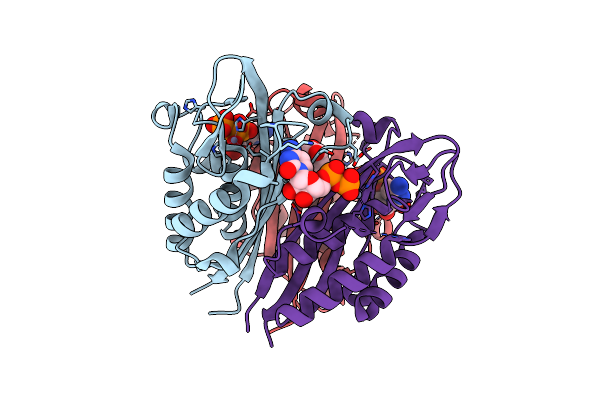 |
Crystal Structure Of 2C-Methyl-D-Erythritol 2,4-Cyclodiphosphate Synthase From Burkholderia Pseudomallei With Bound Ctp And Cdp
Organism: Burkholderia pseudomallei
Method: X-RAY DIFFRACTION Resolution:2.10 Å Release Date: 2009-08-18 Classification: LYASE Ligands: ZN, CDP, CTP, GOL |
Organism: Burkholderia pseudomallei
Method: X-RAY DIFFRACTION
Release Date: 2009-08-18
Ligands: ZN, CDP, CTP, GOL
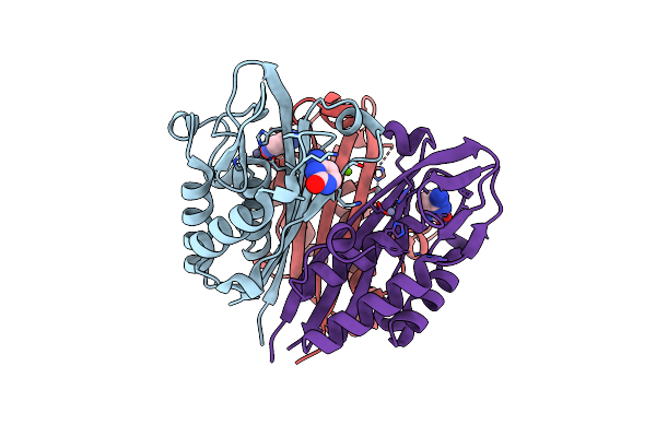 |
Crystal Structure Of 2C-Methyl-D-Erythritol 2,4-Cyclodiphosphate Synthase From Burkholderia Pseudomallei With Cytosine
Organism: Burkholderia pseudomallei
Method: X-RAY DIFFRACTION Resolution:2.30 Å Release Date: 2009-08-18 Classification: LYASE Ligands: ZN, CYT, CL, MG |
Organism: Burkholderia pseudomallei
Method: X-RAY DIFFRACTION
Release Date: 2009-08-18
Ligands: ZN, CYT, CL, MG
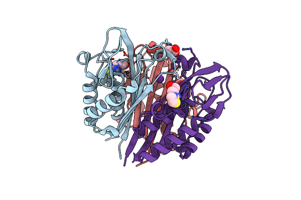 |
Crystal Structure Of 2C-Methyl-D-Erythritol 2,4-Cyclodiphosphate Synthase From Burkholderia Pseudomallei With Fol Fragment 717, Imidazo[2,,1-B][1,3]Thiazol-6-Ylmethanol
Organism: Burkholderia pseudomallei
Method: X-RAY DIFFRACTION Resolution:2.07 Å Release Date: 2009-08-18 Classification: LYASE Ligands: ZN, 717, CL, ACT, K |
Organism: Burkholderia pseudomallei
Method: X-RAY DIFFRACTION
Release Date: 2009-08-18
Ligands: ZN, 717, CL, ACT, K
 |
Crystal Structure Of 2C-Methyl-D-Erythritol 2,4-Cyclodiphosphate Synthase From Burkholderia Pseudomallei With Cytidine
Organism: Burkholderia pseudomallei
Method: X-RAY DIFFRACTION Resolution:2.10 Å Release Date: 2009-08-04 Classification: LYASE Ligands: CTN, ZN, CL, MG |
Organism: Burkholderia pseudomallei
Method: X-RAY DIFFRACTION
Release Date: 2009-08-04
Ligands: CTN, ZN, CL, MG
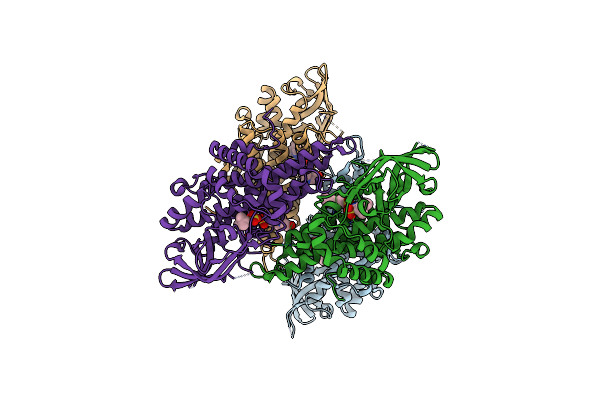 |
Crystal Structure Of Glutaryl-Coa Dehydrogenase From Burkholderia Pseudomallei With Fragment 6421
Organism: Burkholderia pseudomallei
Method: X-RAY DIFFRACTION Resolution:2.15 Å Release Date: 2009-07-07 Classification: OXIDOREDUCTASE Ligands: SO4, QQQ, EPE |
Organism: Burkholderia pseudomallei
Method: X-RAY DIFFRACTION
Release Date: 2009-07-07
Ligands: SO4, QQQ, EPE
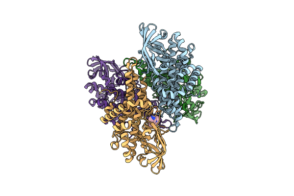 |
Crystal Structure Of Glutaryl-Coa Dehydrogenase From Burkholderia Pseudomallei With Fragment (1,4-Dimethyl-1,2,3,4-Tetrahydroquinoxalin-6-Yl)Methylamine
Organism: Burkholderia pseudomallei
Method: X-RAY DIFFRACTION Resolution:1.99 Å Release Date: 2009-04-28 Classification: OXIDOREDUCTASE Ligands: UFO |
Organism: Burkholderia pseudomallei
Method: X-RAY DIFFRACTION
Release Date: 2009-04-28
Ligands: UFO
 |
Crystal Structure Of Phosphoglyceromutase From Burkholderia Pseudomallei With Vanadate And Glycerol
Organism: Burkholderia pseudomallei
Method: X-RAY DIFFRACTION Resolution:1.93 Å Release Date: 2009-04-07 Classification: ISOMERASE Ligands: VO4, GOL, PG4 |
Organism: Burkholderia pseudomallei
Method: X-RAY DIFFRACTION
Release Date: 2009-04-07
Ligands: VO4, GOL, PG4