

Search Count: 26
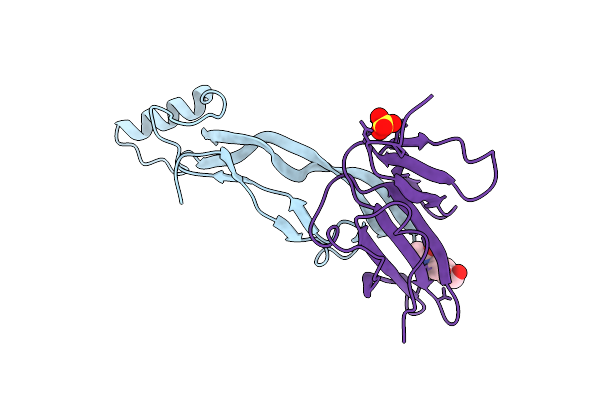 |
Structure Of Amh Bound To Amhr2-Ecd
Organism: Homo sapiens
Method: X-RAY DIFFRACTION Resolution:2.60 Å Release Date: 2021-06-23 Classification: SIGNALING PROTEIN Ligands: NAG, SO4 |
Organism: Homo sapiens
Method: X-RAY DIFFRACTION
Release Date: 2021-06-23
Ligands: NAG, SO4
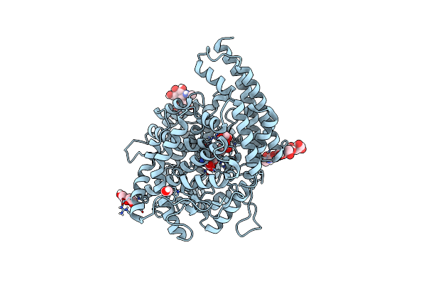 |
Crystal Structure Of Anopheles Gambiae Anoace2 In Complex With Gamma-Polyglutamic Acid.
Organism: Anopheles gambiae
Method: X-RAY DIFFRACTION Resolution:2.20 Å Release Date: 2019-11-13 Classification: HYDROLASE Ligands: PEG, EDO, ZN, KSN |
Organism: Anopheles gambiae
Method: X-RAY DIFFRACTION
Release Date: 2019-11-13
Ligands: PEG, EDO, ZN, KSN
 |
Crystal Structure Of Anopheles Gambiae Anoace2 In Complex With Fosinoprilat
Organism: Anopheles gambiae
Method: X-RAY DIFFRACTION Resolution:2.50 Å Release Date: 2019-11-13 Classification: HYDROLASE Ligands: KS8, ZN |
Organism: Anopheles gambiae
Method: X-RAY DIFFRACTION
Release Date: 2019-11-13
Ligands: KS8, ZN
 |
Crystal Structure Of The Gdf11:Follistatin 288 Complex
Organism: Homo sapiens
Method: X-RAY DIFFRACTION Resolution:2.35 Å Release Date: 2017-03-22 Classification: CYTOKINE/Signaling Protein Ligands: PO4, FLC |
Organism: Homo sapiens
Method: X-RAY DIFFRACTION
Release Date: 2017-03-22
Ligands: PO4, FLC
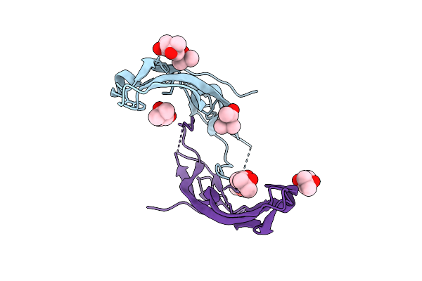 |
Crystal Structure Of Gdf8
Organism: Mus musculus
Method: X-RAY DIFFRACTION Resolution:2.25 Å Release Date: 2017-03-22 Classification: CYTOKINE Ligands: MPD |
Organism: Mus musculus
Method: X-RAY DIFFRACTION
Release Date: 2017-03-22
Ligands: MPD
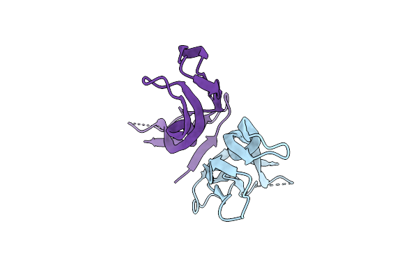 |
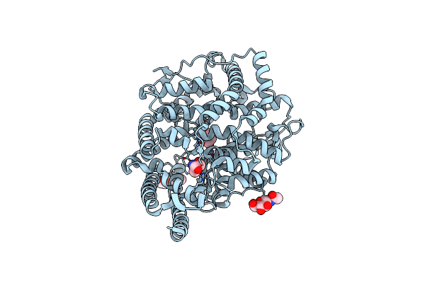
Apo-Structure Of Mature Growth Differentiation Factor 11
Organism: Homo sapiens
Method: X-RAY DIFFRACTION Resolution:1.90 Å Release Date: 2017-03-22 Classification: SIGNALING PROTEIN |
Organism: Homo sapiens
Method: X-RAY DIFFRACTION
Release Date: 2017-03-22
 |
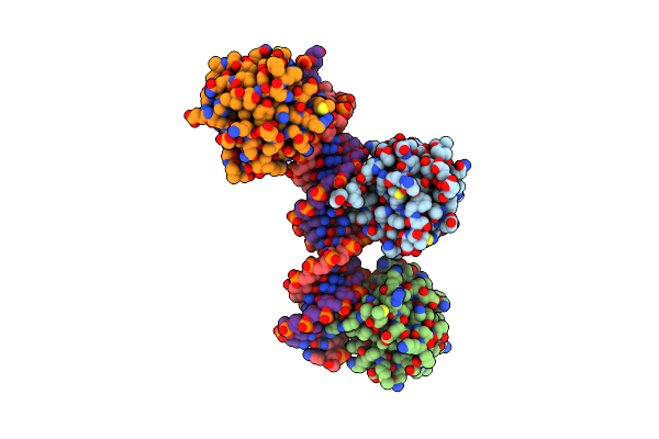
A New Crystal Structure Of The Drosophila Melanogaster Angiotensin Converting Enzyme Homologue Ance.
Organism: Drosophila melanogaster
Method: X-RAY DIFFRACTION Resolution:1.85 Å Release Date: 2015-08-26 Classification: HYDROLASE Ligands: MLT, ZN, NAG, TRS |
Organism: Drosophila melanogaster
Method: X-RAY DIFFRACTION
Release Date: 2015-08-26
Ligands: MLT, ZN, NAG, TRS
 |
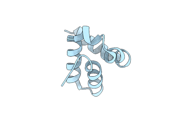
Polyomavirus T-Ag Binds Symmetrical Repeats At The Viral Origin In An Asymmetrical Manner
Organism: Mouse polyomavirus
Method: X-RAY DIFFRACTION Resolution:3.79 Å Release Date: 2013-10-23 Classification: DNA BINDING PROTEIN/DNA |
Organism: Mouse polyomavirus
Method: X-RAY DIFFRACTION
Release Date: 2013-10-23
 |
Crystal Structure Of The Human Spop C-Terminal Domain
Organism: Homo sapiens
Method: X-RAY DIFFRACTION Resolution:1.53 Å Release Date: 2013-09-11 Classification: PROTEIN BINDING |
Organism: Homo sapiens
Method: X-RAY DIFFRACTION
Release Date: 2013-09-11
 |
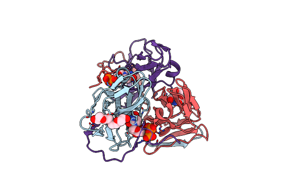
Crystal Structure Of The Human Spop Btb Domain
Organism: Homo sapiens
Method: X-RAY DIFFRACTION Resolution:2.42 Å Release Date: 2013-09-11 Classification: PROTEIN BINDING |
Organism: Homo sapiens
Method: X-RAY DIFFRACTION
Release Date: 2013-09-11
 |
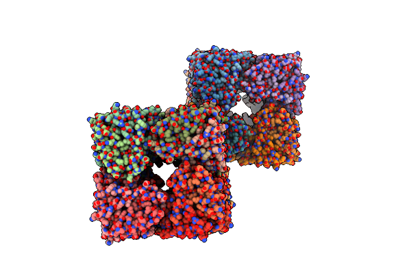
B. Subtilis Dutpase Yncf In Complex With Du, Ppi And Mg B (P212121)
Organism: Bacillus subtilis
Method: X-RAY DIFFRACTION Resolution:1.18 Å Release Date: 2013-05-15 Classification: HYDROLASE Ligands: DUR, CL, PG4, POP, MG |
Organism: Bacillus subtilis
Method: X-RAY DIFFRACTION
Release Date: 2013-05-15
Ligands: DUR, CL, PG4, POP, MG
 |
B. Subtilis Dutpase Yncf In Complex With Du, Ppi And Mg (P212121)
Organism: Bacillus subtilis
Method: X-RAY DIFFRACTION Resolution:2.05 Å Release Date: 2013-05-01 Classification: HYDROLASE Ligands: DUR, POP, MG, ACT, CL |
Organism: Bacillus subtilis
Method: X-RAY DIFFRACTION
Release Date: 2013-05-01
Ligands: DUR, POP, MG, ACT, CL
 |
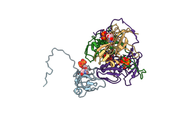
Structure Of B. Subtilis Genomic Dutpase Yncf In Complex With Du, Ppi And Mg In P1
Organism: Bacillus subtilis
Method: X-RAY DIFFRACTION Resolution:2.01 Å Release Date: 2013-04-24 Classification: HYDROLASE Ligands: DUR, POP, MG, PO4 |
Organism: Bacillus subtilis
Method: X-RAY DIFFRACTION
Release Date: 2013-04-24
Ligands: DUR, POP, MG, PO4
 |
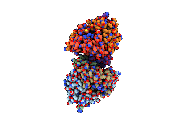
B. Subtilis Dutpase Yncf In Complex With Du Ppi And Mg In H32
Organism: Bacillus subtilis
Method: X-RAY DIFFRACTION Resolution:2.30 Å Release Date: 2013-04-17 Classification: HYDROLASE Ligands: DUR, POP, MG, PO4 |
Organism: Bacillus subtilis
Method: X-RAY DIFFRACTION
Release Date: 2013-04-17
Ligands: DUR, POP, MG, PO4
 |
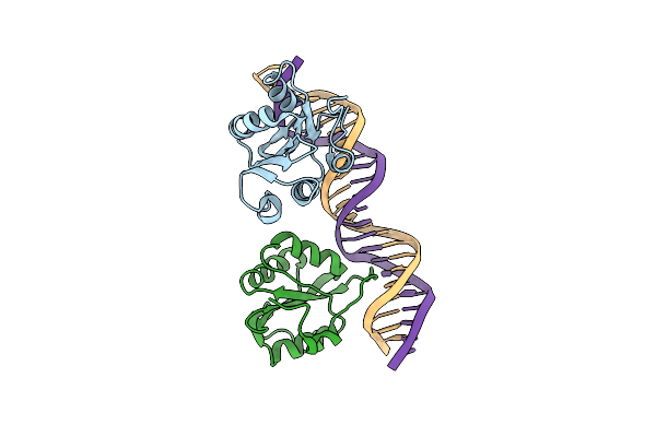
B. Subtilis Prophage Dutpase Yoss In Complex With Dump
Organism: Bacillus subtilis
Method: X-RAY DIFFRACTION Resolution:1.60 Å Release Date: 2013-04-03 Classification: HYDROLASE Ligands: UMP, NA |
Organism: Bacillus subtilis
Method: X-RAY DIFFRACTION
Release Date: 2013-04-03
Ligands: UMP, NA
 |
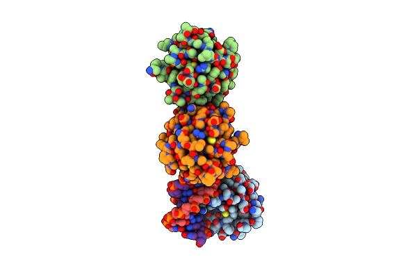
Crystal Structure Of The Sv40 Large T-Antigen Origin Bining Domain Bound To Site I Dna
Organism: Simian virus 40
Method: X-RAY DIFFRACTION Resolution:3.20 Å Release Date: 2013-01-23 Classification: DNA BINDING PROTEIN/DNA |
Organism: Simian virus 40
Method: X-RAY DIFFRACTION
Release Date: 2013-01-23
 |
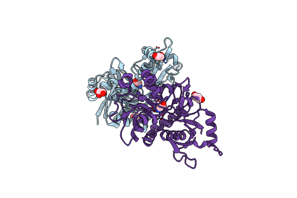
Asymmetric Assembly Of Merkel Cell Polyomavirus Large T-Antigen Origin Binding Domains At The Viral Origin
Organism: Merkel cell polyomavirus
Method: X-RAY DIFFRACTION Resolution:2.90 Å Release Date: 2011-04-27 Classification: DNA BINDING PROTEIN/DNA |
Organism: Merkel cell polyomavirus
Method: X-RAY DIFFRACTION
Release Date: 2011-04-27
 |
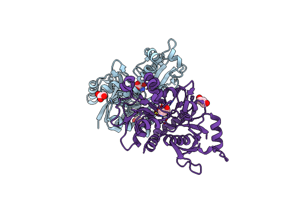
Crystal Structure Of The Nr3A Ligand Binding Core Complex With Glycine At 1.58 Angstrom Resolution
Organism: Rattus norvegicus
Method: X-RAY DIFFRACTION Resolution:1.58 Å Release Date: 2008-08-05 Classification: MEMBRANE PROTEIN Ligands: BR, CL, GOL, GLY |
Organism: Rattus norvegicus
Method: X-RAY DIFFRACTION
Release Date: 2008-08-05
Ligands: BR, CL, GOL, GLY
 |
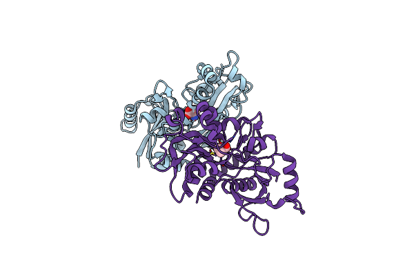
Crystal Structure Of The Nr3A Ligand Binding Core Complex With D-Serine At 1.45 Angstrom Resolution
Organism: Rattus norvegicus
Method: X-RAY DIFFRACTION Resolution:1.45 Å Release Date: 2008-08-05 Classification: MEMBRANE PROTEIN Ligands: CL, DSN, GOL |
Organism: Rattus norvegicus
Method: X-RAY DIFFRACTION
Release Date: 2008-08-05
Ligands: CL, DSN, GOL
 |
Crystal Structure Of The Nr3A Ligand Binding Core Complex With Acpc At 1.96 Angstrom Resolution
Organism: Rattus norvegicus
Method: X-RAY DIFFRACTION Resolution:1.96 Å Release Date: 2008-08-05 Classification: MEMBRANE PROTEIN Ligands: 1AC |
Organism: Rattus norvegicus
Method: X-RAY DIFFRACTION
Release Date: 2008-08-05
Ligands: 1AC