

Search Count: 16
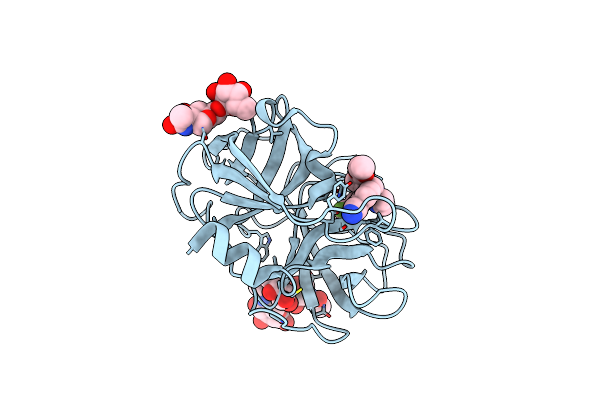 |
Crystal Structure Of Human Neutrophil Elastase In Complex With A Dihydropyrimidone Inhibitor
Organism: Homo sapiens
Method: X-RAY DIFFRACTION Resolution:2.23 Å Release Date: 2016-08-03 Classification: HYDROLASE Ligands: NAG, IUY |
Organism: Homo sapiens
Method: X-RAY DIFFRACTION
Release Date: 2016-08-03
Ligands: NAG, IUY
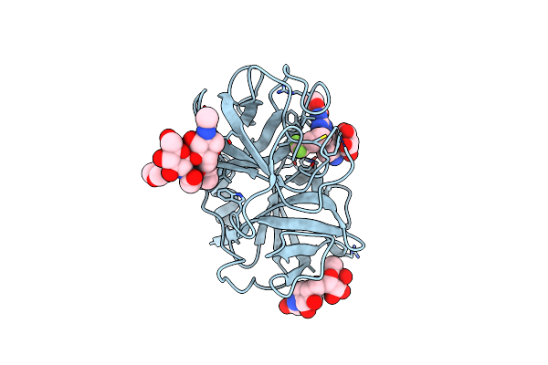 |
Crystal Structure Of Human Neutrophil Elastase In Complex With A Dihydropyrimidone Inhibitor
Organism: Homo sapiens
Method: X-RAY DIFFRACTION Resolution:1.90 Å Release Date: 2016-08-03 Classification: HYDROLASE Ligands: VBM, MES, MLI |
Organism: Homo sapiens
Method: X-RAY DIFFRACTION
Release Date: 2016-08-03
Ligands: VBM, MES, MLI
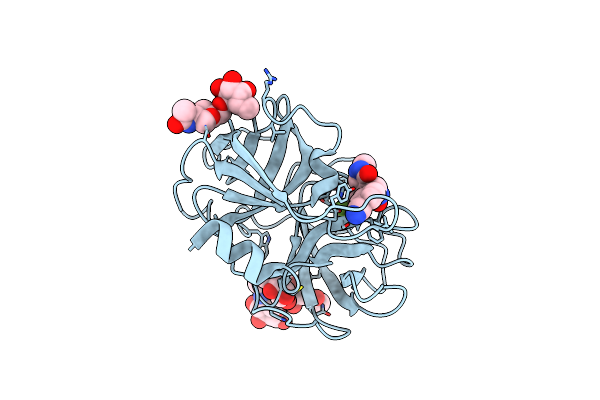 |
Crystal Structure Of Human Neutrophil Elastase In Complex With A Dihydropyrimidone Inhibitor
Organism: Homo sapiens
Method: X-RAY DIFFRACTION Resolution:2.00 Å Release Date: 2016-08-03 Classification: HYDROLASE Ligands: NAG, IUL |
Organism: Homo sapiens
Method: X-RAY DIFFRACTION
Release Date: 2016-08-03
Ligands: NAG, IUL
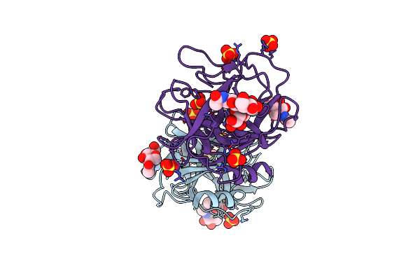 |
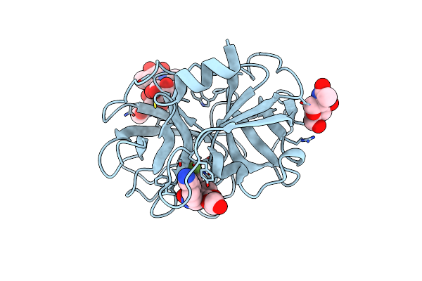
Structure Of Human Neutrophil Elastase (Uncomplexed)
Organism: Homo sapiens
Method: X-RAY DIFFRACTION Resolution:1.86 Å Release Date: 2011-05-11 Classification: HYDROLASE Ligands: NAG, SO4 |
Organism: Homo sapiens
Method: X-RAY DIFFRACTION
Release Date: 2011-05-11
Ligands: NAG, SO4
 |
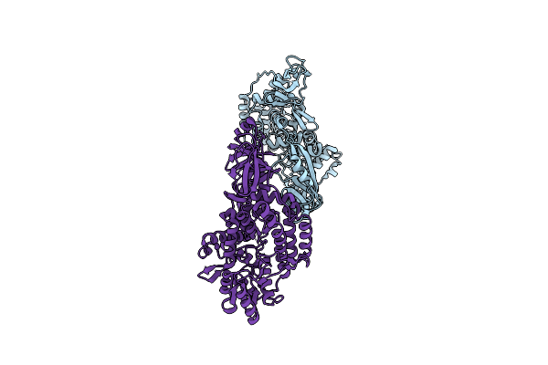
Structure Of Human Neutrophil Elastase In Complex With A Dihydropyrimidone Inhibitor
Organism: Homo sapiens
Method: X-RAY DIFFRACTION Resolution:2.00 Å Release Date: 2011-05-11 Classification: HYDROLASE/HYDROLASE INHIBITOR Ligands: NAG, 2HY |
Organism: Homo sapiens
Method: X-RAY DIFFRACTION
Release Date: 2011-05-11
Ligands: NAG, 2HY
 |
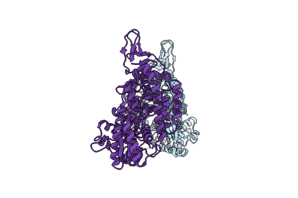
Structure Of Full Length Topoisomerase I From Thermotoga Maritima In Triclinic Crystal Form
Organism: Thermotoga maritima
Method: X-RAY DIFFRACTION Resolution:1.70 Å Release Date: 2006-04-25 Classification: ISOMERASE |
Organism: Thermotoga maritima
Method: X-RAY DIFFRACTION
Release Date: 2006-04-25
 |
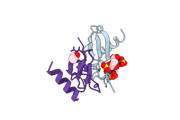
Structure Of Full Length Topoisomerase I From Thermotoga Maritima In Monoclinic Crystal Form
Organism: Thermotoga maritima
Method: X-RAY DIFFRACTION Resolution:1.95 Å Release Date: 2006-04-25 Classification: ISOMERASE |
Organism: Thermotoga maritima
Method: X-RAY DIFFRACTION
Release Date: 2006-04-25
 |
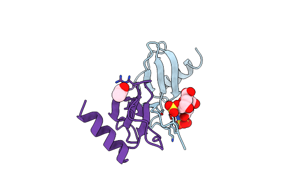
Human Rantes Complexed To Heparin-Derived Disaccharide I-S
Organism: Homo sapiens
Method: X-RAY DIFFRACTION Resolution:2.00 Å Release Date: 2004-11-09 Classification: ATTRACTANT Ligands: ACY |
Organism: Homo sapiens
Method: X-RAY DIFFRACTION
Release Date: 2004-11-09
Ligands: ACY
 |
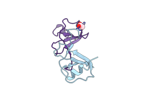
Human Rantes Complexed To Heparin-Derived Disaccharide Iii-S
Organism: Homo sapiens
Method: X-RAY DIFFRACTION Resolution:2.00 Å Release Date: 2004-11-09 Classification: ATTRACTANT Ligands: ACY |
Organism: Homo sapiens
Method: X-RAY DIFFRACTION
Release Date: 2004-11-09
Ligands: ACY
 |
Crystal Structure Of Human Rantes Mutant K45E
Organism: Homo sapiens
Method: X-RAY DIFFRACTION Resolution:1.70 Å Release Date: 2004-11-09 Classification: ATTRACTANT Ligands: ACY |
Organism: Homo sapiens
Method: X-RAY DIFFRACTION
Release Date: 2004-11-09
Ligands: ACY
 |
Crystal Structure Of Human Rantes Mutant 44-Aana-47
Organism: Homo sapiens
Method: X-RAY DIFFRACTION Resolution:2.20 Å Release Date: 2004-11-09 Classification: ATTRACTANT Ligands: SO4 |
Organism: Homo sapiens
Method: X-RAY DIFFRACTION
Release Date: 2004-11-09
Ligands: SO4
 |
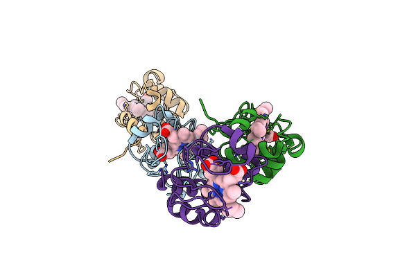
Crystal Structure Of The Unliganded Fv-Fragment Of The Anti-Cytochrome C Oxidase Antibody 7E2
Organism: Mus musculus
Method: X-RAY DIFFRACTION Resolution:1.28 Å Release Date: 2003-04-01 Classification: IMMUNE SYSTEM |
Organism: Mus musculus
Method: X-RAY DIFFRACTION
Release Date: 2003-04-01
 |
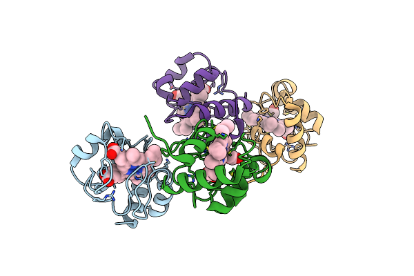
Structure Of The Soluble Domain Of Cytochrome C552 From Paracoccus Denitrificans In The Reduced State
Organism: Paracoccus denitrificans
Method: X-RAY DIFFRACTION Resolution:1.40 Å Release Date: 2000-02-06 Classification: ELECTRON TRANSPORT PROTEIN (CYTOCHROME) Ligands: HEC |
Organism: Paracoccus denitrificans
Method: X-RAY DIFFRACTION
Release Date: 2000-02-06
Ligands: HEC
 |
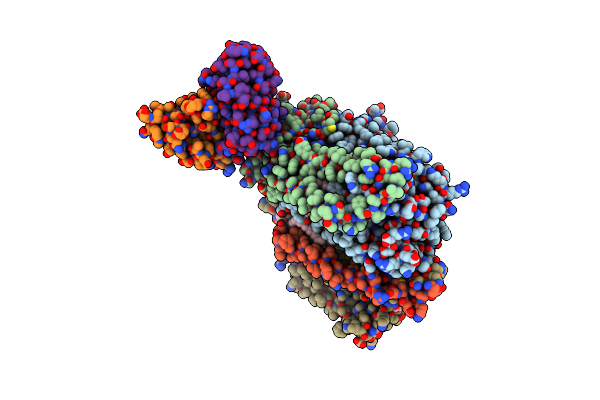
Structure Of The Soluble Domain Of Cytochrome C552 From Paracoccus Denitrificans In The Oxidised State
Organism: Paracoccus denitrificans
Method: X-RAY DIFFRACTION Resolution:1.50 Å Release Date: 2000-02-03 Classification: ELECTRON TRANSPORT PROTEIN (CYTOCHROME) Ligands: HEC |
Organism: Paracoccus denitrificans
Method: X-RAY DIFFRACTION
Release Date: 2000-02-03
Ligands: HEC
 |
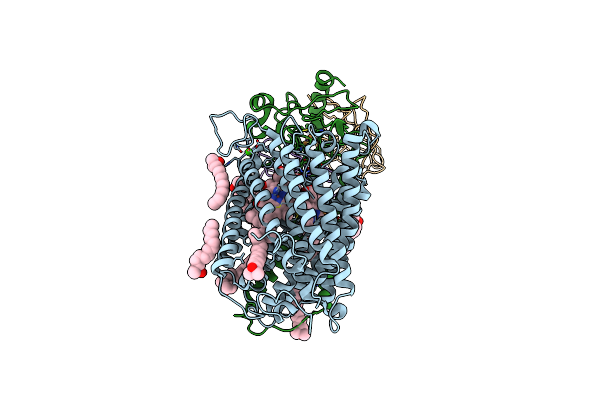
Cryo-Structure Of The Paracoccus Denitrificans Four-Subunit Cytochrome C Oxidase In The Completely Oxidized State Complexed With An Antibody Fv Fragment
Organism: Mus musculus, Paracoccus denitrificans
Method: X-RAY DIFFRACTION Resolution:3.00 Å Release Date: 1999-12-02 Classification: OXIDOREDUCTASE/IMMUNE SYSTEM Ligands: HEA, CU, CA, MN, CUA, PC1 |
Organism: Mus musculus, Paracoccus denitrificans
Method: X-RAY DIFFRACTION
Release Date: 1999-12-02
Ligands: HEA, CU, CA, MN, CUA, PC1
 |
Structure At 2.7 Angstrom Resolution Of The Paracoccus Denitrificans Two-Subunit Cytochrome C Oxidase Complexed With An Antibody Fv Fragment
Organism: Paracoccus denitrificans, Mus musculus
Method: X-RAY DIFFRACTION Resolution:2.70 Å Release Date: 1998-02-11 Classification: COMPLEX (OXIDOREDUCTASE/ANTIBODY) Ligands: CU, MG, CA, HEA, LDA |
Organism: Paracoccus denitrificans, Mus musculus
Method: X-RAY DIFFRACTION
Release Date: 1998-02-11
Ligands: CU, MG, CA, HEA, LDA