Deposition Date
2002-09-16
Release Date
2003-04-01
Last Version Date
2024-11-13
Entry Detail
PDB ID:
1MQK
Keywords:
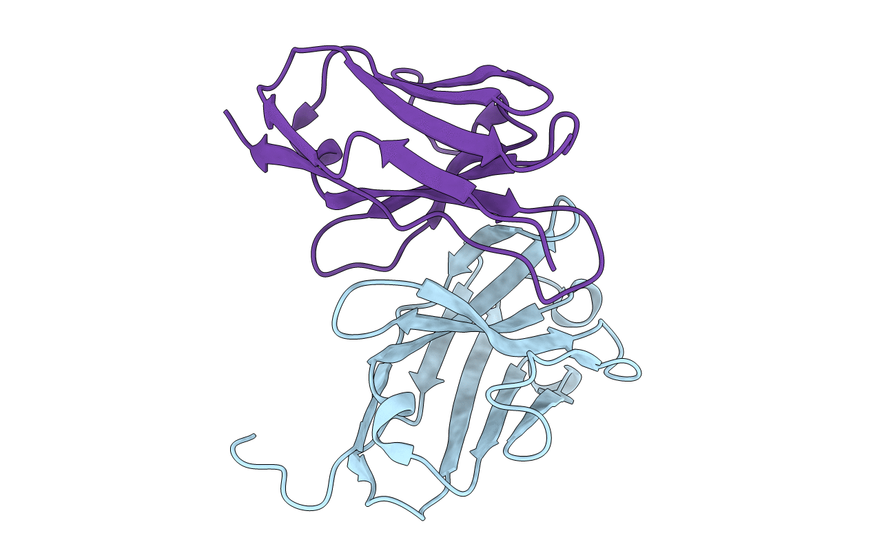
Title:
Crystal structure of the unliganded Fv-fragment of the anti-cytochrome C oxidase antibody 7E2
Biological Source:
Source Organism(s):
Mus musculus (Taxon ID: 10090)
Expression System(s):
Method Details:
Experimental Method:
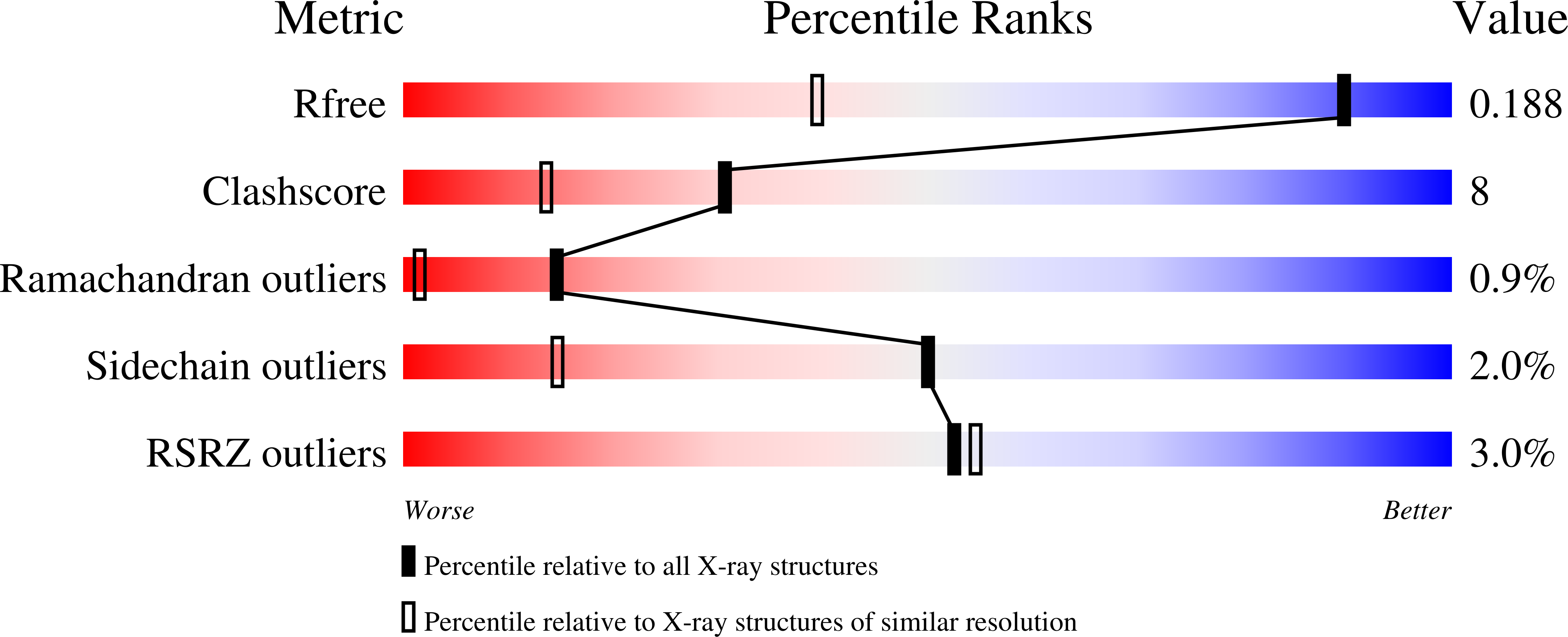
Resolution:
1.28 Å
R-Value Free:
0.19
R-Value Work:
0.13
R-Value Observed:
0.13
Space Group:
P 21 21 21