

Search Count: 47
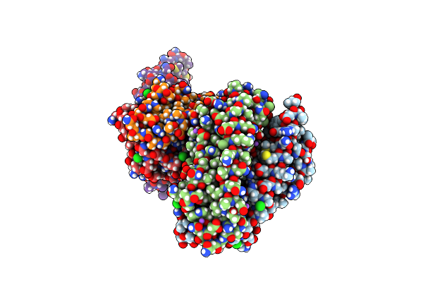 |
Salmonella Enterica Serovar Typhimurium Chemoreceptor Tsr (Taxis To Serine And Repellents) Ligand-Binding Domain In Complex With L-Serine
Organism: Salmonella enterica subsp. enterica serovar typhimurium
Method: X-RAY DIFFRACTION Resolution:2.20 Å Release Date: 2024-05-15 Classification: SIGNALING PROTEIN Ligands: CL, SER, NA |
Organism: Salmonella enterica subsp. enterica serovar typhimurium
Method: X-RAY DIFFRACTION
Release Date: 2024-05-15
Ligands: CL, SER, NA
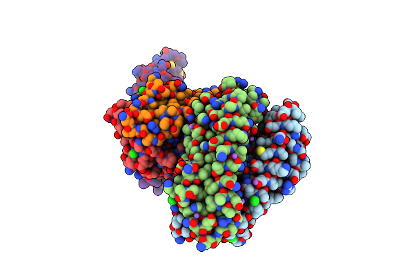 |
Salmonella Enterica Typhimurium Taxis To Serine And Repellents (Tsr) Ligand-Binding Domain With L-Ser, Ph 7
Organism: Salmonella enterica subsp. enterica serovar typhimurium
Method: X-RAY DIFFRACTION Resolution:2.50 Å Release Date: 2024-04-03 Classification: SIGNALING PROTEIN |
Organism: Salmonella enterica subsp. enterica serovar typhimurium
Method: X-RAY DIFFRACTION
Release Date: 2024-04-03
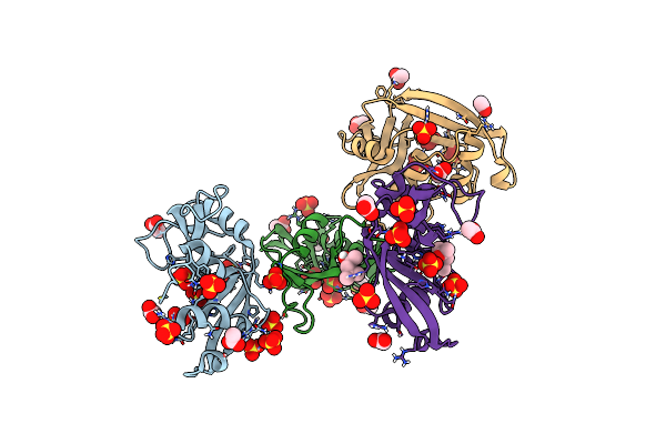 |
Crystal Structure Of A Resurrected Ancestor (Ancrnase) Of The Pancreatic-Type Rnases 2 And 3 Sub-Families
Organism: Synthetic construct
Method: X-RAY DIFFRACTION Resolution:2.03 Å Release Date: 2024-02-28 Classification: HYDROLASE Ligands: SO4, ACT, MPD |
Organism: Synthetic construct
Method: X-RAY DIFFRACTION
Release Date: 2024-02-28
Ligands: SO4, ACT, MPD
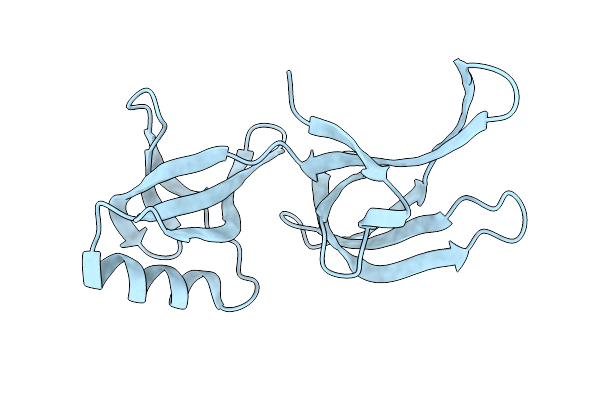 |
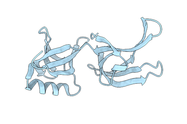
Structure Of Hex-1 From N. Crassa Crystallized In Cellulo (Cytosol), Diffracted At 100K And Resolved Using Crystfel
Organism: Neurospora crassa
Method: X-RAY DIFFRACTION Resolution:1.83 Å Release Date: 2024-02-21 Classification: STRUCTURAL PROTEIN |
Organism: Neurospora crassa
Method: X-RAY DIFFRACTION
Release Date: 2024-02-21
 |
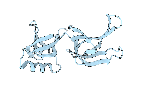
Structure Of Hex-1 From N. Crassa Crystallized In Cellulo, Diffracted At 100K And Resolved Using Crystfel
Organism: Neurospora crassa
Method: X-RAY DIFFRACTION Resolution:1.56 Å Release Date: 2024-02-21 Classification: STRUCTURAL PROTEIN |
Organism: Neurospora crassa
Method: X-RAY DIFFRACTION
Release Date: 2024-02-21
 |
Structure Of Hex-1 (Cyto V2) From N. Crassa Grown In Living Insect Cells, Diffracted At 100K And Resolved Using Crystfel
Organism: Neurospora crassa
Method: X-RAY DIFFRACTION Resolution:1.85 Å Release Date: 2024-02-21 Classification: STRUCTURAL PROTEIN |
Organism: Neurospora crassa
Method: X-RAY DIFFRACTION
Release Date: 2024-02-21
 |
Structure Of Hex-1 From N. Crassa Crystallized In Cellulo, Diffracted At 100K And Resolved Using Xds
Organism: Neurospora crassa
Method: X-RAY DIFFRACTION Resolution:1.85 Å Release Date: 2024-02-21 Classification: STRUCTURAL PROTEIN |
Organism: Neurospora crassa
Method: X-RAY DIFFRACTION
Release Date: 2024-02-21
 |
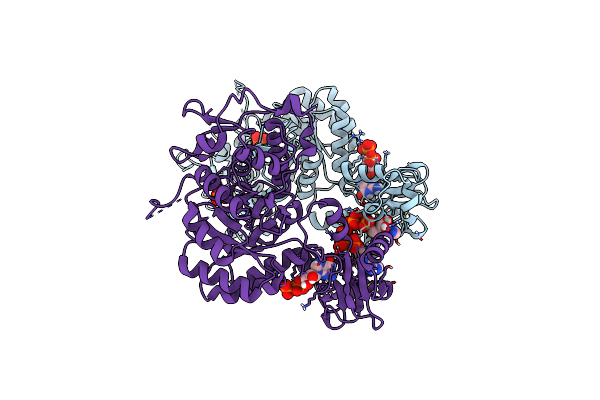
Trypanosoma Brucei Imp Dehydrogenase (Ori) Crystallized In High Five Cells Reveals Native Ligands Atp, Gdp And Phosphate. Diffraction Data Collection At 100 K In Cellulo; Xds Processing
Organism: Trypanosoma brucei
Method: X-RAY DIFFRACTION Resolution:3.00 Å Release Date: 2024-02-21 Classification: TRANSFERASE Ligands: ATP, GDP, PO4 |
Organism: Trypanosoma brucei
Method: X-RAY DIFFRACTION
Release Date: 2024-02-21
Ligands: ATP, GDP, PO4
 |
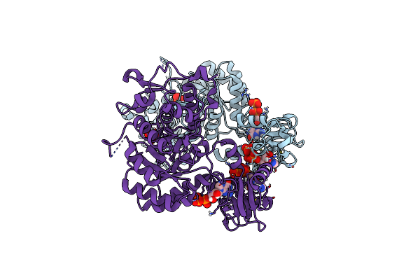
Trypanosoma Brucei Imp Dehydrogenase (Cyto) Crystallized In High Five Cells Revealing Native Ligands Atp, Gdp And Phosphate. Diffraction Data Collection At 100 K In Cellulo
Organism: Trypanosoma brucei
Method: X-RAY DIFFRACTION Resolution:2.40 Å Release Date: 2024-01-17 Classification: TRANSFERASE Ligands: GDP, ATP, PO4 |
Organism: Trypanosoma brucei
Method: X-RAY DIFFRACTION
Release Date: 2024-01-17
Ligands: GDP, ATP, PO4
 |
Trypanosoma Brucei Imp Dehydrogenase (Ori) Crystallized In High Five Cells Reveals Native Ligands Atp, Gdp And Phosphate. Diffraction Data Collection At 100 K In Cellulo; Crystfel Processing
Organism: Trypanosoma brucei
Method: X-RAY DIFFRACTION Resolution:2.30 Å Release Date: 2024-01-17 Classification: TRANSFERASE Ligands: GDP, ATP, PO4 |
Organism: Trypanosoma brucei
Method: X-RAY DIFFRACTION
Release Date: 2024-01-17
Ligands: GDP, ATP, PO4
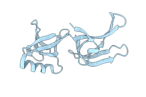 |
Hex-1 (In Cellulo, In Situ) Crystallized And Diffracted In High Five Cells. Growth And Sx Data Collection At 296 K On Crystaldirect Plates
Organism: Neurospora crassa or74a
Method: X-RAY DIFFRACTION Resolution:2.16 Å Release Date: 2024-01-17 Classification: STRUCTURAL PROTEIN |
Organism: Neurospora crassa or74a
Method: X-RAY DIFFRACTION
Release Date: 2024-01-17
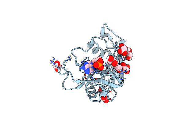 |
Crystal Structure Of Human Eosinophil-Derived Neurotoxin (Edn, Ribonuclease 2) In Complex With 5'-Adenosine Monophosphate (Amp)
Organism: Homo sapiens
Method: X-RAY DIFFRACTION Resolution:1.70 Å Release Date: 2023-11-29 Classification: HYDROLASE Ligands: SIN, EDO, AMP |
Organism: Homo sapiens
Method: X-RAY DIFFRACTION
Release Date: 2023-11-29
Ligands: SIN, EDO, AMP
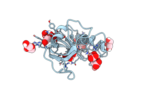 |
Crystal Structure Of Apo Eosinophil Cationic Protein (Ribonuclease 3) From Macaca Fascicularis (Mfecp)
Organism: Macaca fascicularis
Method: X-RAY DIFFRACTION Resolution:1.80 Å Release Date: 2023-08-16 Classification: HYDROLASE Ligands: GOL, CIT, PEG |
Organism: Macaca fascicularis
Method: X-RAY DIFFRACTION
Release Date: 2023-08-16
Ligands: GOL, CIT, PEG
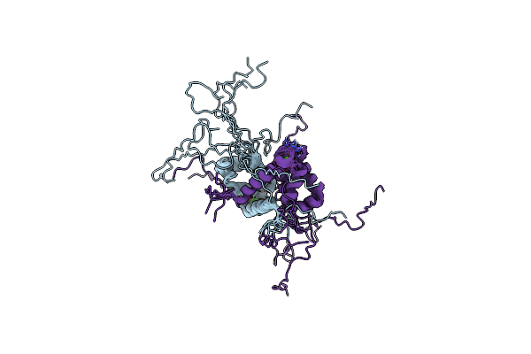 |
Evolution Avoids A Pathological Stabilizing Interaction In The Immune Protein S100A9
Organism: Homo sapiens
Method: SOLUTION NMR Release Date: 2022-10-26 Classification: SIGNALING PROTEIN Ligands: CA |
Organism: Homo sapiens
Method: SOLUTION NMR
Release Date: 2022-10-26
Ligands: CA
 |
Alpha-Lytic Protease Homolog N4
Organism: Streptomyces monomycini
Method: X-RAY DIFFRACTION Resolution:1.25 Å Release Date: 2021-01-20 Classification: UNKNOWN FUNCTION Ligands: SO4 |
Organism: Streptomyces monomycini
Method: X-RAY DIFFRACTION
Release Date: 2021-01-20
Ligands: SO4
 |
Homo Sapiens S100A5
Organism: Homo sapiens
Method: X-RAY DIFFRACTION Resolution:1.25 Å Release Date: 2020-09-30 Classification: METAL BINDING PROTEIN Ligands: CA |
Organism: Homo sapiens
Method: X-RAY DIFFRACTION
Release Date: 2020-09-30
Ligands: CA
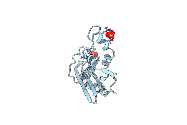 |
Ancestral Rnase H
Organism: Unidentified
Method: X-RAY DIFFRACTION Resolution:1.36 Å Release Date: 2014-11-19 Classification: HYDROLASE Ligands: SO4 |
Organism: Unidentified
Method: X-RAY DIFFRACTION
Release Date: 2014-11-19
Ligands: SO4
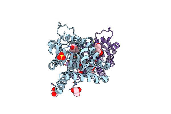 |
X-Ray Crystal Structure Of The Ancestral 3-Keto Steroid Receptor - Progesterone Complex
Method: X-RAY DIFFRACTION
Resolution:3.00 Å Release Date: 2012-12-26 Classification: Steroid-Binding Protein Ligands: STR, GOL, MES, CPS, SO4 |
Method: X-RAY DIFFRACTION
Release Date: 2012-12-26
Ligands: STR, GOL, MES, CPS, SO4
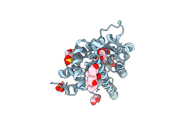 |
X-Ray Crystal Structure Of The Ancestral 3-Keto Steroid Receptor - Doc Complex
Method: X-RAY DIFFRACTION
Resolution:2.78 Å Release Date: 2012-12-12 Classification: Steroid-Binding Protein Ligands: 1CA, SO4, GOL, MES, CPS |
Method: X-RAY DIFFRACTION
Release Date: 2012-12-12
Ligands: 1CA, SO4, GOL, MES, CPS
 |
Crystal Structure Of Staphylococcal Nuclease Variant D+Phs/V66K At Ph 9 Determined At 100 K
Organism: Staphylococcus aureus
Method: X-RAY DIFFRACTION Resolution:2.00 Å Release Date: 2010-05-19 Classification: HYDROLASE Ligands: CA, THP |
Organism: Staphylococcus aureus
Method: X-RAY DIFFRACTION
Release Date: 2010-05-19
Ligands: CA, THP