

Search Count: 26
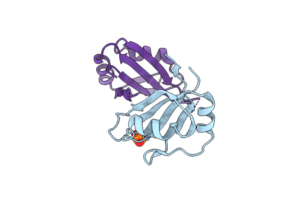 |
2.1 A Resolution Structure Of The Musashi-2 (Msi2) Rna Recognition Motif 1 (Rrm1) Domain
Organism: Homo sapiens
Method: X-RAY DIFFRACTION Resolution:2.10 Å Release Date: 2019-10-23 Classification: RNA BINDING PROTEIN Ligands: PO4 |
Organism: Homo sapiens
Method: X-RAY DIFFRACTION
Release Date: 2019-10-23
Ligands: PO4
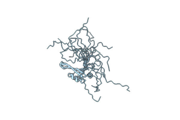 |
Solution Structure Of Musashi2 Rrm1
Organism: Homo sapiens
Method: SOLUTION NMR Release Date: 2019-01-30 Classification: RNA BINDING PROTEIN |
Organism: Homo sapiens
Method: SOLUTION NMR
Release Date: 2019-01-30
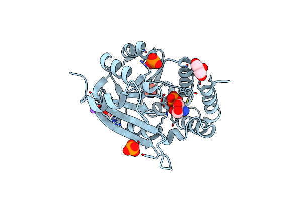 |
1.50A Resolution Structure Of Catechol O-Methyltransferase (Comt) From Nannospalax Galili
Organism: Nannospalax galili
Method: X-RAY DIFFRACTION Resolution:1.50 Å Release Date: 2018-09-12 Classification: TRANSFERASE Ligands: GOL, NA, TRS, PO4 |
Organism: Nannospalax galili
Method: X-RAY DIFFRACTION
Release Date: 2018-09-12
Ligands: GOL, NA, TRS, PO4
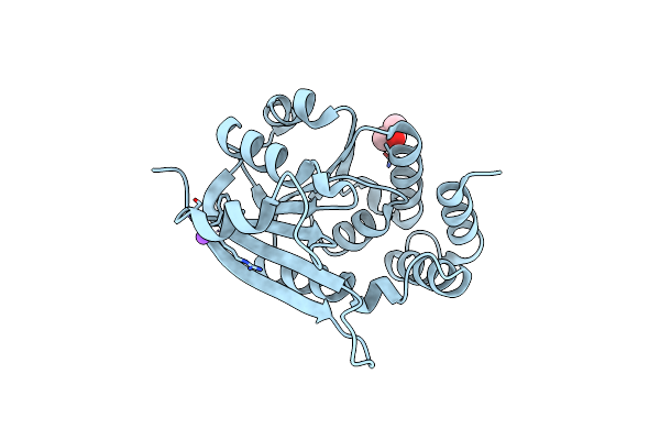 |
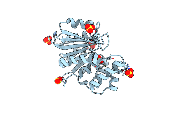
1.90A Resolution Structure Of Catechol O-Methyltransferase (Comt) L136M (Hexagonal Form) From Nannospalax Galili
Organism: Nannospalax galili
Method: X-RAY DIFFRACTION Resolution:1.90 Å Release Date: 2018-09-12 Classification: TRANSFERASE |
Organism: Nannospalax galili
Method: X-RAY DIFFRACTION
Release Date: 2018-09-12
 |
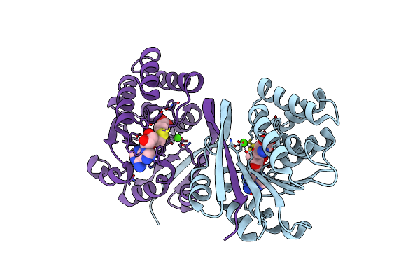
1.70A Resolution Structure Of Catechol O-Methyltransferase (Comt) L136M (Rhombohedral Form) From Nannospalax Galili
Organism: Nannospalax galili
Method: X-RAY DIFFRACTION Resolution:1.70 Å Release Date: 2018-09-12 Classification: TRANSFERASE Ligands: SO4 |
Organism: Nannospalax galili
Method: X-RAY DIFFRACTION
Release Date: 2018-09-12
Ligands: SO4
 |
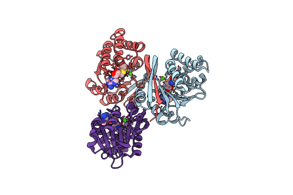
2.15A Resolution Structure Of Sah Bound Catechol O-Methyltransferase (Comt) From Nannospalax Galili
Organism: Nannospalax galili
Method: X-RAY DIFFRACTION Resolution:2.15 Å Release Date: 2018-09-12 Classification: TRANSFERASE Ligands: CA, SAH |
Organism: Nannospalax galili
Method: X-RAY DIFFRACTION
Release Date: 2018-09-12
Ligands: CA, SAH
 |
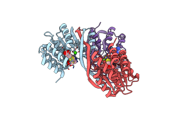
2.25A Resolution Domain Swapped Dimer Structure Of Sah Bound Catechol O-Methyltransferase (Comt) From Nannospalax Galili
Organism: Nannospalax galili
Method: X-RAY DIFFRACTION Resolution:2.25 Å Release Date: 2018-09-12 Classification: TRANSFERASE Ligands: CA, SAH |
Organism: Nannospalax galili
Method: X-RAY DIFFRACTION
Release Date: 2018-09-12
Ligands: CA, SAH
 |
2.55A Resolution Structure Of Sah Bound Catechol O-Methyltransferase (Comt) L136M From Nannospalax Galili
Organism: Nannospalax galili
Method: X-RAY DIFFRACTION Resolution:2.55 Å Release Date: 2018-09-12 Classification: TRANSFERASE Ligands: CA, SAH |
Organism: Nannospalax galili
Method: X-RAY DIFFRACTION
Release Date: 2018-09-12
Ligands: CA, SAH
 |
Crystal Structure Of Apo C-Cbl Tkbd Refined To 2.25 A Resolution
Organism: Homo sapiens
Method: X-RAY DIFFRACTION Resolution:2.25 Å Release Date: 2017-01-18 Classification: LIGASE |
Organism: Homo sapiens
Method: X-RAY DIFFRACTION
Release Date: 2017-01-18
 |
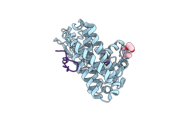
Crystal Structure Of C-Cbl Tkbd-Ring Domains (Y371E Mutant) Refined To 1.85 A Resolution
Organism: Homo sapiens
Method: X-RAY DIFFRACTION Resolution:1.85 Å Release Date: 2017-01-18 Classification: LIGASE Ligands: EDO, ZN, NA |
Organism: Homo sapiens
Method: X-RAY DIFFRACTION
Release Date: 2017-01-18
Ligands: EDO, ZN, NA
 |
Crystal Structure Of C-Cbl Tkbd Domain In Complex With Spry2 Peptide (36-60, Py55) Refined To 1.8A Resolution (P6 Form)
Organism: Homo sapiens
Method: X-RAY DIFFRACTION Resolution:1.80 Å Release Date: 2017-01-18 Classification: LIGASE Ligands: CL, NA, 1PE |
Organism: Homo sapiens
Method: X-RAY DIFFRACTION
Release Date: 2017-01-18
Ligands: CL, NA, 1PE
 |
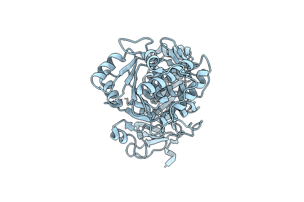
Crystal Structure Of C-Cbl Tkbd In Complex With Spry2 Peptide (36-60, Py55) Refined To 1.8 A Resolution (P21 Form)
Organism: Homo sapiens
Method: X-RAY DIFFRACTION Resolution:1.80 Å Release Date: 2017-01-18 Classification: LIGASE |
Organism: Homo sapiens
Method: X-RAY DIFFRACTION
Release Date: 2017-01-18
 |
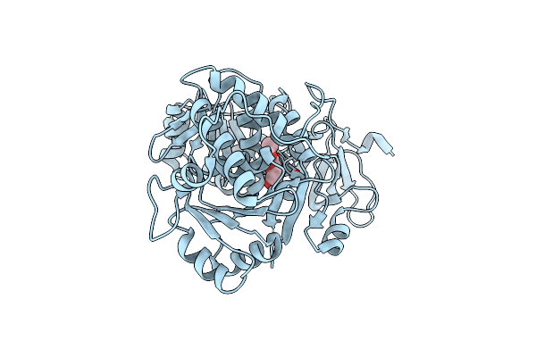
Firefly Luciferase Apo Structure (P41 Form)
Organism: Photinus pyralis
Method: X-RAY DIFFRACTION Resolution:2.10 Å Release Date: 2010-02-16 Classification: OXIDOREDUCTASE |
Organism: Photinus pyralis
Method: X-RAY DIFFRACTION
Release Date: 2010-02-16
 |
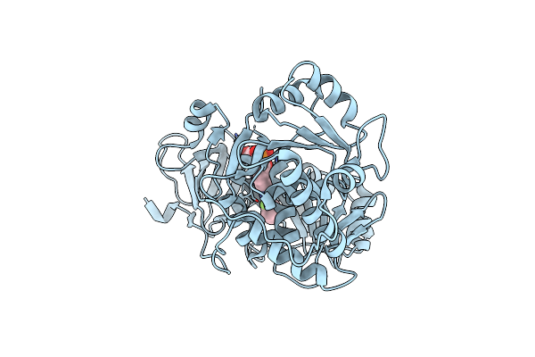
Firefly Luciferase Apo Structure (P41 Form) With Peg 400 Bound
Organism: Photinus pyralis
Method: X-RAY DIFFRACTION Resolution:2.05 Å Release Date: 2010-02-16 Classification: OXIDOREDUCTASE Ligands: PG4 |
Organism: Photinus pyralis
Method: X-RAY DIFFRACTION
Release Date: 2010-02-16
Ligands: PG4
 |
Firefly Luciferase Inhibitor Complex
Organism: Photinus pyralis
Method: X-RAY DIFFRACTION Resolution:2.00 Å Release Date: 2010-02-16 Classification: OXIDOREDUCTASE Ligands: M24 |
Organism: Photinus pyralis
Method: X-RAY DIFFRACTION
Release Date: 2010-02-16
Ligands: M24
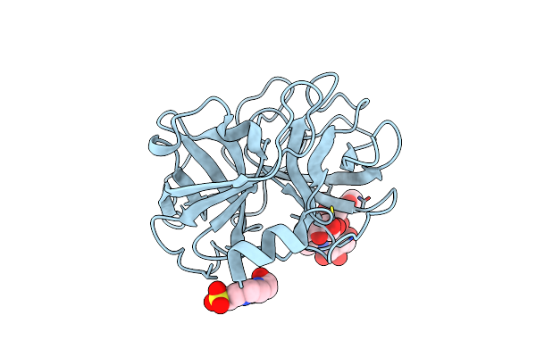 |
Covalent Complex Structure Of Elastase
Organism: Homo sapiens
Method: X-RAY DIFFRACTION Resolution:1.80 Å Release Date: 2008-07-01 Classification: HYDROLASE Ligands: EPE |
Organism: Homo sapiens
Method: X-RAY DIFFRACTION
Release Date: 2008-07-01
Ligands: EPE
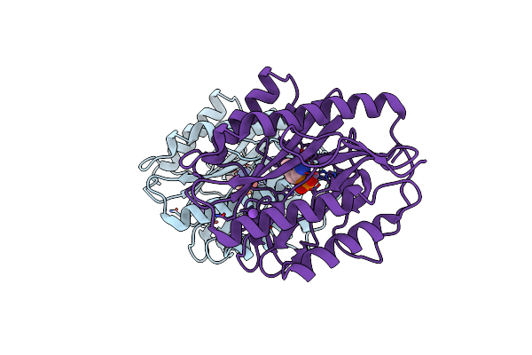 |
Structural Basis Of Catalysis By Mononuclear Methionine Aminopeptidase
Organism: Escherichia coli
Method: X-RAY DIFFRACTION Resolution:2.00 Å Release Date: 2006-07-04 Classification: HYDROLASE Ligands: MN, NA, NLP |
Organism: Escherichia coli
Method: X-RAY DIFFRACTION
Release Date: 2006-07-04
Ligands: MN, NA, NLP
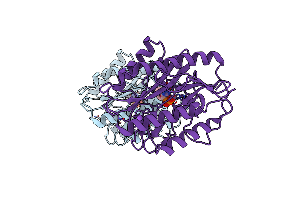 |
E. Coli Methionine Aminopeptidase In Complex With Nlep, 1: 0.5, Di-Metalated
Organism: Escherichia coli
Method: X-RAY DIFFRACTION Resolution:1.80 Å Release Date: 2006-07-04 Classification: HYDROLASE Ligands: MN, NA, NLP |
Organism: Escherichia coli
Method: X-RAY DIFFRACTION
Release Date: 2006-07-04
Ligands: MN, NA, NLP
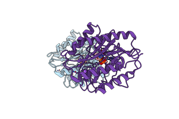 |
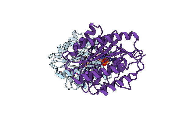
E. Coli Methionine Aminopeptidase In Complex With Nlep, 1: 1, Di-Metalated
Organism: Escherichia coli
Method: X-RAY DIFFRACTION Resolution:1.60 Å Release Date: 2006-07-04 Classification: HYDROLASE Ligands: MN, NA, NLP |
Organism: Escherichia coli
Method: X-RAY DIFFRACTION
Release Date: 2006-07-04
Ligands: MN, NA, NLP
 |
E. Coli Methionine Aminopeptidase In Complex With Nlep, 1: 2, Di-Metalated
Organism: Escherichia coli
Method: X-RAY DIFFRACTION Resolution:1.70 Å Release Date: 2006-07-04 Classification: HYDROLASE Ligands: MN, NA, NLP |
Organism: Escherichia coli
Method: X-RAY DIFFRACTION
Release Date: 2006-07-04
Ligands: MN, NA, NLP