
Search Count: 35
 |
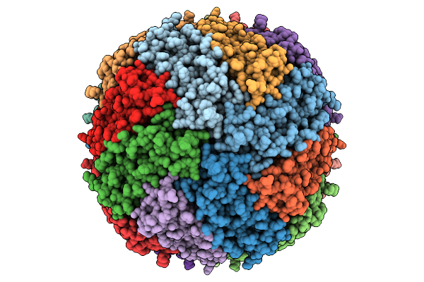
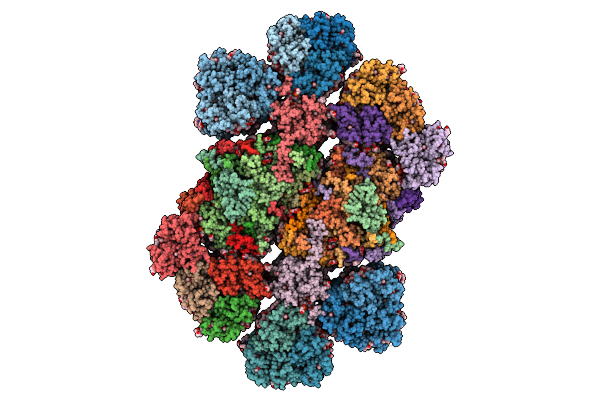
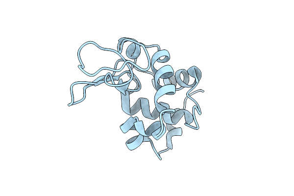
Cryo-Em Structure Of Human Apoferritin At Ph 3.5
Organism: Homo sapiens
Method: ELECTRON MICROSCOPY Release Date: 2026-02-25 Classification: METAL BINDING PROTEIN Ligands: FE, MG |
 |
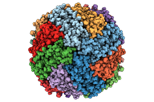
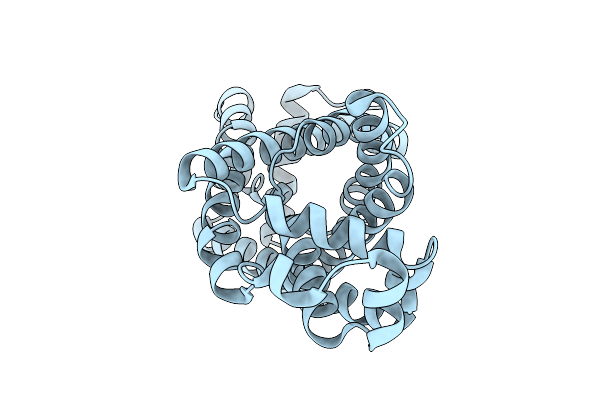
Cryo-Em Structure Of Human Apoferritin At Ph 4
Organism: Homo sapiens
Method: ELECTRON MICROSCOPY Release Date: 2026-02-25 Classification: METAL BINDING PROTEIN Ligands: FE, MG |
 |
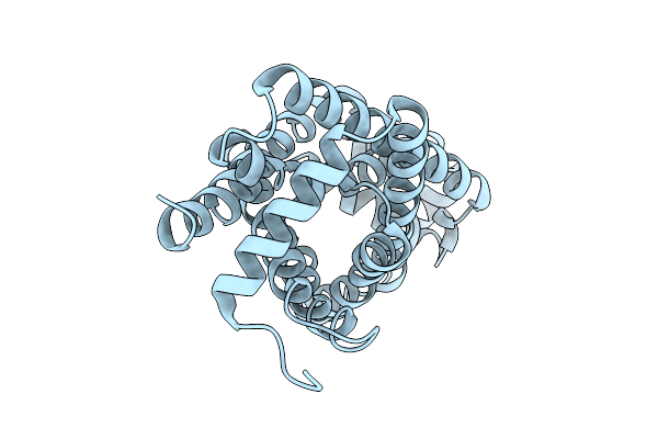
Cryo-Em Structure Of Human Apoferritin At Ph 5
Organism: Homo sapiens
Method: ELECTRON MICROSCOPY Release Date: 2026-02-25 Classification: METAL BINDING PROTEIN Ligands: FE, MG |
 |
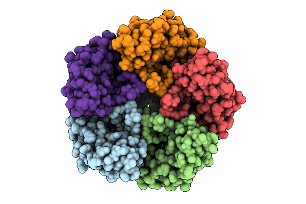
Cryo-Em Structure Of Human Apoferritin At Ph 7
Organism: Homo sapiens
Method: ELECTRON MICROSCOPY Release Date: 2026-02-25 Classification: METAL BINDING PROTEIN Ligands: FE, MG |
 |
Cryo-Em Structure Of Human Apoferritin At Ph 9
Organism: Homo sapiens
Method: ELECTRON MICROSCOPY Release Date: 2026-02-25 Classification: METAL BINDING PROTEIN Ligands: FE, MG |
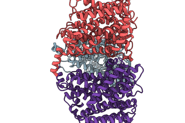 |
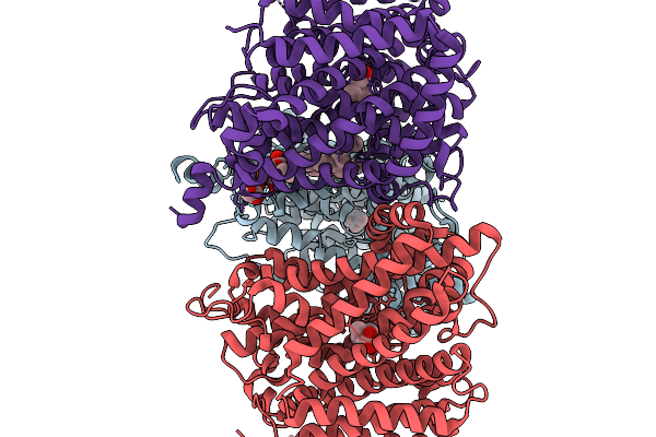
Upregulated State Of Betp In Potassium
Organism: Corynebacterium glutamicum
Method: ELECTRON MICROSCOPY Resolution:3.09 Å Release Date: 2026-02-18 Classification: TRANSPORT PROTEIN |
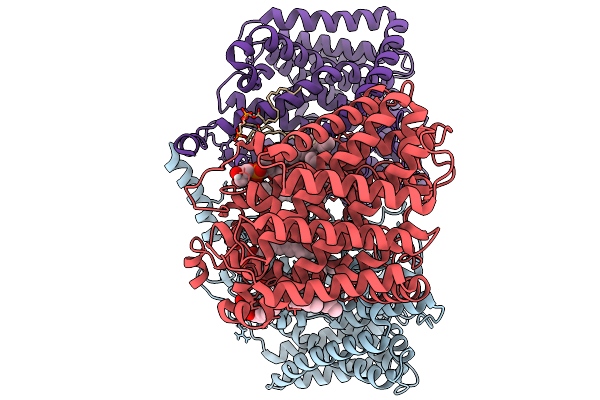 |
Transient Activated State Of Betp
Organism: Corynebacterium glutamicum
Method: ELECTRON MICROSCOPY Resolution:3.31 Å Release Date: 2026-02-18 Classification: TRANSPORT PROTEIN Ligands: PGT, CDL |
 |
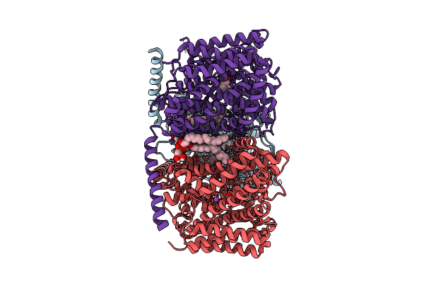
Transient Activated State Of Betp In Complex With Betaine
Organism: Corynebacterium glutamicum
Method: ELECTRON MICROSCOPY Resolution:3.25 Å Release Date: 2026-02-18 Classification: TRANSPORT PROTEIN Ligands: PGT, BET |
 |
Downregulated State Of The Betaine Transporter Betp
Organism: Corynebacterium glutamicum
Method: ELECTRON MICROSCOPY Resolution:3.57 Å Release Date: 2026-02-18 Classification: TRANSPORT PROTEIN Ligands: PGT, CDL |
 |
Upregulated State Of Betp With Potassium In Amphipol A8-35
Organism: Corynebacterium glutamicum
Method: ELECTRON MICROSCOPY Resolution:3.33 Å Release Date: 2026-02-18 Classification: TRANSPORT PROTEIN Ligands: K |
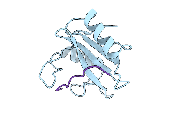 |
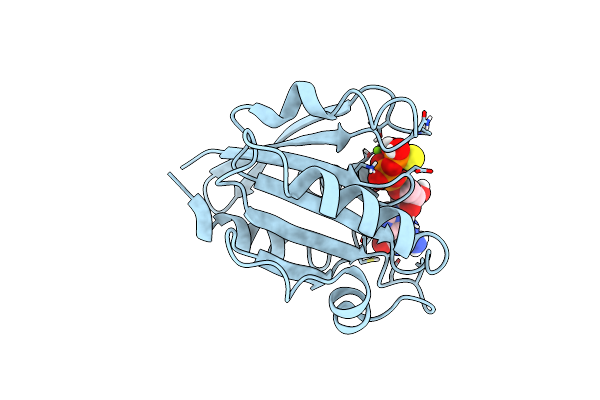
Structure Of The N-Sh2 Domain Of Shp2 In Complex With The Phosphoy627-Gab1 (613-651) Peptide
Organism: Homo sapiens
Method: X-RAY DIFFRACTION Resolution:2.08 Å Release Date: 2025-12-31 Classification: SIGNALING PROTEIN |
 |
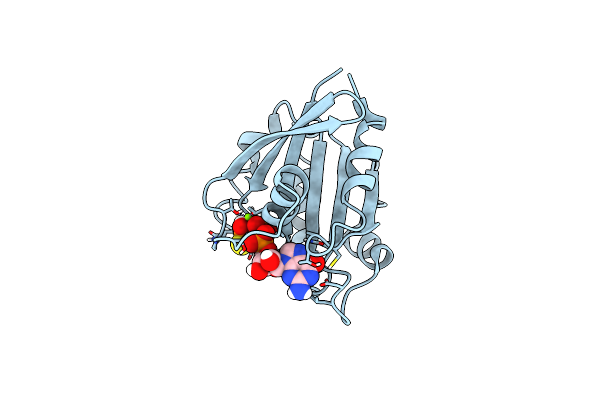
Micro-Ed Structure Of The Nsh2-Csh2 Tandem Domain Of Shp2 In Complex With The Bis-Phosphorylated Py627-Py659-Gab1 (613-694) Peptide
Organism: Homo sapiens
Method: ELECTRON CRYSTALLOGRAPHY Resolution:3.20 Å Release Date: 2025-12-31 Classification: SIGNALING PROTEIN |
 |
Downregulated Closed State Of Betp In Complex With Betaine
Organism: Corynebacterium glutamicum
Method: ELECTRON MICROSCOPY Release Date: 2025-12-17 Classification: TRANSPORT PROTEIN Ligands: PGT, BET, NA |
 |
Cryo-Em Structure Of Photosystem Ii C2S2M2L2 Supercomplex From The Green Alga Chlorella Ohadii
|
 |
Cryo-Em Reconstruction Of The Full-Length E. Coli Transmembrane Formate Transporter Foca
Organism: Escherichia coli k-12
Method: ELECTRON MICROSCOPY Release Date: 2025-10-22 Classification: MEMBRANE PROTEIN Ligands: HOH |
 |
Cryo-Em Reconstruction Of The Full-Length H209N Mutant E. Coli Transmembrane Formate Transporter Foca
Organism: Escherichia coli k-12
Method: ELECTRON MICROSCOPY Release Date: 2025-10-22 Classification: MEMBRANE PROTEIN |
 |
Asymmetric Cryo-Em Reconstruction Of The Full-Length E. Coli Transmembrane Formate Transporter Foca
Organism: Escherichia coli k-12
Method: ELECTRON MICROSCOPY Release Date: 2025-10-22 Classification: MEMBRANE PROTEIN |
 |
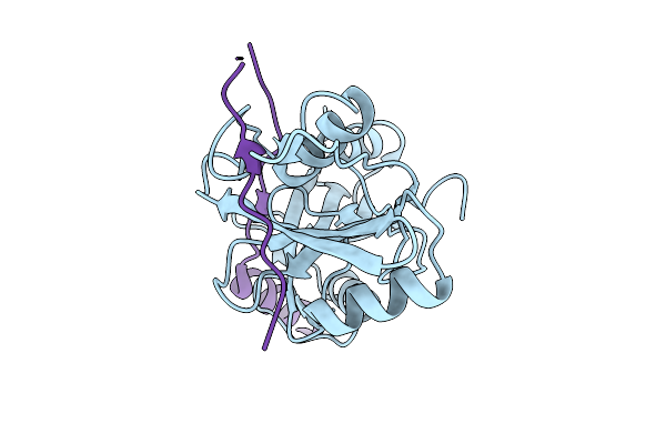
Hen Egg-White Lysozyme Structure Determined By 3Ded/Microed On A 200 Kev Microscope
Organism: Gallus gallus
Method: ELECTRON CRYSTALLOGRAPHY Resolution:2.80 Å Release Date: 2025-03-19 Classification: HYDROLASE |
 |
Cryo-Em Structure Of Arf1-Decorated Membrane Tubules
Organism: Saccharomyces cerevisiae
Method: ELECTRON MICROSCOPY Release Date: 2025-01-22 Classification: TRANSPORT PROTEIN Ligands: MG, GSP |
 |
Cryo-Em Structure Of Arf1-Decorated Membrane Tubules
Organism: Saccharomyces cerevisiae
Method: ELECTRON MICROSCOPY Release Date: 2025-01-22 Classification: TRANSPORT PROTEIN Ligands: MG, GSP |