
Search Count: 155
All
Selected
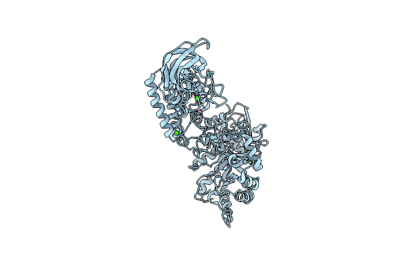 |
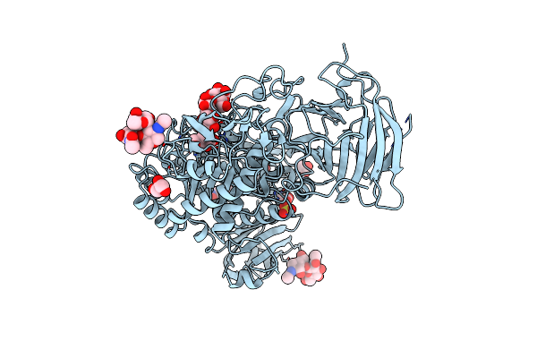
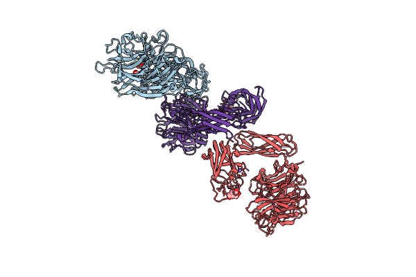
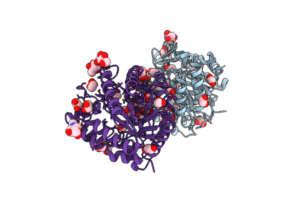
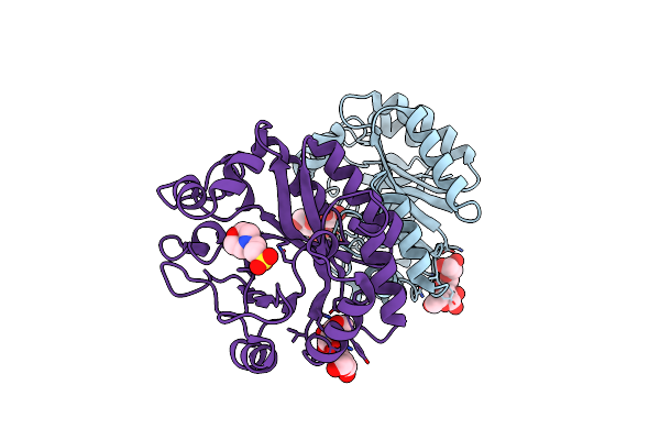
Structure Of A Family 84 Glycoside Hydrolase And A Family 32 Carbohydrate-Binding Module In Tandem From Clostridium Perfringens.
Organism: Clostridium perfringens
Method: X-RAY DIFFRACTION Resolution:3.30 Å Release Date: 2009-01-27 Classification: HYDROLASE Ligands: CA |
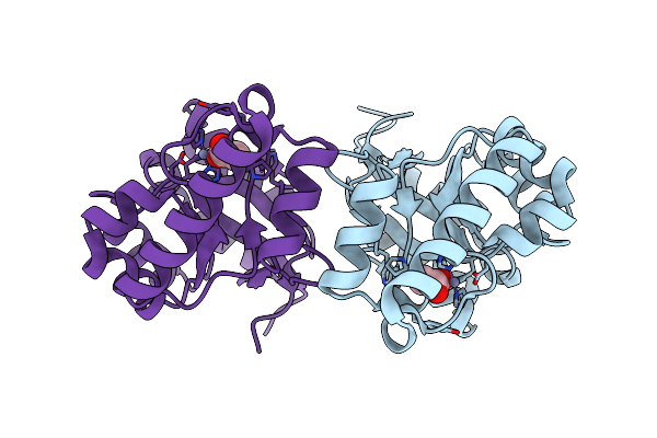 |
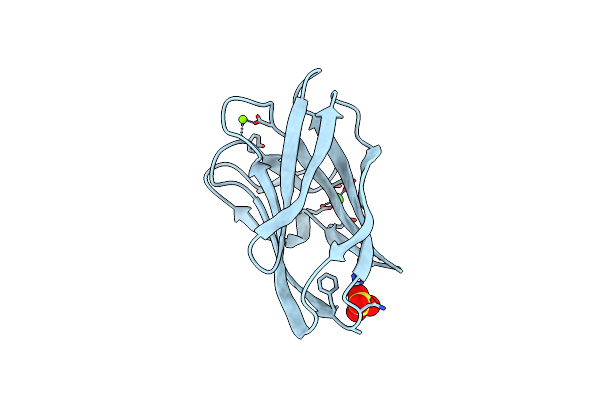
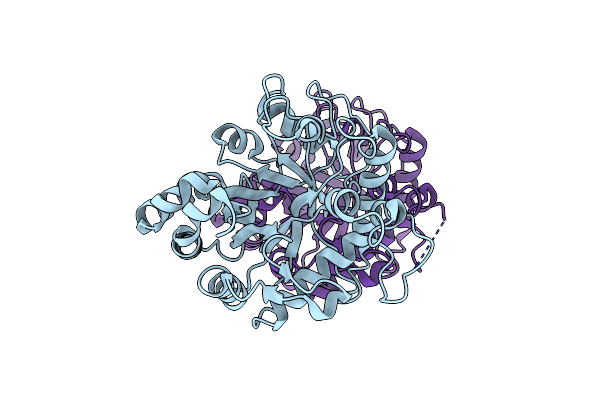
Family 4 Carbohydrate Esterase From Streptomyces Lividans In Complex With Acetate
Organism: Streptomyces lividans
Method: X-RAY DIFFRACTION Resolution:1.60 Å Release Date: 2006-01-23 Classification: HYDROLASE Ligands: ZN, ACT |
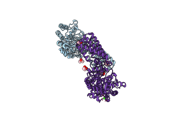 |
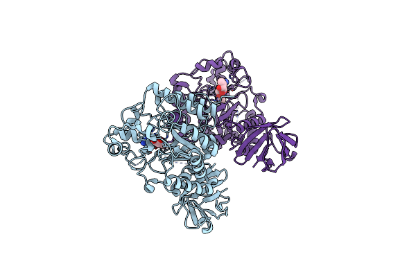
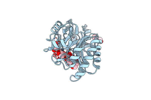
Family 84 Glycoside Hydrolase From Clostridium Perfringens, 2.1 Angstrom Structure
Organism: Clostridium perfringens
Method: X-RAY DIFFRACTION Resolution:2.10 Å Release Date: 2009-01-27 Classification: HYDROLASE Ligands: CA, CAC, NA |
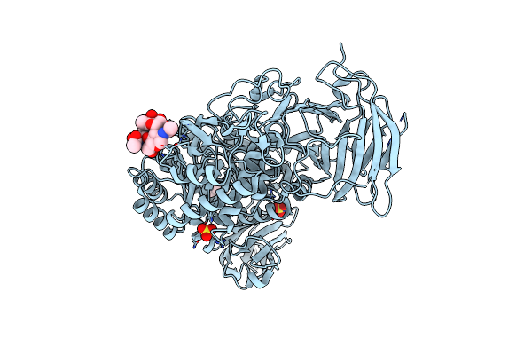 |
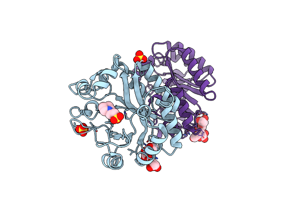
Organism: Beta vulgaris
Method: X-RAY DIFFRACTION Resolution:2.79 Å Release Date: 2013-05-29 Classification: HYDROLASE, CARBOHYDRATE Ligands: NAG, SO4 |
 |
Organism: Beta vulgaris
Method: X-RAY DIFFRACTION Resolution:1.70 Å Release Date: 2013-05-29 Classification: HYDROLASE, CARBOHYDRATE Ligands: ACY, GOL, SO4, NAG |
 |
Organism: Paenibacillus polymyxa
Method: X-RAY DIFFRACTION Resolution:0.80 Å Release Date: 2004-10-27 Classification: HYDROLASE Ligands: CA, MG, SO4 |
 |
Carbohydrate-Binding Of The Starch Binding Domain Of Rhizopus Oryzae Glucoamylase In Complex With Beta-Cyclodextrin And Maltoheptaose
Organism: Rhizopus oryzae
Method: X-RAY DIFFRACTION Resolution:1.80 Å Release Date: 2008-08-19 Classification: HYDROLASE Ligands: ZN |
 |
Carbohydrate-Binding Of The Starch Binding Domain Of Rhizopus Oryzae Glucoamylase In Complex With Beta-Cyclodextrin And Maltoheptaose
Organism: Rhizopus oryzae
Method: X-RAY DIFFRACTION Resolution:2.30 Å Release Date: 2008-08-19 Classification: HYDROLASE Ligands: SO4 |
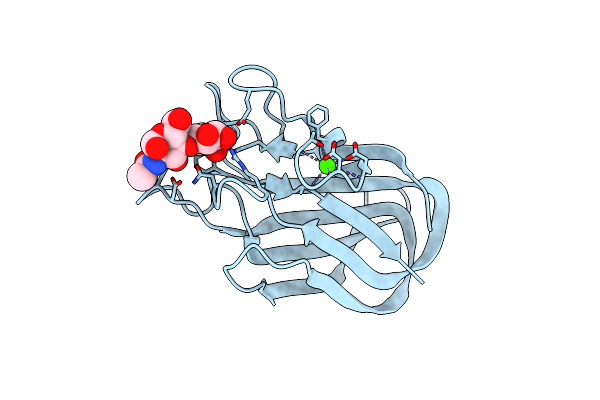 |
High Resolution Crystal Structure Of Cbm32 From A N-Acetyl-Beta- Hexosaminidase In Complex With Lacnac
Organism: Clostridium perfringens
Method: X-RAY DIFFRACTION Resolution:2.40 Å Release Date: 2006-09-20 Classification: HYDROLASE Ligands: CA |
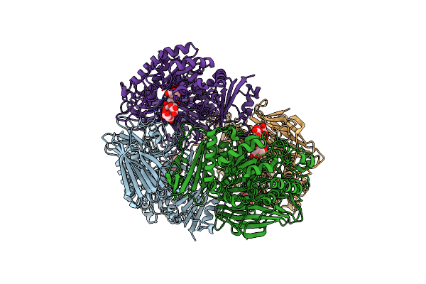 |
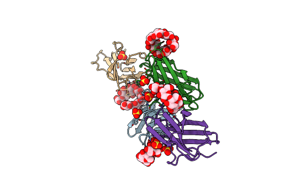
Crystal Structure Of Gh36 Alpha-Galactosidase Agaa A355E D478A From Geobacillus Stearothermophilus In Complex With Stachyose
Organism: Geobacillus stearothermophilus
Method: X-RAY DIFFRACTION Resolution:3.60 Å Release Date: 2012-10-03 Classification: HYDROLASE |
 |
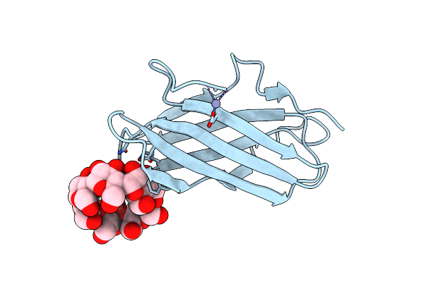
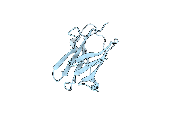
Galactose Recognition By The Carbohydrate-Binding Module Of A Bacterial Sialidase.
Organism: Micromonospora viridifaciens
Method: X-RAY DIFFRACTION Resolution:2.00 Å Release Date: 2005-08-19 Classification: HYDROLASE |
 |
Carbohydrate-Binding Of The Starch Binding Domain Of Rhizopus Oryzae Glucoamylase In Complex With Beta-Cyclodextrin And Maltoheptaose
Organism: Rhizopus oryzae
Method: X-RAY DIFFRACTION Resolution:1.25 Å Release Date: 2009-07-07 Classification: HYDROLASE |
 |
Organism: Aspergillus niger
Method: X-RAY DIFFRACTION Resolution:1.27 Å Release Date: 2020-01-29 Classification: HYDROLASE Ligands: NAG, EDO |
 |
Organism: Solanum tuberosum
Method: X-RAY DIFFRACTION Resolution:1.26 Å Release Date: 2012-05-30 Classification: HYDROLASE |
 |
Organism: Aspergillus terreus
Method: X-RAY DIFFRACTION Resolution:1.38 Å Release Date: 2018-11-14 Classification: HYDROLASE Ligands: NAG, CA, 1PE, PG4, PGE, PG0, EDO, DTU, ACT, MPO, BGC |
 |
Thermotoga Maritima Alpha-L-Fucosynthase, Tmd224G, In Complex With Alpha-L-Fuc-(1-2)-Beta-L-Fuc-N3
Organism: Thermotoga maritima
Method: X-RAY DIFFRACTION Resolution:2.65 Å Release Date: 2010-01-19 Classification: HYDROLASE |
 |
Organism: Bacillus subtilis
Method: X-RAY DIFFRACTION Resolution:2.68 Å Release Date: 2009-04-28 Classification: HYDROLASE |
 |
Crystal Structure Of A Gh128 (Subgroup Iv) Endo-Beta-1,3-Glucanase From Lentinula Edodes (Legh128_Iv) With Laminaribiose At The Surface-Binding Site
Organism: Lentinula edodes
Method: X-RAY DIFFRACTION Resolution:1.85 Å Release Date: 2020-05-20 Classification: HYDROLASE Ligands: CL, MES, SO4 |
 |
Crystal Structure Of A Gh128 (Subgroup I) Endo-Beta-1,3-Glucanase (E199A Mutant) From Amycolatopsis Mediterranei (Amgh128_I) In Complex With Laminaripentaose
Organism: Amycolatopsis mediterranei
Method: X-RAY DIFFRACTION Resolution:1.91 Å Release Date: 2020-05-20 Classification: HYDROLASE Ligands: BGC, ZN, PEG |
 |
Crystal Structure (C2 Form) Of A Gh128 (Subgroup Iv) Endo-Beta-1,3-Glucanase From Lentinula Edodes (Legh128_Iv) In Complex With Laminaritriose
Organism: Lentinula edodes
Method: X-RAY DIFFRACTION Resolution:1.60 Å Release Date: 2020-05-20 Classification: HYDROLASE Ligands: CL, MES |