

Search Count: 59
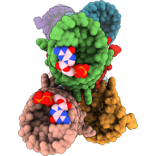 |
Crystal Structure Of Xanthine/Guanine Rna Aptamer (P1-12Bp) With Hypoxanthine
Organism: Synthetic construct
Method: X-RAY DIFFRACTION Resolution:2.95 Å Release Date: 2026-09-16 Classification: RNA Ligands: GTP, HPA, GDP, NA |
Organism: Synthetic construct
Method: X-RAY DIFFRACTION
Release Date: 2026-09-16
Ligands: GTP, HPA, GDP, NA
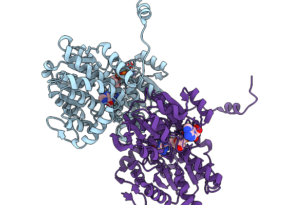 |
Crystal Structure Of S-Adenosyl-L-Homocysteine Hydrolase From Pyrococcus Furiosus In Complex With Hypoxanthine
Organism: Pyrococcus furiosus
Method: X-RAY DIFFRACTION Resolution:2.06 Å Release Date: 2026-03-04 Classification: HYDROLASE Ligands: NAD, HPA |
Organism: Pyrococcus furiosus
Method: X-RAY DIFFRACTION
Release Date: 2026-03-04
Ligands: NAD, HPA
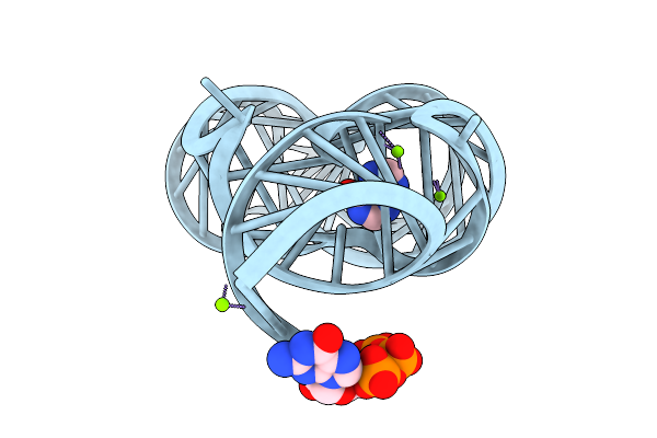 |
Crystal Structure Of Guanine-Ii Riboswitch In Complex With Hypoxanthine
Organism: Paenibacillus sp. fsl e2-0178
Method: X-RAY DIFFRACTION Resolution:2.26 Å Release Date: 2025-11-26 Classification: RNA Ligands: HPA, GTP, MG |
Organism: Paenibacillus sp. fsl e2-0178
Method: X-RAY DIFFRACTION
Release Date: 2025-11-26
Ligands: HPA, GTP, MG
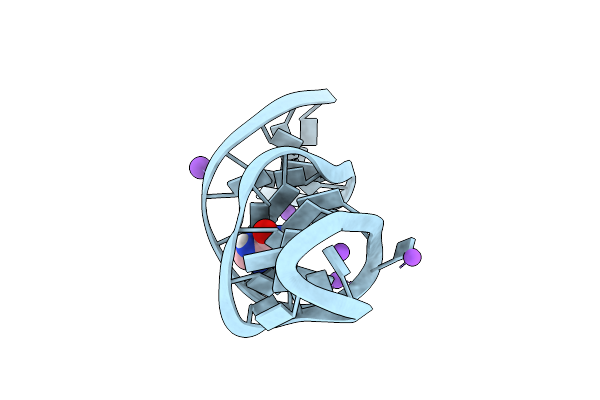 |
Crystal Structure Of Theophylline Dna Aptamer Bound To Hypoxanthine
Organism: Synthetic construct
Method: X-RAY DIFFRACTION Resolution:1.52 Å Release Date: 2025-04-16 Classification: DNA Ligands: HPA, NA, K |
Organism: Synthetic construct
Method: X-RAY DIFFRACTION
Release Date: 2025-04-16
Ligands: HPA, NA, K
 |
Crystal Structure Of Ptmb In Complex With Cyclo-(L-Trp-L-Trp) And Hypoxanthine
Organism: Kitasatospora mediocidica kctc 9733
Method: X-RAY DIFFRACTION Resolution:2.10 Å Release Date: 2024-11-20 Classification: OXIDOREDUCTASE Ligands: UYM, HPA, HEM |
Organism: Kitasatospora mediocidica kctc 9733
Method: X-RAY DIFFRACTION
Release Date: 2024-11-20
Ligands: UYM, HPA, HEM
 |
Crystal Structure Of The Ligand Binding Domain Of The Halomonas Titanicae Chemoreceptor Htc10 In Complex With Hypoxanthine
Organism: Halomonas titanicae
Method: X-RAY DIFFRACTION Resolution:3.63 Å Release Date: 2024-11-20 Classification: SIGNALING PROTEIN Ligands: HPA |
Organism: Halomonas titanicae
Method: X-RAY DIFFRACTION
Release Date: 2024-11-20
Ligands: HPA
 |
Crystal Structure Of Cytochrome P450 Gymb5 From Streptomyces Katrae In Complex With Cyy And Hypoxanthine
Organism: Streptomyces katrae
Method: X-RAY DIFFRACTION Resolution:1.81 Å Release Date: 2024-06-26 Classification: OXIDOREDUCTASE Ligands: HEM, YTT, HPA, EDO |
Organism: Streptomyces katrae
Method: X-RAY DIFFRACTION
Release Date: 2024-06-26
Ligands: HEM, YTT, HPA, EDO
 |
Structure Of K. Lactis Pnp Bound To Hypoxanthine
Organism: Kluyveromyces lactis nrrl y-1140
Method: X-RAY DIFFRACTION Resolution:2.10 Å Release Date: 2023-10-18 Classification: TRANSFERASE Ligands: HPA, ACT |
Organism: Kluyveromyces lactis nrrl y-1140
Method: X-RAY DIFFRACTION
Release Date: 2023-10-18
Ligands: HPA, ACT
 |
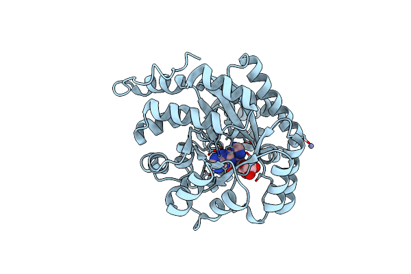
Crystal Structure Of Human Aprt Wild Type In Complex With Hypoxanthine, Prpp And Mg2+
Organism: Homo sapiens
Method: X-RAY DIFFRACTION Resolution:1.90 Å Release Date: 2019-07-31 Classification: TRANSFERASE Ligands: GOL, MG, PRP, HPA |
Organism: Homo sapiens
Method: X-RAY DIFFRACTION
Release Date: 2019-07-31
Ligands: GOL, MG, PRP, HPA
 |
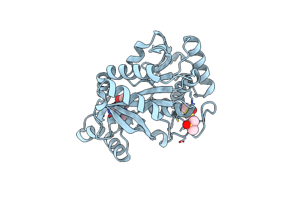
Crystal Structure Of Plasmodium Falciparum Adenosine Deaminase C27Q+L227I Mutant Co-Complexed With Zn Ion, Hypoxanthine And Inosine
Organism: Plasmodium falciparum 3d7
Method: X-RAY DIFFRACTION Resolution:2.48 Å Release Date: 2019-05-01 Classification: HYDROLASE Ligands: NOS, HPA, ZN |
Organism: Plasmodium falciparum 3d7
Method: X-RAY DIFFRACTION
Release Date: 2019-05-01
Ligands: NOS, HPA, ZN
 |
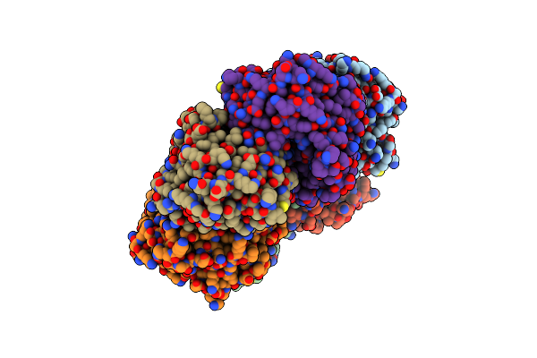
Crystal Structure Of Isoform 2 Of Purine Nucleoside Phosphorylase Complexed With Hypoxanthine
Organism: Schistosoma mansoni
Method: X-RAY DIFFRACTION Resolution:2.10 Å Release Date: 2017-10-11 Classification: TRANSFERASE Ligands: HPA, DMS |
Organism: Schistosoma mansoni
Method: X-RAY DIFFRACTION
Release Date: 2017-10-11
Ligands: HPA, DMS
 |
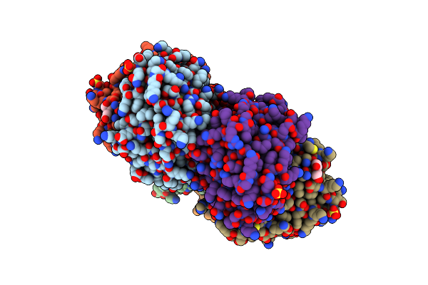
Crystal Structure Of H. Pylori Purine Nucleoside Phosphorylase From Clinical Isolate Hppnp-1
Organism: Helicobacter pylori r018c
Method: X-RAY DIFFRACTION Resolution:2.31 Å Release Date: 2017-04-05 Classification: TRANSFERASE Ligands: HPA, PO4 |
Organism: Helicobacter pylori r018c
Method: X-RAY DIFFRACTION
Release Date: 2017-04-05
Ligands: HPA, PO4
 |
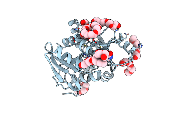
Crystal Structure Of H. Pylori Purine Nucleoside Phosphorylase From Clinical Isolate Hppnp-2
Organism: Helicobacter pylori
Method: X-RAY DIFFRACTION Resolution:2.41 Å Release Date: 2017-04-05 Classification: TRANSFERASE Ligands: SO4, HPA, ACT, PEG, PO4 |
Organism: Helicobacter pylori
Method: X-RAY DIFFRACTION
Release Date: 2017-04-05
Ligands: SO4, HPA, ACT, PEG, PO4
 |
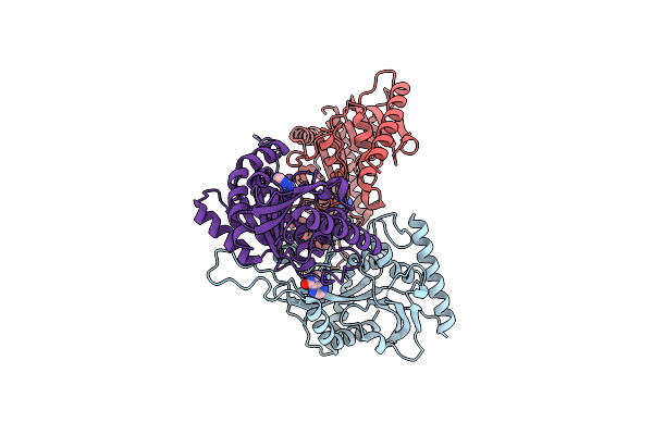
Crystal Structure Of H. Pylori Purine Nucleoside Phosphorylase From Clinical Isolate Hppnp-3
Organism: Helicobacter pylori
Method: X-RAY DIFFRACTION Resolution:2.40 Å Release Date: 2017-04-05 Classification: TRANSFERASE Ligands: HPA, PO4, PG4 |
Organism: Helicobacter pylori
Method: X-RAY DIFFRACTION
Release Date: 2017-04-05
Ligands: HPA, PO4, PG4
 |
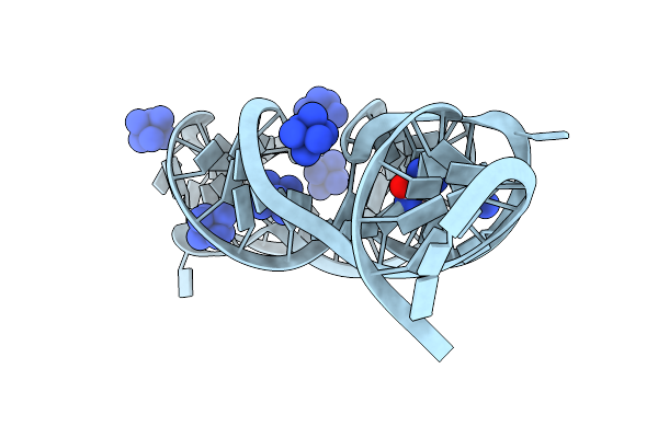
Purine Nucleoside Phosphorylase
Organism: Kluyveromyces lactis (strain atcc 8585 / cbs 2359 / dsm 70799 / nbrc 1267 / nrrl y-1140 / wm37)
Method: X-RAY DIFFRACTION Resolution:1.97 Å Release Date: 2017-03-01 Classification: TRANSFERASE Ligands: HPA |
Organism: Kluyveromyces lactis (strain atcc 8585 / cbs 2359 / dsm 70799 / nbrc 1267 / nrrl y-1140 / wm37)
Method: X-RAY DIFFRACTION
Release Date: 2017-03-01
Ligands: HPA
 |
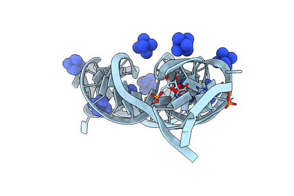
5'-Monophosphate Wt Guanine Riboswitch Bound To Hypoxanthine.
Organism: Bacillus subtilis subsp. subtilis str. 168
Method: X-RAY DIFFRACTION Resolution:3.05 Å Release Date: 2015-08-12 Classification: RNA Ligands: NCO, HPA |
Organism: Bacillus subtilis subsp. subtilis str. 168
Method: X-RAY DIFFRACTION
Release Date: 2015-08-12
Ligands: NCO, HPA
 |
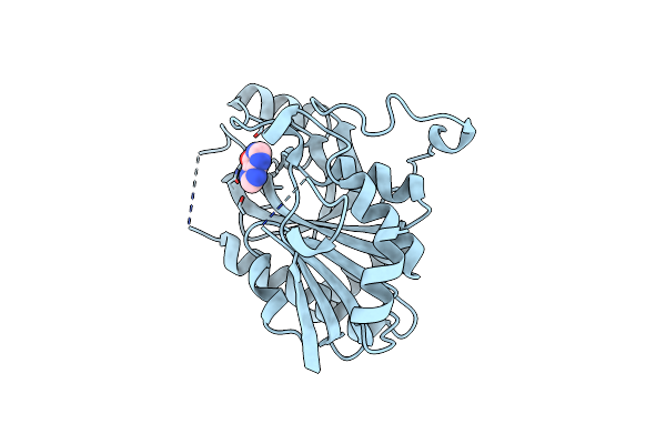
5'-Monophosphate Z:P Guanine Riboswitch Bound To Hypoxanthine.
Organism: Synthetic construct
Method: X-RAY DIFFRACTION Resolution:3.22 Å Release Date: 2015-08-12 Classification: RNA Ligands: NCO, HPA |
Organism: Synthetic construct
Method: X-RAY DIFFRACTION
Release Date: 2015-08-12
Ligands: NCO, HPA
 |
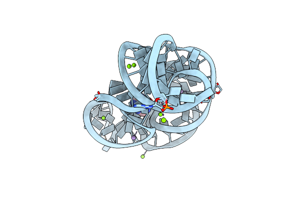
Crystal Structure Of A Purine Nucleoside Phosphorylase (Psi-Nysgrc-029736) From Agrobacterium Vitis
Organism: Agrobacterium vitis
Method: X-RAY DIFFRACTION Resolution:2.40 Å Release Date: 2014-10-08 Classification: TRANSFERASE Ligands: HPA |
Organism: Agrobacterium vitis
Method: X-RAY DIFFRACTION
Release Date: 2014-10-08
Ligands: HPA
 |
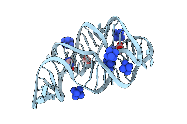
Crystal Structure Of The E. Coli Thim Riboswitch In Complex With Hypoxanthine
Organism: Escherichia coli
Method: X-RAY DIFFRACTION Resolution:2.90 Å Release Date: 2014-06-04 Classification: RNA Ligands: HPA, MN, MG |
Organism: Escherichia coli
Method: X-RAY DIFFRACTION
Release Date: 2014-06-04
Ligands: HPA, MN, MG
 |
Crystal Structure Of The A24U Mutant Xpt-Pbux Guanine Riboswitch Aptamer Domain In Complex With Hypoxanthine
Method: X-RAY DIFFRACTION
Resolution:1.50 Å Release Date: 2013-02-27 Classification: RNA Ligands: HPA, ACT, NCO |
Method: X-RAY DIFFRACTION
Release Date: 2013-02-27
Ligands: HPA, ACT, NCO