

Search Count: 13
 |
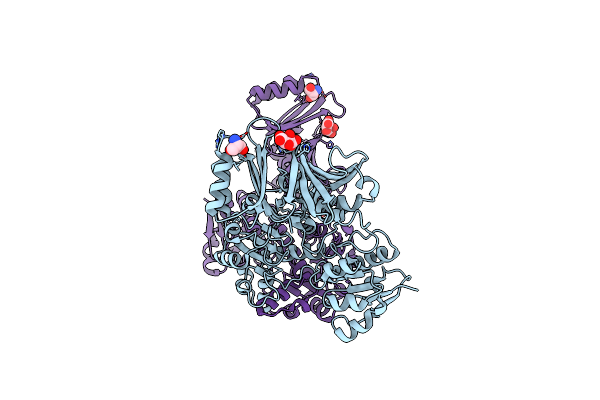
Crystal Structure Of L-Serine Dehydratase From Legionella Pneumophila
Organism: Legionella pneumophila subsp. pneumophila str. thunder bay
Method: X-RAY DIFFRACTION Resolution:2.25 Å Release Date: 2014-11-19 Classification: LYASE Ligands: SF4, CL |
Organism: Legionella pneumophila subsp. pneumophila str. thunder bay
Method: X-RAY DIFFRACTION
Release Date: 2014-11-19
Ligands: SF4, CL
 |
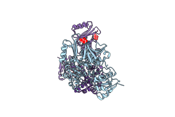
Crystal Structure Of Serine Bound D-3-Phosphoglycerate Dehydrogenase From Mycobacterium Tuberculosis
Organism: Mycobacterium tuberculosis
Method: X-RAY DIFFRACTION Resolution:2.70 Å Release Date: 2008-07-22 Classification: OXIDOREDUCTASE Ligands: SER, TLA |
Organism: Mycobacterium tuberculosis
Method: X-RAY DIFFRACTION
Release Date: 2008-07-22
Ligands: SER, TLA
 |
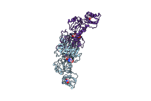
Crystal Structure Of Hydroxypyruvic Acid Phosphate Bound D-3-Phosphoglycerate Dehydrogenase In Mycobacterium Tuberculosis
Organism: Mycobacterium tuberculosis
Method: X-RAY DIFFRACTION Resolution:2.40 Å Release Date: 2008-07-22 Classification: OXIDOREDUCTASE Ligands: TLA, HPV |
Organism: Mycobacterium tuberculosis
Method: X-RAY DIFFRACTION
Release Date: 2008-07-22
Ligands: TLA, HPV
 |
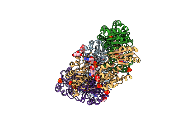
Crystal Structure Of Serine Bound G336V Mutant Of E.Coli Phosphoglycerate Dehydrogenase
Organism: Escherichia coli
Method: X-RAY DIFFRACTION Resolution:2.46 Å Release Date: 2007-04-24 Classification: OXIDOREDUCTASE Ligands: NAI, SER |
Organism: Escherichia coli
Method: X-RAY DIFFRACTION
Release Date: 2007-04-24
Ligands: NAI, SER
 |
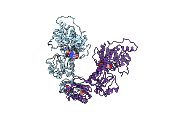
Crystal Structure Of G336V Mutant Of E.Coli Phosphoglycerate Dehydrogenase
Organism: Escherichia coli
Method: X-RAY DIFFRACTION Resolution:2.60 Å Release Date: 2007-04-24 Classification: OXIDOREDUCTASE Ligands: PO4, NAI, CIT, SO4 |
Organism: Escherichia coli
Method: X-RAY DIFFRACTION
Release Date: 2007-04-24
Ligands: PO4, NAI, CIT, SO4
 |
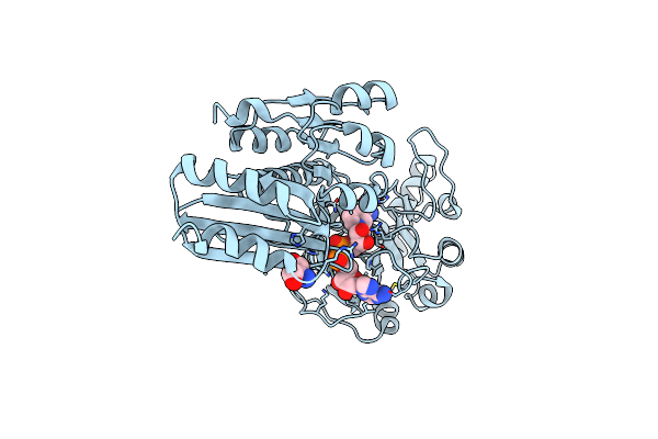
Crystal Structure Of Serine Bound G336V,G337V Double Mutant Of E.Coli Phosphoglycerate Dehydrogenase
Organism: Escherichia coli
Method: X-RAY DIFFRACTION Resolution:2.80 Å Release Date: 2007-04-24 Classification: OXIDOREDUCTASE Ligands: NAI, SER |
Organism: Escherichia coli
Method: X-RAY DIFFRACTION
Release Date: 2007-04-24
Ligands: NAI, SER
 |
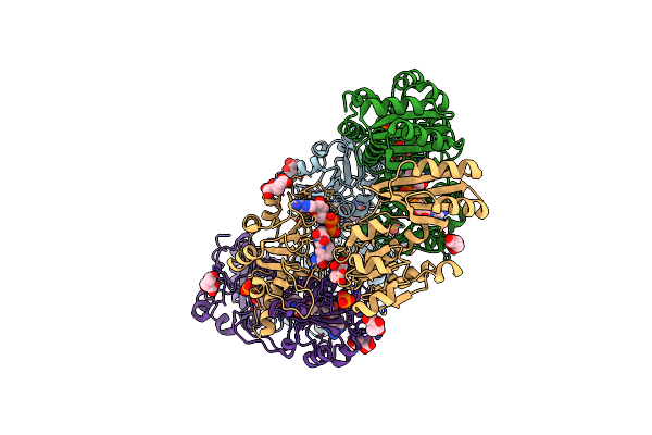
Crystal Structure Of Serine Bound G336V Mutant Of E.Coli Phosphoglycerate Dehydrogenase
Organism: Escherichia coli
Method: X-RAY DIFFRACTION Resolution:2.74 Å Release Date: 2007-04-24 Classification: OXIDOREDUCTASE Ligands: SER, NAI |
Organism: Escherichia coli
Method: X-RAY DIFFRACTION
Release Date: 2007-04-24
Ligands: SER, NAI
 |
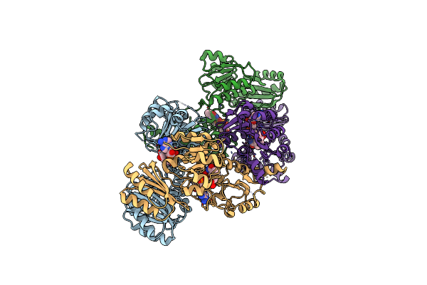
The Active Form Of Phosphoglycerate Dehydrogenase
Organism: Escherichia coli
Method: X-RAY DIFFRACTION Resolution:2.24 Å Release Date: 2005-04-26 Classification: OXIDOREDUCTASE Ligands: PO4, AKG, UNL, NAD |
Organism: Escherichia coli
Method: X-RAY DIFFRACTION
Release Date: 2005-04-26
Ligands: PO4, AKG, UNL, NAD
 |
Crystal Structure Of W139G D-3-Phosphoglycerate Dehydrogenase Complexed With Nad+
Organism: Escherichia coli
Method: X-RAY DIFFRACTION Resolution:2.09 Å Release Date: 2005-02-22 Classification: OXIDOREDUCTASE Ligands: NAD |
Organism: Escherichia coli
Method: X-RAY DIFFRACTION
Release Date: 2005-02-22
Ligands: NAD
 |
Crystal Structure Of D-3-Phosphoglycerate Dehydrogenase From Mycobacterium Tuberculosis
Organism: Mycobacterium tuberculosis
Method: X-RAY DIFFRACTION Resolution:2.30 Å Release Date: 2005-01-25 Classification: OXIDOREDUCTASE Ligands: TLA |
Organism: Mycobacterium tuberculosis
Method: X-RAY DIFFRACTION
Release Date: 2005-01-25
Ligands: TLA
 |
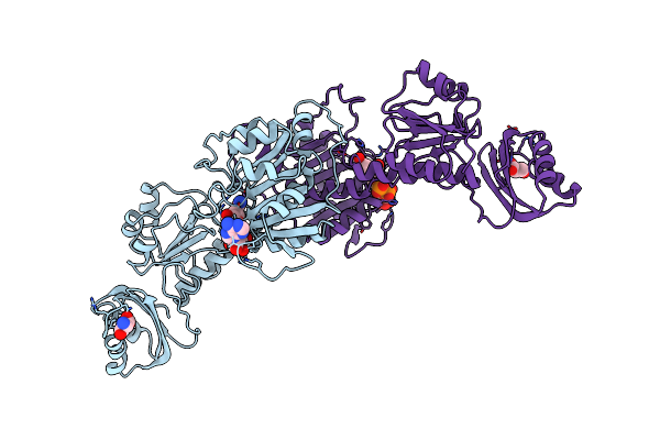
Long Fatty Acid Chain Enoyl-Acp Reductase (Inha) In Complex With An Isonicotinic-Acyl-Nadh Inhibitor
Organism: Mycobacterium tuberculosis
Method: X-RAY DIFFRACTION Resolution:2.70 Å Release Date: 1998-03-25 Classification: OXIDOREDUCTASE Ligands: ZID |
Organism: Mycobacterium tuberculosis
Method: X-RAY DIFFRACTION
Release Date: 1998-03-25
Ligands: ZID
 |
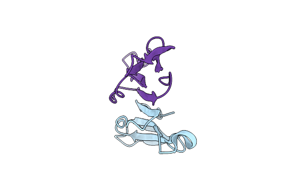
The Allosteric Ligand Site In The Vmax-Type Cooperative Enzyme Phosphoglycerate Dehydrogenase
Organism: Escherichia coli
Method: X-RAY DIFFRACTION Resolution:2.75 Å Release Date: 1995-07-31 Classification: OXIDOREDUCTASE (NAD(A)) Ligands: NAD, SER |
Organism: Escherichia coli
Method: X-RAY DIFFRACTION
Release Date: 1995-07-31
Ligands: NAD, SER
 |
Crystal Structure Of Kappa-Bungarotoxin At 2.3-Angstrom Resolution
Organism: Bungarus multicinctus
Method: X-RAY DIFFRACTION Resolution:2.30 Å Release Date: 1994-12-20 Classification: TOXIN |
Organism: Bungarus multicinctus
Method: X-RAY DIFFRACTION
Release Date: 1994-12-20