

Search Count: 18
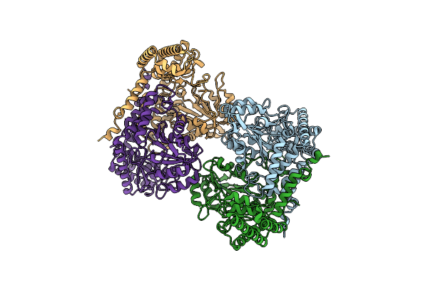 |
Cryo-Em Structure Of Human Shmt1
Organism: Homo sapiens
Method: ELECTRON MICROSCOPY Release Date: 2024-07-24 Classification: TRANSFERASE |
Organism: Homo sapiens
Method: ELECTRON MICROSCOPY
Release Date: 2024-07-24
 |
Human Pyridoxine-5'-Phosphate Oxidase In Complex With Plp
Organism: Homo sapiens
Method: X-RAY DIFFRACTION Resolution:1.69 Å Release Date: 2024-02-07 Classification: OXIDOREDUCTASE Ligands: BME, FMN, PLP, NA |
Organism: Homo sapiens
Method: X-RAY DIFFRACTION
Release Date: 2024-02-07
Ligands: BME, FMN, PLP, NA
 |
Human Pyridoxine-5'-Phosphate Oxidase Mutant R225H
Organism: Homo sapiens
Method: X-RAY DIFFRACTION Resolution:2.75 Å Release Date: 2024-02-07 Classification: OXIDOREDUCTASE Ligands: PO4, FMN |
Organism: Homo sapiens
Method: X-RAY DIFFRACTION
Release Date: 2024-02-07
Ligands: PO4, FMN
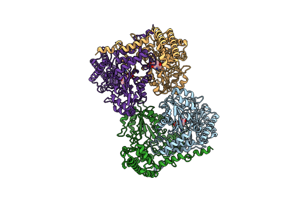 |
Cryo-Em Structure Of The Human Shmt1-Rna Complex
Organism: Homo sapiens
Method: ELECTRON MICROSCOPY Release Date: 2023-06-14 Classification: TRANSFERASE Ligands: PLP |
Organism: Homo sapiens
Method: ELECTRON MICROSCOPY
Release Date: 2023-06-14
Ligands: PLP
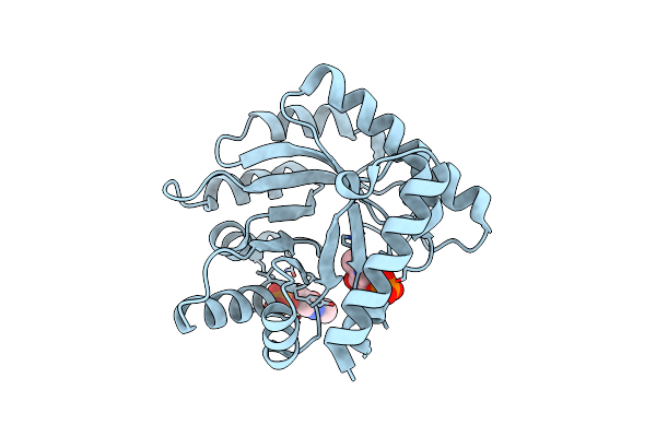 |
The Crystal Structure Of The K36A/K38A/K233A/K234A Quadruple Mutant Of E. Coli Yggs In Complex With Plp
Organism: Escherichia coli
Method: X-RAY DIFFRACTION Resolution:2.40 Å Release Date: 2022-03-30 Classification: PROTEIN TRANSPORT Ligands: PLP |
Organism: Escherichia coli
Method: X-RAY DIFFRACTION
Release Date: 2022-03-30
Ligands: PLP
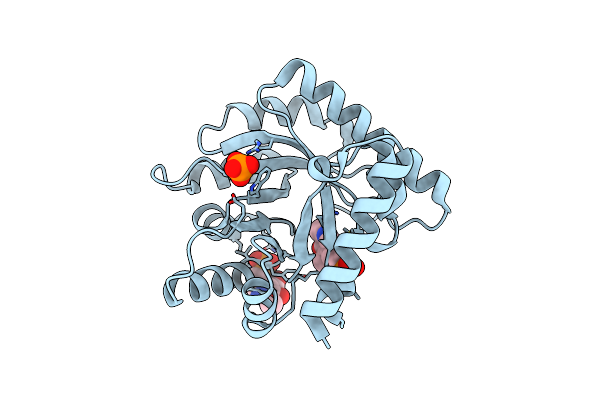 |
Crystal Structure Of The Wild Type Escherichia Coli Pyridoxal 5'-Phosphate Homeostasis Protein (Yggs)
Organism: Escherichia coli
Method: X-RAY DIFFRACTION Resolution:2.10 Å Release Date: 2022-03-23 Classification: PROTEIN TRANSPORT Ligands: PLP, PO4 |
Organism: Escherichia coli
Method: X-RAY DIFFRACTION
Release Date: 2022-03-23
Ligands: PLP, PO4
 |
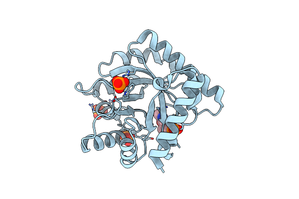
Crystal Structure Of Escherichia Coli Apo Pyridoxal 5'-Phosphate Homeostasis Protein (Yggs)
Organism: Escherichia coli
Method: X-RAY DIFFRACTION Resolution:2.00 Å Release Date: 2022-03-23 Classification: PROTEIN TRANSPORT Ligands: SO4 |
Organism: Escherichia coli
Method: X-RAY DIFFRACTION
Release Date: 2022-03-23
Ligands: SO4
 |
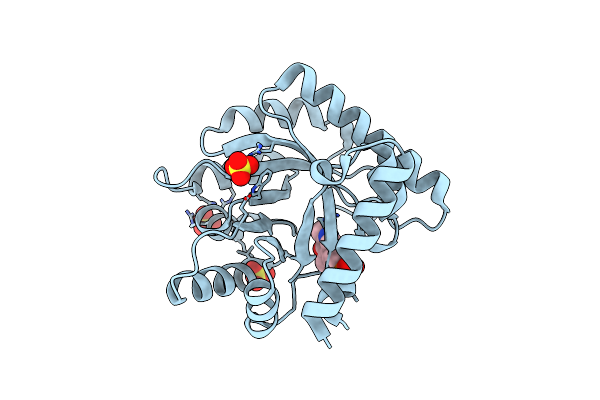
The Crystal Structure Of The K36A Mutant Of E. Coli Yggs In Complex With Plp
Organism: Escherichia coli
Method: X-RAY DIFFRACTION Resolution:2.00 Å Release Date: 2022-03-23 Classification: PROTEIN TRANSPORT Ligands: PLP, PO4 |
Organism: Escherichia coli
Method: X-RAY DIFFRACTION
Release Date: 2022-03-23
Ligands: PLP, PO4
 |
The Crystal Structure Of The K137A Mutant Of E. Coli Yggs In Complex With Plp
Organism: Escherichia coli
Method: X-RAY DIFFRACTION Resolution:2.10 Å Release Date: 2022-03-23 Classification: PROTEIN TRANSPORT Ligands: PLP, SO4 |
Organism: Escherichia coli
Method: X-RAY DIFFRACTION
Release Date: 2022-03-23
Ligands: PLP, SO4
 |
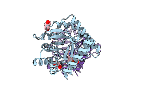
The Crystal Structure Of The K36A/K38A Double Mutant Of E. Coli Yggs In Complex With Plp
Organism: Escherichia coli
Method: X-RAY DIFFRACTION Resolution:2.07 Å Release Date: 2022-03-23 Classification: PROTEIN TRANSPORT Ligands: PO4 |
Organism: Escherichia coli
Method: X-RAY DIFFRACTION
Release Date: 2022-03-23
Ligands: PO4
 |
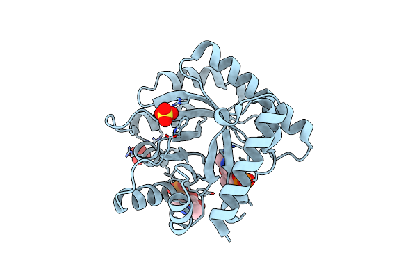
The Crystal Structure Of The K38A/K137A/K233A/K234A Quadruple Mutant Of E. Coli Yggs In Complex With Plp
Organism: Escherichia coli
Method: X-RAY DIFFRACTION Resolution:2.30 Å Release Date: 2022-03-23 Classification: PROTEIN TRANSPORT Ligands: PLP, BU1 |
Organism: Escherichia coli
Method: X-RAY DIFFRACTION
Release Date: 2022-03-23
Ligands: PLP, BU1
 |
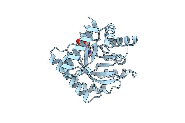
The Crystal Structure Of The K36A/K137A Double Mutant Of E. Coli Yggs In Complex With Plp
Organism: Escherichia coli
Method: X-RAY DIFFRACTION Resolution:2.30 Å Release Date: 2022-03-23 Classification: PROTEIN TRANSPORT Ligands: PLP, SO4 |
Organism: Escherichia coli
Method: X-RAY DIFFRACTION
Release Date: 2022-03-23
Ligands: PLP, SO4
 |
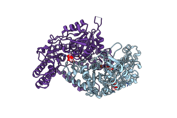
The Crystal Structure Of The Wild-Type Of E. Coli Yggs In Complex With Pnp
Organism: Escherichia coli
Method: X-RAY DIFFRACTION Resolution:2.60 Å Release Date: 2022-03-23 Classification: PROTEIN TRANSPORT Ligands: PXP |
Organism: Escherichia coli
Method: X-RAY DIFFRACTION
Release Date: 2022-03-23
Ligands: PXP
 |
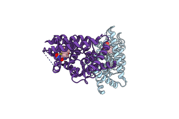
Crystal Structure Of Homo Sapiens Holo Serine Hydroxymethyltransferase 2 (Mitochondrial) (Shmt2), Isoform 3, Transcript Variant 5, 483 Aa, At 2.6 Ang. Resolution
Organism: Homo sapiens
Method: X-RAY DIFFRACTION Resolution:2.60 Å Release Date: 2015-01-28 Classification: TRANSFERASE Ligands: GOL, PEG |
Organism: Homo sapiens
Method: X-RAY DIFFRACTION
Release Date: 2015-01-28
Ligands: GOL, PEG
 |
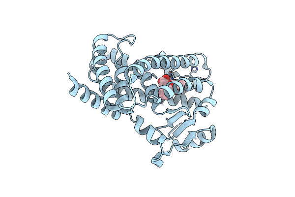
Crystal Structure Of The Complex Between Ppargamma Mutant R357A And Rosiglitazone
Organism: Homo sapiens
Method: X-RAY DIFFRACTION Resolution:2.60 Å Release Date: 2014-07-23 Classification: TRANSCRIPTION/TRANSCRIPTION inhibitor Ligands: BRL |
Organism: Homo sapiens
Method: X-RAY DIFFRACTION
Release Date: 2014-07-23
Ligands: BRL
 |
Structure Of The Complex Between The F360L Ppargamma Mutant And The Ligand Lt175 (Space Group I222)
Organism: Homo sapiens
Method: X-RAY DIFFRACTION Resolution:2.38 Å Release Date: 2014-06-25 Classification: TRANSCRIPTION/TRANSCRIPTION inhibitor Ligands: LRG |
Organism: Homo sapiens
Method: X-RAY DIFFRACTION
Release Date: 2014-06-25
Ligands: LRG
 |
Crystal Structure Of The Complex Of F360L Ppargamma Mutant With The Ligand Lt175
Organism: Homo sapiens
Method: X-RAY DIFFRACTION Resolution:2.28 Å Release Date: 2014-06-25 Classification: TRANSCRIPTION/TRANSCRIPTION inhibitor Ligands: LRG |
Organism: Homo sapiens
Method: X-RAY DIFFRACTION
Release Date: 2014-06-25
Ligands: LRG
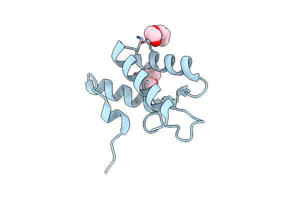 |
Crystal Structure Of The Periplasmic Domain Of Ccmh From Pseudomonas Aeruginosa
Organism: Pseudomonas aeruginosa
Method: X-RAY DIFFRACTION Resolution:1.70 Å Release Date: 2007-07-10 Classification: OXIDOREDUCTASE Ligands: PG4 |
Organism: Pseudomonas aeruginosa
Method: X-RAY DIFFRACTION
Release Date: 2007-07-10
Ligands: PG4