

Search Count: 42
 |
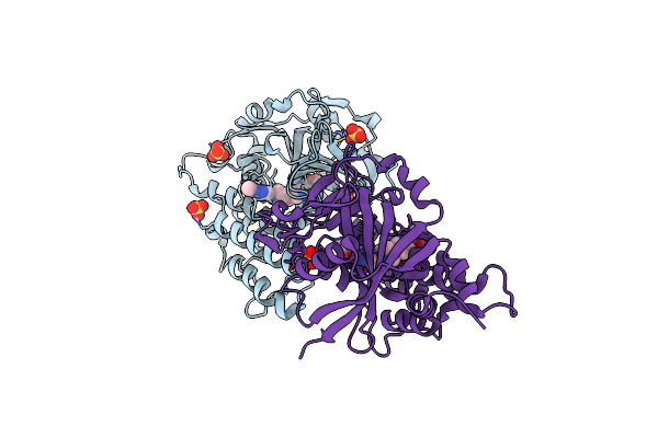
Crystal Structure Of The Catalytic Domain From The Bont-Like Toxin Complex Pg1 Of Paeniclostridium Ghonii
Organism: Paraclostridium ghonii
Method: X-RAY DIFFRACTION Resolution:1.80 Å Release Date: 2025-10-15 Classification: TOXIN Ligands: ZN, SO4, ACT, GOL |
Organism: Paraclostridium ghonii
Method: X-RAY DIFFRACTION
Release Date: 2025-10-15
Ligands: ZN, SO4, ACT, GOL
 |
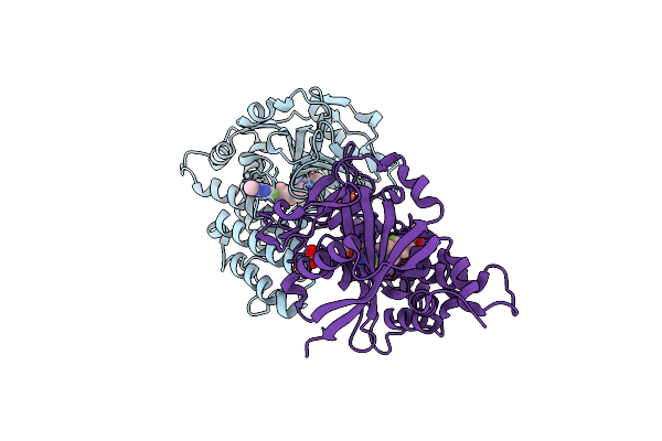
Crystal Structure Of Parp1 Catalytic Domain Bound To Azd5305 (Saruparib)
Organism: Homo sapiens
Method: X-RAY DIFFRACTION Resolution:1.59 Å Release Date: 2024-12-04 Classification: TRANSFERASE Ligands: A1H63, SO4 |
Organism: Homo sapiens
Method: X-RAY DIFFRACTION
Release Date: 2024-12-04
Ligands: A1H63, SO4
 |
Crystal Structure Of Parp1 Catalytic Domain Bound To Azd9574
Organism: Homo sapiens
Method: X-RAY DIFFRACTION Resolution:1.82 Å Release Date: 2024-12-04 Classification: TRANSFERASE Ligands: A1H64, SO4 |
Organism: Homo sapiens
Method: X-RAY DIFFRACTION
Release Date: 2024-12-04
Ligands: A1H64, SO4
 |
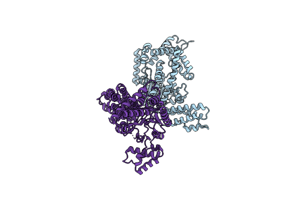
Crystal Structure Of Chelona Toxin, A Diphtheria Toxin Homolog, From Austwickia Chelonae
Organism: Austwickia chelonae
Method: X-RAY DIFFRACTION Resolution:2.50 Å Release Date: 2024-08-07 Classification: TOXIN Ligands: CA |
Organism: Austwickia chelonae
Method: X-RAY DIFFRACTION
Release Date: 2024-08-07
Ligands: CA
 |
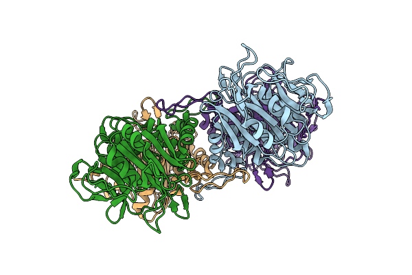
Crystal Structure Of Glucosyltransferase (Gtd) Domain Of Tpel
Organism: Clostridium perfringens
Method: X-RAY DIFFRACTION Resolution:2.22 Å Release Date: 2024-08-07 Classification: TOXIN |
Organism: Clostridium perfringens
Method: X-RAY DIFFRACTION
Release Date: 2024-08-07
 |
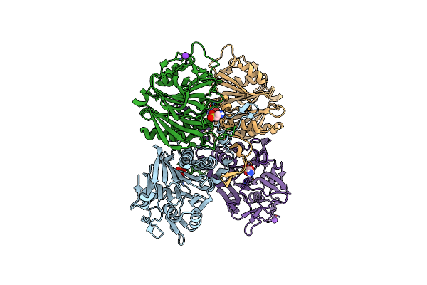
Bile Salt Hydrolase A From Lactobacillus Acidophilus
Organism: Lactobacillus acidophilus
Method: X-RAY DIFFRACTION Resolution:2.15 Å Release Date: 2023-01-25 Classification: HYDROLASE |
Organism: Lactobacillus acidophilus
Method: X-RAY DIFFRACTION
Release Date: 2023-01-25
 |
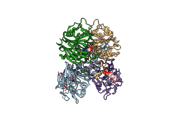
Bile Salt Hydrolase A From Lactobacillus Gasseri With Taurine Bound
Organism: Lactobacillus gasseri
Method: X-RAY DIFFRACTION Resolution:2.05 Å Release Date: 2023-01-25 Classification: HYDROLASE Ligands: TAU, K |
Organism: Lactobacillus gasseri
Method: X-RAY DIFFRACTION
Release Date: 2023-01-25
Ligands: TAU, K
 |
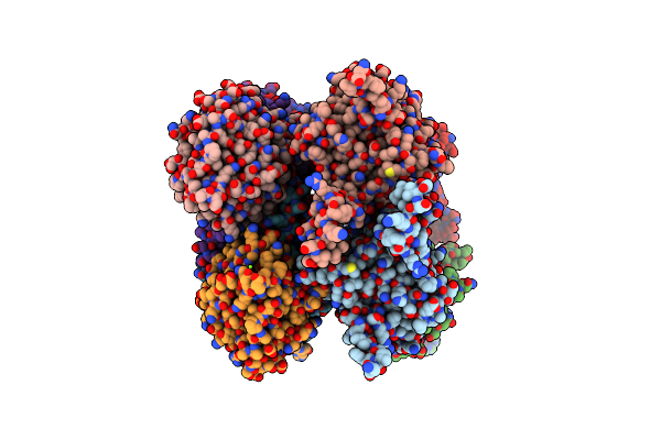
Bile Salt Hydrolase A From Lactobacillus Gasseri With Chenodeoxycholate And Taurine Bound
Organism: Lactobacillus gasseri
Method: X-RAY DIFFRACTION Resolution:1.35 Å Release Date: 2023-01-25 Classification: HYDROLASE Ligands: TAU, JN3 |
Organism: Lactobacillus gasseri
Method: X-RAY DIFFRACTION
Release Date: 2023-01-25
Ligands: TAU, JN3
 |
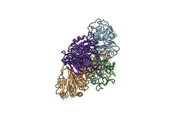
Bile Salt Hydrolase B From Lactobacillus Gasseri
Organism: Lactobacillus gasseri
Method: X-RAY DIFFRACTION Resolution:1.56 Å Release Date: 2023-01-25 Classification: HYDROLASE Ligands: MG |
Organism: Lactobacillus gasseri
Method: X-RAY DIFFRACTION
Release Date: 2023-01-25
Ligands: MG
 |
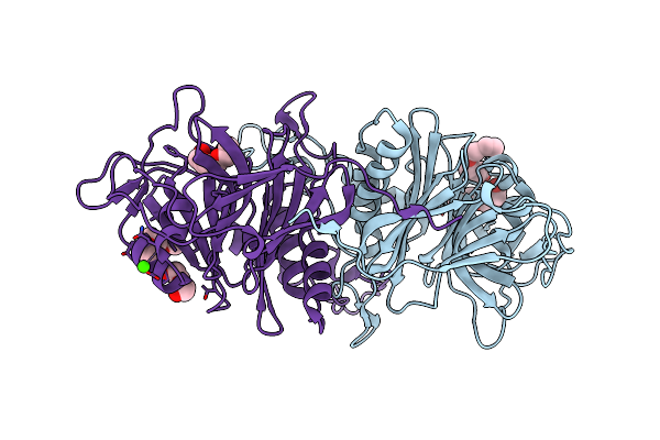
Bile Salt Hydrolase C From Lactobacillus Johnsonii
Organism: Lactobacillus johnsonii
Method: X-RAY DIFFRACTION Resolution:1.45 Å Release Date: 2023-01-25 Classification: HYDROLASE |
Organism: Lactobacillus johnsonii
Method: X-RAY DIFFRACTION
Release Date: 2023-01-25
 |
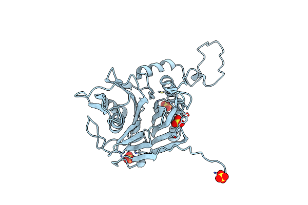
Bile Salt Hydrolase From Lactobacillus Ingluviei
Organism: Lactobacillus ingluviei
Method: X-RAY DIFFRACTION Resolution:1.43 Å Release Date: 2023-01-25 Classification: HYDROLASE Ligands: PG4, PEG, PGE, CA |
Organism: Lactobacillus ingluviei
Method: X-RAY DIFFRACTION
Release Date: 2023-01-25
Ligands: PG4, PEG, PGE, CA
 |
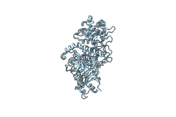
Bile Salt Hydrolase From Lactobacillus Reuteri
Organism: Lactobacillus reuteri
Method: X-RAY DIFFRACTION Resolution:1.66 Å Release Date: 2023-01-25 Classification: HYDROLASE Ligands: SO4 |
Organism: Lactobacillus reuteri
Method: X-RAY DIFFRACTION
Release Date: 2023-01-25
Ligands: SO4
 |
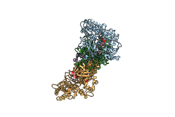
Crystal Structure Of Peptono Toxin, A Diphtheria Toxin Homolog, From Seinonella Peptonophila
Organism: Seinonella peptonophila
Method: X-RAY DIFFRACTION Resolution:2.19 Å Release Date: 2022-04-20 Classification: TOXIN Ligands: IOD |
Organism: Seinonella peptonophila
Method: X-RAY DIFFRACTION
Release Date: 2022-04-20
Ligands: IOD
 |
Crystal Structure Of Albireti Toxin, A Diphtheria Toxin Homolog, From Streptomyces Albireticuli
Organism: Streptomyces albireticuli
Method: X-RAY DIFFRACTION Resolution:2.69 Å Release Date: 2022-04-20 Classification: TOXIN Ligands: PG4, ACT, CA |
Organism: Streptomyces albireticuli
Method: X-RAY DIFFRACTION
Release Date: 2022-04-20
Ligands: PG4, ACT, CA
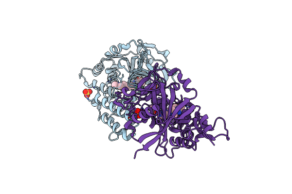 |
Parp1 Catalytic Domain In Complex With 8-Chloroquinazolinone-Based Inhibitor (Compound 9)
Organism: Homo sapiens
Method: X-RAY DIFFRACTION Resolution:2.05 Å Release Date: 2021-09-15 Classification: TRANSFERASE Ligands: VKW, SO4 |
Organism: Homo sapiens
Method: X-RAY DIFFRACTION
Release Date: 2021-09-15
Ligands: VKW, SO4
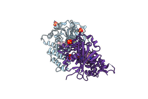 |
Parp1 Catalytic Domain In Complex With Isoquinolone-Based Inhibitor (Compound 16)
Organism: Homo sapiens
Method: X-RAY DIFFRACTION Resolution:1.97 Å Release Date: 2021-09-15 Classification: TRANSFERASE Ligands: VKT, SO4 |
Organism: Homo sapiens
Method: X-RAY DIFFRACTION
Release Date: 2021-09-15
Ligands: VKT, SO4
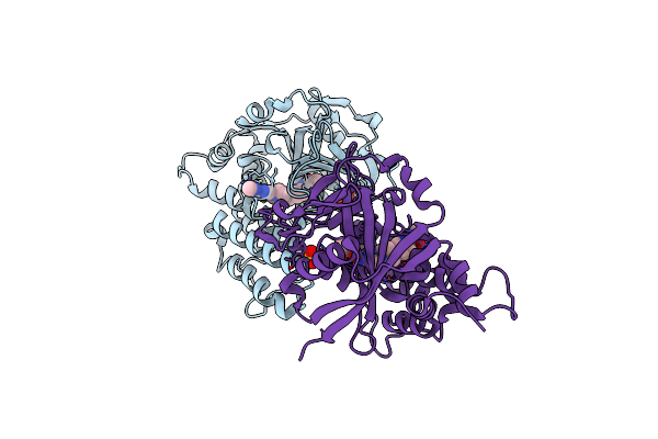 |
Parp1 Catalytic Domain In Complex With A Selective Pyridine Carboxamide-Based Inhibitor (Compound 22)
Organism: Homo sapiens
Method: X-RAY DIFFRACTION Resolution:1.85 Å Release Date: 2021-09-15 Classification: TRANSFERASE Ligands: VKQ, SO4 |
Organism: Homo sapiens
Method: X-RAY DIFFRACTION
Release Date: 2021-09-15
Ligands: VKQ, SO4
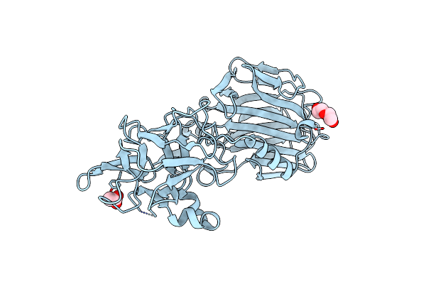 |
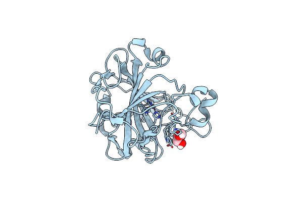
Crystal Structure Of The Binding Domain Of Paraclostridial Mosquitocidal Protein 1
Organism: Paraclostridium bifermentans
Method: X-RAY DIFFRACTION Resolution:1.95 Å Release Date: 2019-07-10 Classification: TOXIN Ligands: PEG, ACT |
Organism: Paraclostridium bifermentans
Method: X-RAY DIFFRACTION
Release Date: 2019-07-10
Ligands: PEG, ACT
 |
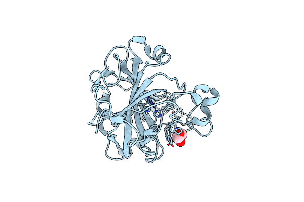
The Structural Implications Of The Secondary Co2 Binding Pocket In Human Carbonic Anhydrase Ii
Organism: Homo sapiens
Method: X-RAY DIFFRACTION Resolution:2.05 Å Release Date: 2014-04-30 Classification: LYASE Ligands: ZN, GOL |
Organism: Homo sapiens
Method: X-RAY DIFFRACTION
Release Date: 2014-04-30
Ligands: ZN, GOL
 |
The Structural Implications Of The Secondary Co2 Binding Pocket In Human Carbonic Anhydrase Ii
Organism: Homo sapiens
Method: X-RAY DIFFRACTION Resolution:1.63 Å Release Date: 2014-04-30 Classification: LYASE Ligands: ZN, GOL |
Organism: Homo sapiens
Method: X-RAY DIFFRACTION
Release Date: 2014-04-30
Ligands: ZN, GOL